Evidence and Consensus Based Imaging Guidelines in Cytomegalovirus retinitis. Multimodal imaging in Uveitis (MUV) Taskforce Report 15
https://t.co/qJ2tCfpH5o
@aniruddha9#ophthalmology#retina
A 16-year-old girl with visual acuity of 20/20 in the right eye (OD) and 20/32 in the left eye (OS) was referred to assess “anomalous retinal vessels” OS. Multimodal imaging confirmed an arteriovenous malformation (AVM) OS. Magnetic resonance imaging of the brain was within normal limits. A, Color fundus photography is normal OD and shows a retinal AVM of the central macula OS. B, Ultra-widefield fluorescein angiography confirms direct artery to venous filling and an associated abnormal capillary bed OS. C, OCT of the macula OS shows the hyperreflective vessels without fluid or exudation. D, En face OCT of the outer retina OS precisely highlights the AVM.
https://t.co/77ws1sbIah
#ophthalmology #retina
New Ophthalmology Journal Podcast! @DrewCareyMD interviews Dr. Avner Hostovsky on his study evaluating the diagnostic yield of a structured systemic workup in patients presenting with acute visual symptoms who were diagnosed with isolated paracentral acute middle maculopathy.
Listen here:
https://t.co/MwDvDTMOMw
#ophthalmology #ophthalmologyjournal #podcast
Disease and Participant-Related Correlates of Genetic Testing Completion for Hereditary Eye Disorders in a Cohort of Over 1400 Patients
https://t.co/cGn7AWivC6
#ophthalmology
Macular Neovascularization in Neovascular AMD: A Systematic Review of OCTA-Based Assessments and Challenges in Standardization
https://t.co/zbgq2yDIwY
#ophthalmology#retina
A 9-year-old girl presented with sudden painless visual loss (20/40) after a febrile illness. Fundus images show multiple discrete, round whitish retinal lesions in both eyes, more numerous in the right eye (A), involving the posterior-pole and midperiphery. The left eye shows fewer, smaller lesions predominantly in the midperiphery (B). OCT demonstrates hyperreflective inner retinal lesions with extension into the vitreous and surrounding vitritis (C). Lasiodiplodia theobromae, a plant-associated filamentous dematiaceous fungus, was identified on panfungal polymerase chain reaction sequencing of the vitreous sample. The bilateral multifocal pattern possibly reflects hematogenous spread after recent systemic infection, with significant resolution after intravitreal voriconazole, systemic amphotericin-B, and fluconazole (D).
https://t.co/1BibCKWh4E
#ophthalmology #retina
Anatomical and Functional Outcomes After Pars Plana Vitrectomy for Giant Retinal Tear–Related Detachment: The Manchester GRT Study
https://t.co/FJoOJlWsH8
#ophthalmology#retina
A 43-year-old woman presented in 2019 with photopsias and a visual field index (VFI) of 60%. Fundus autofluorescence and OCT (A) revealed hyper-autofluorescence and ellipsoid zone (EZ) loss in the nasal, superior, and inferior parafoveal regions; the temporal sector was spared. Fundus photography remained normal. Six years later, after another viral illness, VFI decreased to 48% despite stable best-corrected visual acuity (20/30). Fundus autofluorescence and OCT (B) showed a new temporal area of involvement. Notably, the previously damaged nasal, superior, and inferior zones all demonstrated partial EZ restoration with some persistence of field loss. Testing for syphilis and human immunodeficiency virus was negative. This case illustrates recurrent acute zonal occult outer retinopathy and the potential for segmental outer retinal repair.
https://t.co/JZyk7wld3U
#ophthalmology #retina
The June issue of Ophthalmology Retina is out now! Featuring articles on:
✅Eyes Achieving Absence of Diabetic Macular Edema and Very Good Vision: A Post Hoc Analysis of DRCR Network Protocol AC
✅Diagnostic Pars Plana Vitrectomy in Undifferentiated Uveitis: Systematic Review and Meta-Analysis
✅Standardizing Measurement of cRORA (Complete Retinal Pigment Epithelial and Outer Retinal Atrophy)- CAM Report 8
✅Global Validation of the Postnatal Growth and Retinopathy of Prematurity Screening Model: A Systematic Review and Meta-analysis
And much more!
https://t.co/HyfC0C1Tua
#ophthalmology #retina
Highly Myopic Macular Hole Surgery by the Internal Limiting Membrane Flap with No Gas Tamponade Technique: A Prospective Interventional Case Series
https://t.co/jemRIWccNF
@simonkhszeto#ophthalmology#retina
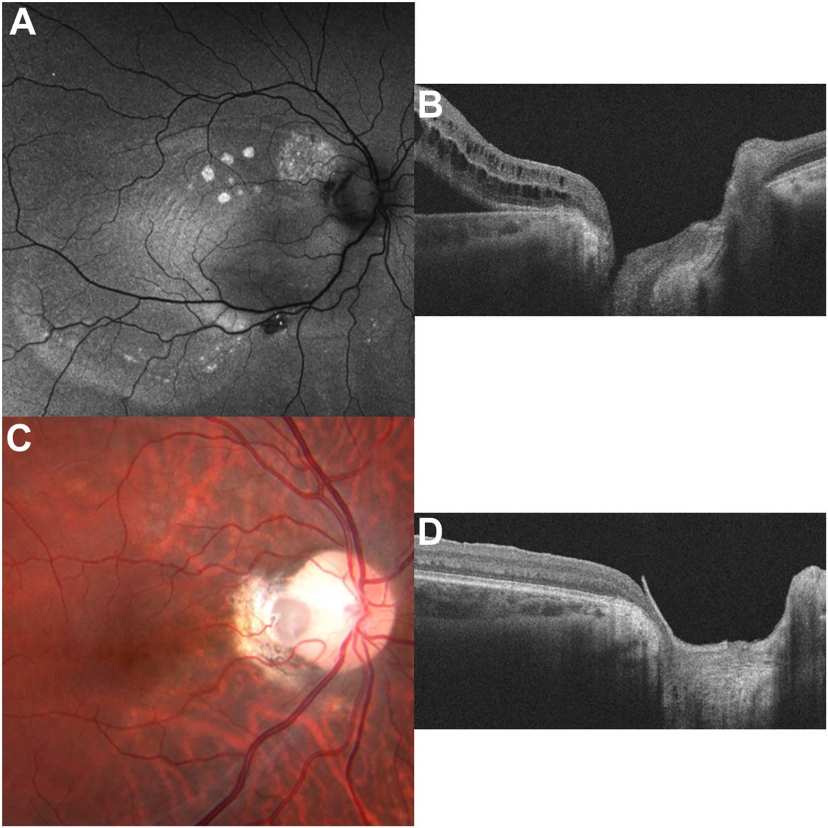
A 19-year-old man presented with decreased vision after 3 prior vitrectomies for optic nerve pit maculopathy at another hospital including such treatments as internal limiting membrane peel, endolaser to optic nerve pit edge, and C3F8 gas use. Best-corrected visual acuity was 20/100. Fundus autofluorescence showed multiple areas of prior and current subretinal fluid (A), and OCT showed optic disc excavation with intraretinal fluid and subretinal fluid (B). He underwent pars plana vitrectomy with amniotic membrane (AMT) graft to cover the pit and SF6 gas. Postoperative imaging showed fluid resolution and good graft position; best-corrected visual acuity improved to 20/50. One-year postoperative OCT confirmed stable AMT position without recurrent fluid (C, D).
https://t.co/H4oS8qQpLP
#ophthalmology #retina
Effects of Time From Diagnosis to Treatment and Baseline Vision on Retinal Vein Occlusion Outcomes in Aflibercept 2 mg Phase 3 Trials
https://t.co/e6XXe2oa2L
#ophthalmology#retina