Have you integrated APOL1genetic testing into your practice?
🔬✨Discover the No-Cost APOL1 Genotyping Program for eligible patients sponsored by Vertex Pharmaceuticals — helping you deliver precision care without added cost.
Learn more today! ⬇️
#GeneticTesting #KidneyHealth #SCM25
ANSWER: A
Histologic evidence of acute tissue injury in AABMR includes 1or more of the following: microvascular inflammation (glomerulitis and/or peritubular capillaritis), intimal or transmural arteritis, and acute TMA and acute tubular injury (in the absence of any other apparent cause)
#pathology #nephx #renal #renalpath
#KidneyQuiz Acute tubular injury, in the absence of any apparent cause, can be included as histologic evidence for acute tissue injury in Active Antibody Mediated Rejection (AABMR). Is this statement true or false?
#nephX#kidneypath#pathX#nephtwitter
#KidneyQuiz Acute tubular injury, in the absence of any apparent cause, can be included as histologic evidence for acute tissue injury in Active Antibody Mediated Rejection (AABMR). Is this statement true or false?
#nephX#kidneypath#pathX#nephtwitter
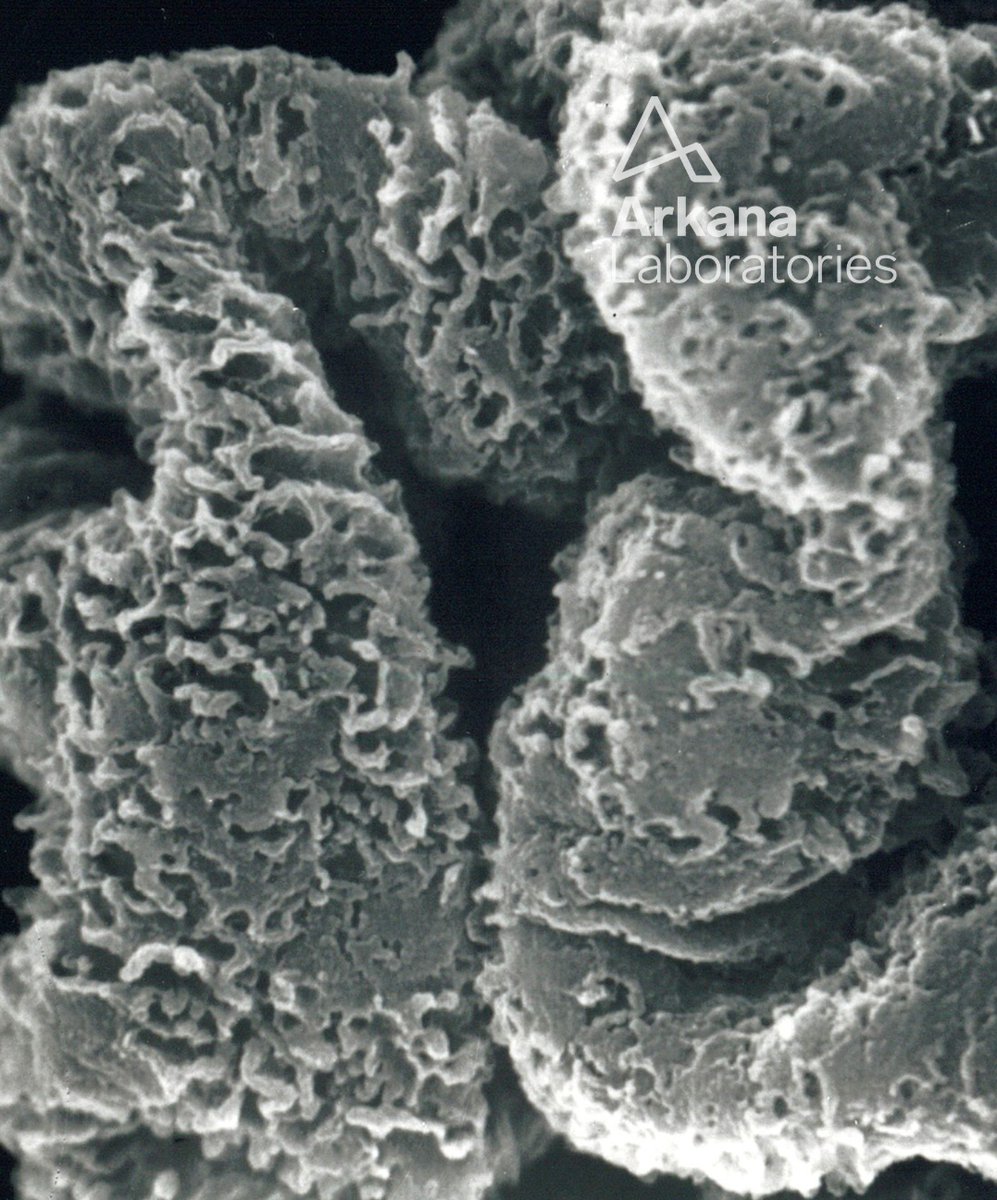
Here is today’s #eyeSCANdy! Acellular scanning EM from a biopsy with membranous glomerulonephritis showing depressions and larger "craters" along the subepithelial aspect of the basement membrane.
Photo courtesy of Dr. Stephen Bonsib. #renal#pathology#kidneypath
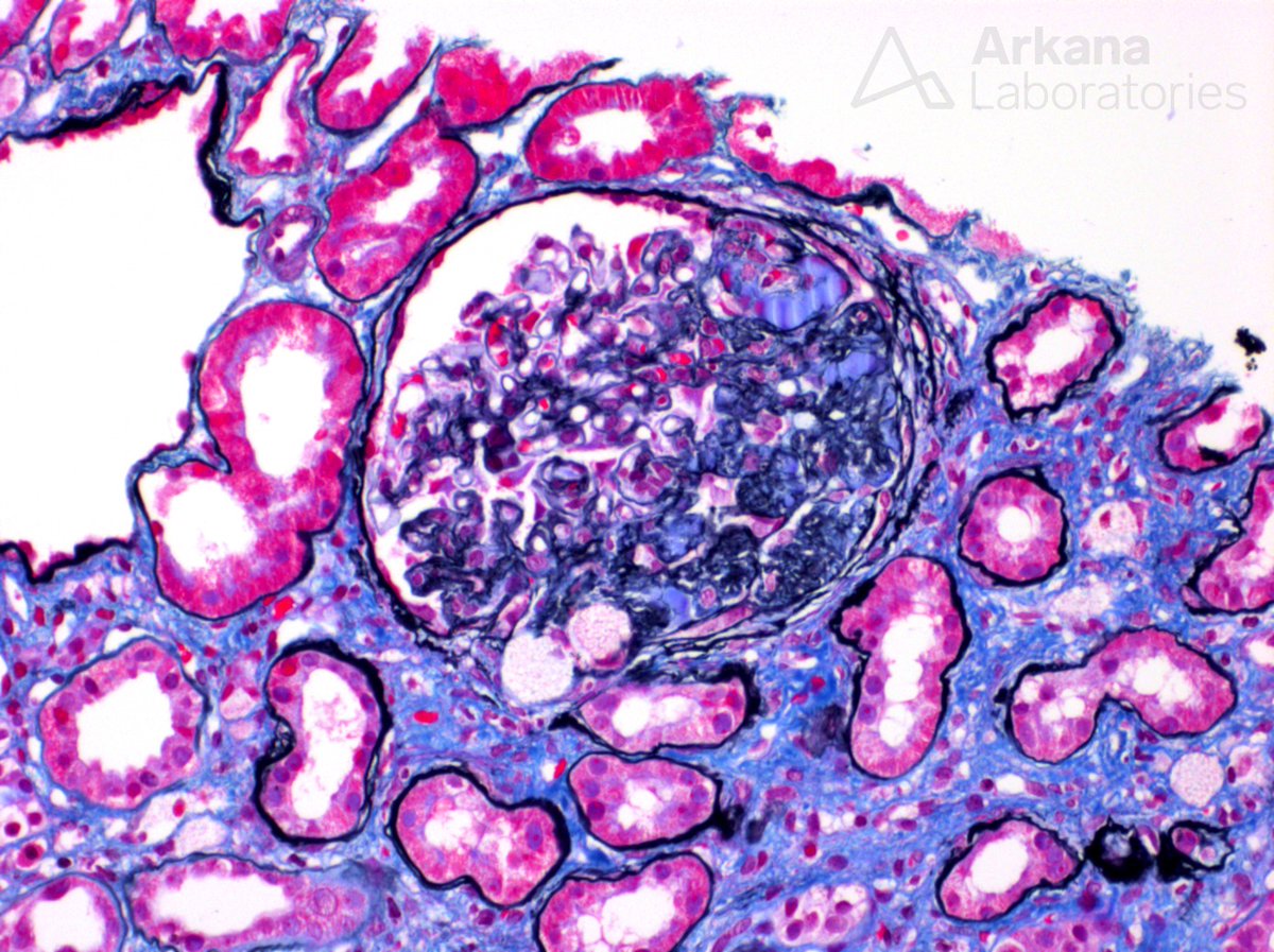
The photomicrograph shows a focal segmental glomerulosclerotic, NOS lesion. In this setting electron microscopy to evaluate epithelial foot processes is imperative and is shown below. The absence of significant foot process effacement makes a primary podocytopathy unlikely and suggests a secondary etiology. Per history, this 32 y/o Hispanic male presents with nephrotic range proteinuria and mildly depressed serum albumin without full nephrotic syndrome. He has no previous, significant medical history; however he has a family history of end-stage kidney disease requiring renal replacement therapy in several family members. This history, in concert with the extensive chronicity seen in the biopsy at the patient’s young age is unusual and raises the possibility of an underlying genetic etiology of focal segmental glomerulosclerosis. Further genetic testing was suggested as was correlation to exclude known causes of secondary focal segmental glomerulosclerosis.
#renalpath #kidneypath #pathology #renal #pathtwitter
Collapsing glomerulopathy is characterized by glomerular tuft collapse with overlying epithelial hypertrophy and hyperplasia in Bowman’s space. This pattern of glomerular injury is most commonly seen in African Americans and has been shown to be strongly associated with the presence of APOL1 risk variants in a number of associated diseases including HIV infection, lupus nephritis, interferon therapy, PLA2R-positive membranous glomerulopathy, and idiopathic cases.
#TeachingPoints #kidneypath #renal #pathology
This year's ASP conference at Arkana was a huge success! We learned so much and loved hearing from all of our presenters. Thank you to everyone who attended, it was great having you. 🔬💙
#arkpaths#pathology
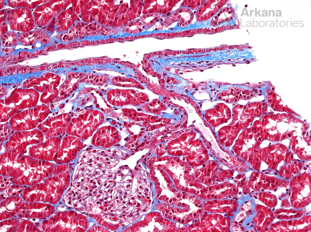
The photomicrograph is a trichrome stain showing normal, back-to-back tubules, wispy unremarkable interstitium, arteries and an arteriole with no abnormality and a normal glomerulus with patent capillary loops and appropriate cellularity. In essence this photomicrograph shows no pathologic abnormalities and just normal renal tissue.
#renalpath #kidneypath #pathology #renal #pathtwitter
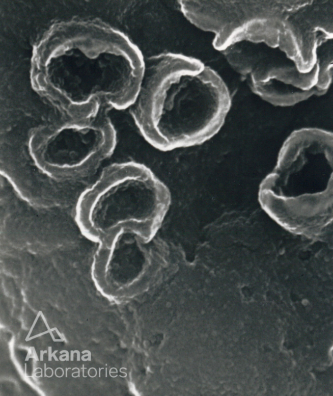
Here is today’s #eyeSCANdy! Acellular scanning EM showing "craters" at high power on the subepithelial aspect of the glomerular basement membranes from a biopsy with membranous glomerulonephritis
Photo courtesy of Dr. Stephen Bonsib. #renal#pathology#kidneypath
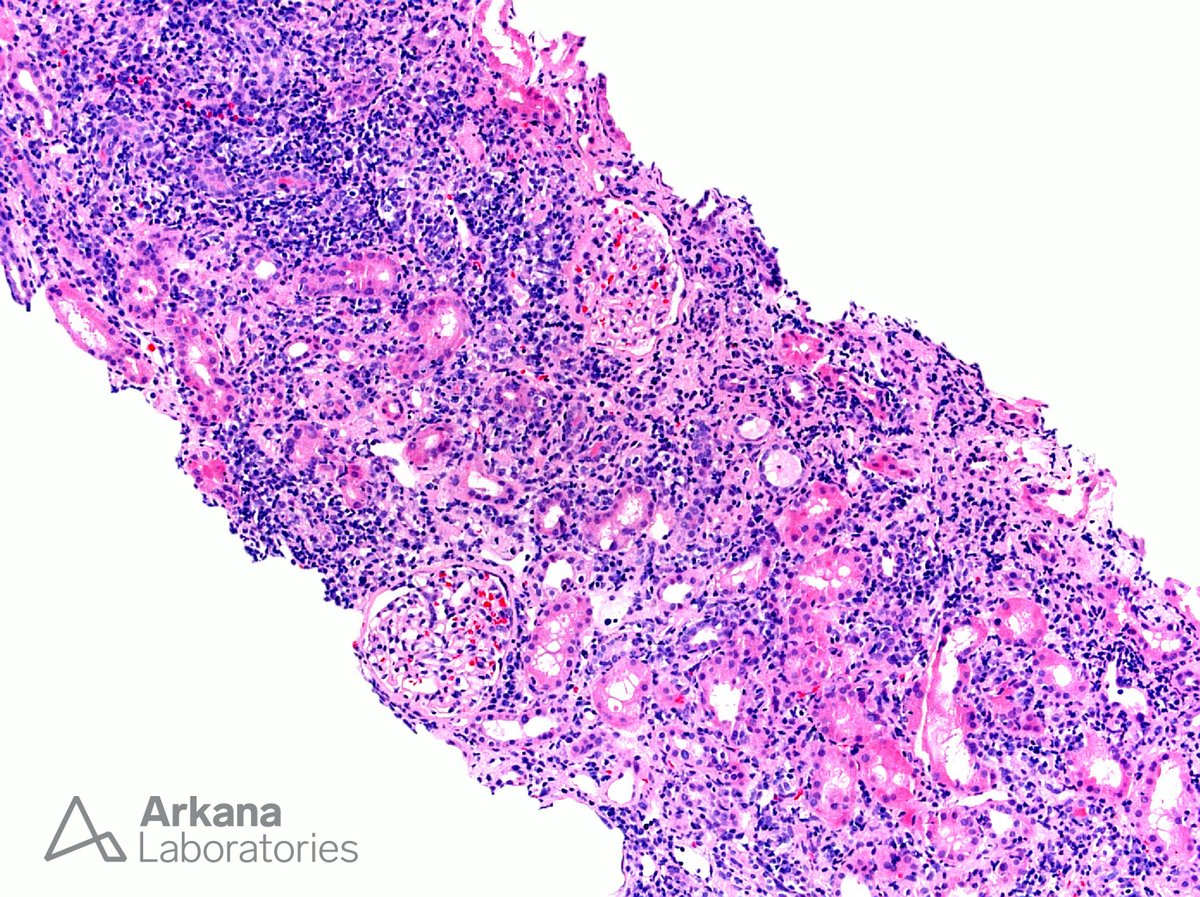
Chronic interstitial nephritis is a diagnosis made on renal biopsy when interstitial inflammation is present in a background of fibrosis (as depicted here). The differential diagnosis is broad but consists primarily of autoimmune-related and drug-induced etiologies. As opposed to acute interstitial nephritis, which is of relatively recent onset, chronic interstitial nephritis results from a long-standing inflammatory process. The distinction between these two morphologic patterns is made based on the appearance of the background interstitium. If the inflammation is located in areas of fibrosis, as is seen in this case of CIN due to Sjögren’s syndrome, the process is designated CIN whereas if the background is edematous it is designated AIN. The composition of the interstitial inflammatory infiltrate can be identical in both AIN and CIN. Specifically, the inflammatory cells in AIN can be primarily mononuclear (lymphocytes and plasma cells) and the inflammatory cells comprising the infiltrate in CIN can be mixed and even include numerous eosinophils.
#TeachingPoints #kidneypath #renal #pathology
The answer is A: Acid alpha-glucosidase
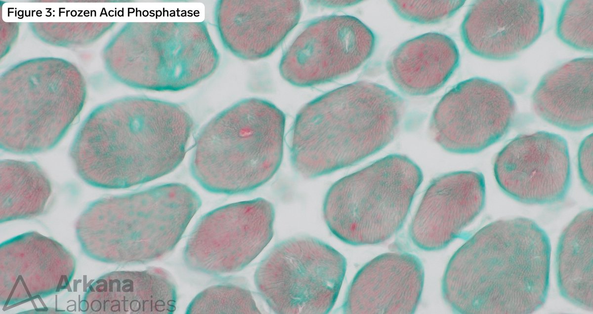
The patient has compound heterozygous mutations in the GAA gene which codes for the acid alpha-glucosidase enzyme. The patient has Pompe disease, aka glycogen storage disease (GSD) type II and is on enzyme replacement therapy and is demonstrating improvement on Hex4 urine testing. The biopsy demonstrates findings of punctate lysosomes on acid phosphatase staining with small PAS positive vacuoles. Ultrastructural evaluation reveals rare, small, membrane bound collections of glycogen. Together the findings are supportive of a treatment response.
Glycogen phosphorylase deficiency can be caused by the PYGL gene for liver or PYGM gene for skeletal muscle. PYGM codes for myophosphorylase and when deficient is the cause for type V GSD, McArdle disease
Glucose-6-phosphate translocase – encoded by the SLC37A4 gene and causes GSD Type Ib when deficient.
https://t.co/aPEvRm1eTQ
Clinical History:
The patient is a young child a year prior to the current biopsy to have compound heterozygous GAA variants. Hex4 levels were initially elevated but since initiating enzyme replacement therapy, the levels have dropped within normal limits.
What enzyme is deficient in this patient’s disease?
A. Acid alpha-glucosidase
B. Glycogen phosphorylase
C. Glucose-6-phosphate translocase
D. Myophosphorylase
#NeuroNotes #neuropath #pathology #neuromuscularpath #PathX #PathTwitter
The answer is A: Acid alpha-glucosidase
The patient has compound heterozygous mutations in the GAA gene which codes for the acid alpha-glucosidase enzyme. The patient has Pompe disease, aka glycogen storage disease (GSD) type II and is on enzyme replacement therapy and is demonstrating improvement on Hex4 urine testing. The biopsy demonstrates findings of punctate lysosomes on acid phosphatase staining with small PAS positive vacuoles. Ultrastructural evaluation reveals rare, small, membrane bound collections of glycogen. Together the findings are supportive of a treatment response.
Glycogen phosphorylase deficiency can be caused by the PYGL gene for liver or PYGM gene for skeletal muscle. PYGM codes for myophosphorylase and when deficient is the cause for type V GSD, McArdle disease
Glucose-6-phosphate translocase – encoded by the SLC37A4 gene and causes GSD Type Ib when deficient.
https://t.co/aPEvRm1eTQ
Clinical History:
The patient is a young child a year prior to the current biopsy to have compound heterozygous GAA variants. Hex4 levels were initially elevated but since initiating enzyme replacement therapy, the levels have dropped within normal limits.
What enzyme is deficient in this patient’s disease?
A. Acid alpha-glucosidase
B. Glycogen phosphorylase
C. Glucose-6-phosphate translocase
D. Myophosphorylase
#NeuroNotes #neuropath #pathology #neuromuscularpath #PathX #PathTwitter
Here is today’s #eyeSCANdy! Acellular scanning EM of a glomerulus with membranous glomerulonephritis, stage II showing diffuse reticular appearance of GBM elaboration.
Photo courtesy of Dr. Stephen Bonsib. #renal#pathology#kidneypath
The photomicrograph shows endocapillary proliferation with exuberant intracapillary neutrophils. And, by immunofluorescence IgG and C3 were noted (see below). This pattern of injury is most commonly found in infection-associated glomerulonephritis which was the case here. Of note, neutrophil rich intracapillary infiltrates, while most commonly associated with infection-associated glomerulonephritis, are not specific and can sometimes be seen in other diseases such as cryoglobulinemic glomerulonephritis, paraprotein-associated glomerulonephritis, and C3 glomerulonephritis as well as others.
#renalpath #kidneypath #pathology #renal #pathtwitter
A PAS section shows an atheroembolus occluding an artery (arrow) with an adjacent ischemic appearing glomerulus. Atheroemboli appear as slit-like spaces in sections cut from formalin fixed paraffin-embedded sections due to the fact that the cholesterol crystals are dissolved by the lipid solvents during processing. However, as demonstrated here, the crystals can be visualized by their birefringence under polarized light in the cryosections for immunofluorescence evaluation. Atheroembolization is often preceded by in an invasive vascular procedure such as coronary artery bypass or aortic aneurysm repair though they can also be precipitated by trauma and are often idiopathic. Systemic manifestations that sometimes accompany atheroembolic disease including transient eosinophilia, skin lesions (livedo reticularis), and hypocomplementemia.
#TeachingPoints #kidneypath #renal #pathology