@Sarah_Vgb, one of our PhD students, did a fantastic job in explaining what contrast-enhanced CT (CE-CT) is all about! Watch the video below to learn the basics of contrast agents and their use in CE-CT 🦴
Wat hebben beenmerg, de lever & een tumor gemeen? Ze bestaan uit zgn 'zacht' #weefsel dat zich niet gemakkelijk via CT-scan laat visualiseren. @Sarah_Vgb@KU_Leuven @UCLouvain_be ontwikkelt nieuwe #contraststoffen om zacht weefsel 'in beeld te brengen' 🎥👉https://t.co/X8aIbSZihU
A new article co-authored by members of the Contrast Team (Maïté Pétré and @GreetKerckhofs) unravelling the link between aortic microstructure and mechanical behaviour using CECT for more flexible modelling
https://t.co/2GXljLU5OD
A new article co-authored by members of the Contrast Team (Tim Balcaen and @GreetKerckhofs) on yet another application for CECT: imaging of biofilm for water treatment. https://t.co/Sz0mNOqtqY
A new review article authored by members of the Contrast Team (Lara Mazy and @GreetKerckhofs) on combined in situ testing and X-ray microfocus computed tomography for biological application.
https://t.co/joQNwvozT5
A new article co-authored by members of the Contrast Team (Delia Hoffmann, Tim Balcaen, Sarah Vangrunderbeeck, Arne Maes, Grzegorz Pyka and @GreetKerckhofs) on 3D histology of Soft Tissues by Contrast-Enhanced X-Ray Microfocus Computed Tomography.
https://t.co/lvqyfwzaQE
A new article co-authored by members of the ContrasTTeam (Pierre Schneidewind, Grzegorz Pyka, and @GreetKerckhofs ) on Unravelling fascia lata intrinsic vascular architecture using microfocus X-ray Computed Tomography.https://t.co/N6S9MDoVtU
A new review article by members of the contrasTTeam (Tim Balcaen, Sarah Vangrunderbeeck, @GreetKerckhofs) on contrast-enhancing staining agents (CESAs) for ex vivo Contrast-Enhanced Computed Tomography. https://t.co/jQxWB5xmtX
Another article, co-authored by a member of the Contrast Team (@GreetKerckhofs), where CECT was used to analyze the vasculature in the bone marrow of murine bones. https://t.co/NoGWg0RjwZ
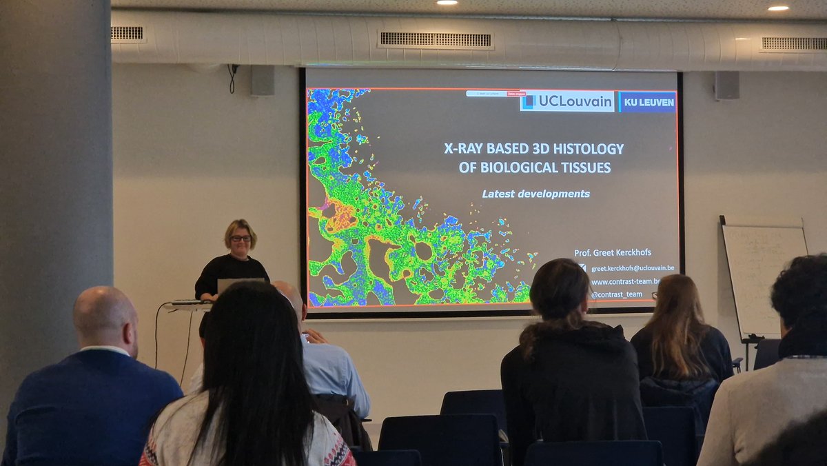
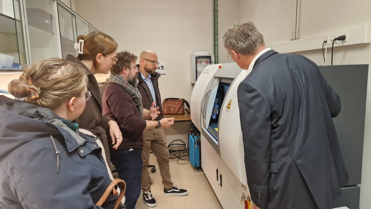
During our annual symposium, fascinating seminars on X-ray based 3D histology of tissues were presented. Various types of biomedical research enabled by the X-ray microfocus computed tomography (microCT) machine were showcased. The event concluded with a visit of our lab.
Another publication with co-authors from the Contrast Team exploring the biological effects of copper alloying in Zn-based biodegradable arterial implants. https://t.co/jywYiqKaqO
Another publication from the Contrast Team (Lisa Leyssens, Grzegorz Pyka, @GreetKerckhofs ), Exploring the biodegradability of metallic intravascular stent materials using microCT. https://t.co/9St5P1tpm2
Another publication, co-authored by members of the Contrast Team (Maïté Pétré, Tim Balcaen, Pierre Schneidewind, Lara Mazy, Grzegorz Pyka,@GreetKerckhofs), assessing the effect on microstructural and mechanical properties of six different staining agents. https://t.co/VKpDRHj4xK
A new publication from the ContrasT Team (Tim Balcaen, @GreetKerckhofs ), where several contrast-enhancing staining agents have been explored for studying adipose tissue through contrast-enhanced computed tomography.https://t.co/aGgPxdLpqR
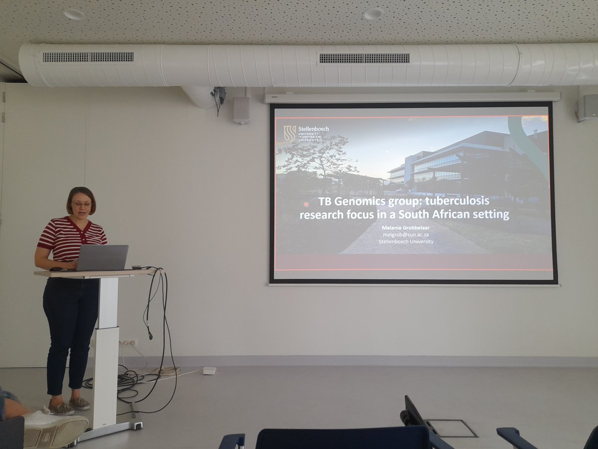
This morning, Dr. Melanie Grobbelaar presented a nice seminar on tuberculosis research focused on a South African setting, which is conducted within the Tuberculosis Genomics (TB Genomics) research group.
And yet another publication with co-authors from the ContrasT Team (@GreetKerckhofs , Arne Maes) showing how X-ray-based 3D histology on human biopsies could reveal blood vessel ingrowth in the donor fascia.
https://t.co/BGhFYJJ0QL
Another publication, co-authored by members of the Contrast team (@GreetKerckhofs, Grzegorz Pyka), focusing on the use of micro-CT for imaging large ex-vivo human samples in anatomical and forensic research : https://t.co/lP1VN52Znn
Another publication featuring a co-author from the ContrasT Team (@GreetKerckhofs ) on the use of CECT imaging to measure cranial development 🎉 https://t.co/RswLhfMDTv
Another publication featuring a co-author from the ContrasT Team (@GreetKerckhofs ) on the use of the staining Hf-WD POM to visualize bone marrow vasculature with Contrast-Enhanced Computer Tomography. 🥳https://t.co/81BDRlxodw
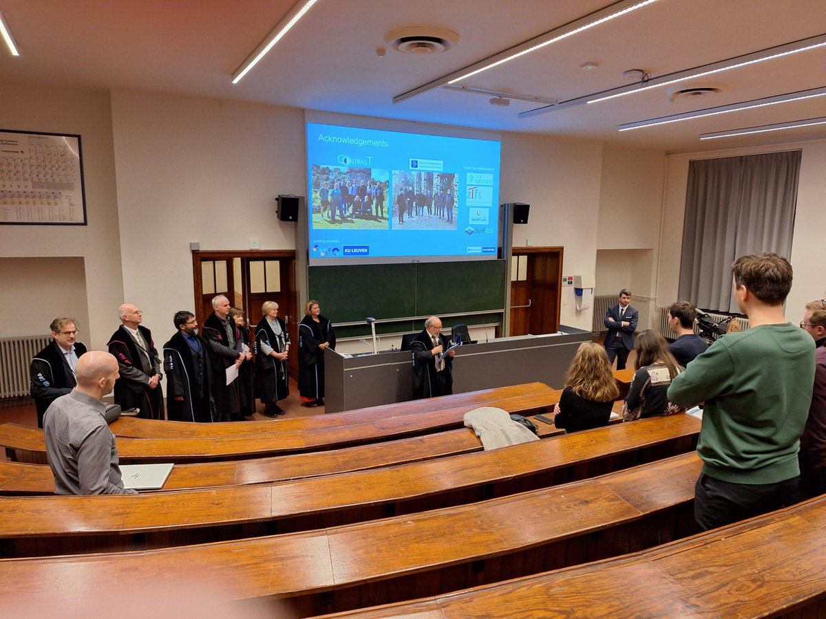
Yesterday, Arne Maes successfully defended his PhD thesis, introducing a novel technique called cryogenic contrast-enhanced microCT,
Congratulations, Dr. Maes!