🔬This week’s theme: Placental #Pathology
➡️Day 2: What’s your #DailyDx of these placental finding?
➡️What clinical scenario(s) might you expect to see this?
➡️What gross findings might you see in the placenta in association with this finding?
#PlacentalPath#UMichPath
Dividing membrane shows a Diamniotic Dichorionic Twin Placenta (Di-Di Placenta). Two chorions plastered together. It makes the likelihood of dizygous twinning (fraternal) much more probable than monozigous (identical).
A, B, & C are evidence of maternal vascular malperfusion
Evidence of fetal vascular malperfusion include thombi & stem vessel obliteration (large vessels) as well as avascular villi & villous stromal-vascular karyorrhexis (distal vessels)
#PlacentaPath #Placenta #PediPath #PathTwitter #PathResidents
📽️🙏Dr. Drucilla Roberts https://t.co/pHIH9JrLvu
✅D
Which of the following is evidence of fetal vascular malperfusion?
A) Decidual arteriopathy
B) Distal villous hypoplasia
C) Hypertensive infarct
D) Villous stromal-vascular karyorrhexis
#PlacentaPath#Placenta#PediPath#PathTwitter#PathResidents
🔬 This week’s theme: Gynecologic #Pathology
➡️ Day 1: What’s your #DailyDx of this ovary? Similar findings were present in both ovaries.
➡️ What could be associated with this finding?
➡️ We’ll tweet the answers & some quick facts tomorrow!
#GYNPath
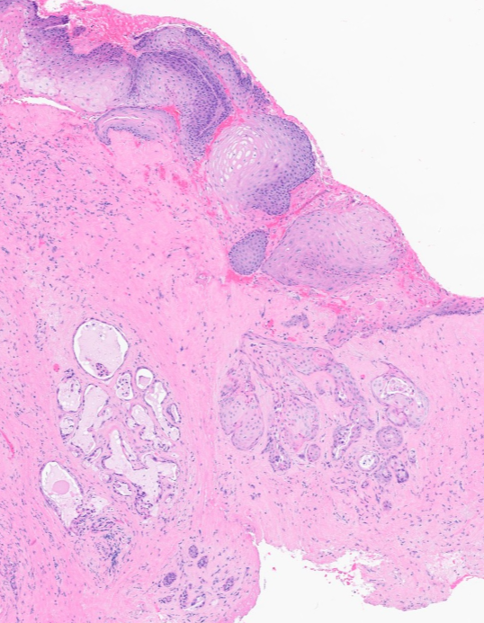
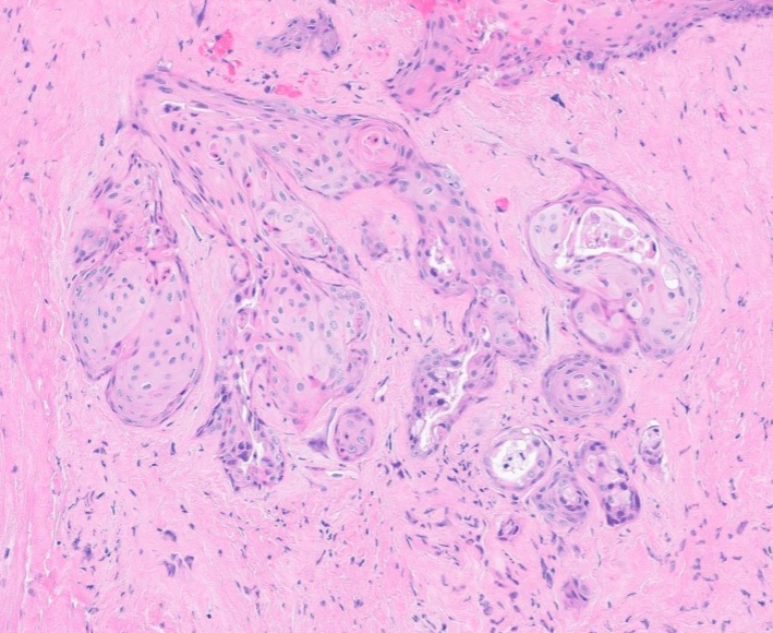
67 yo male
History of supraglottic/glottic SCC s/p radiation
Now presenting with glottic stenosis
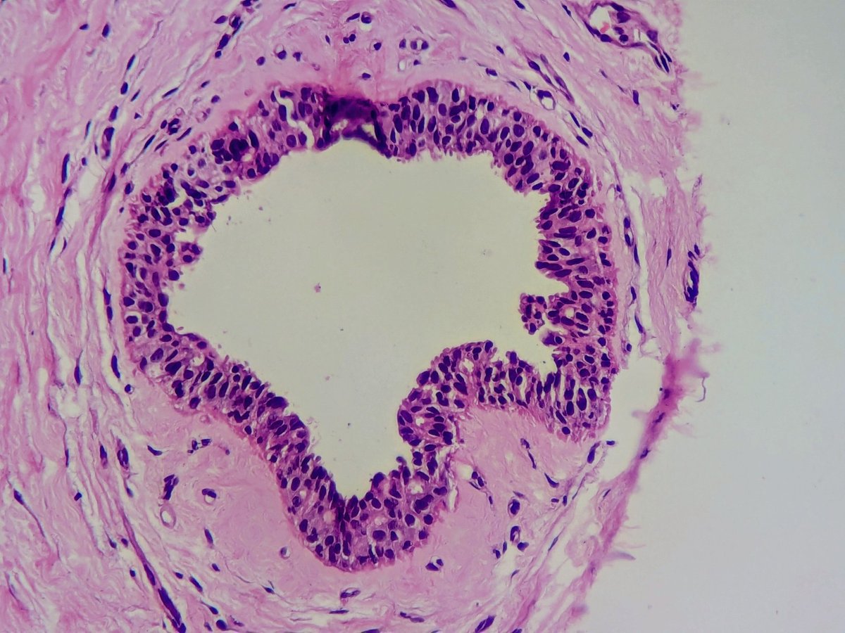
Biopsy shows sialometaplasia: a benign reactive process that is frequently misdiagnosed as recurrent SCC
- Commonly occurs after radiation or chemoradiation for head and neck SCC
- Can appear on ANY mucosal site
- Preserves lobular architecture
- Smooth borders with minimal atypia
- Myoepithelial layer sometimes present (pic 4)
Dr. Cipriani #USCAP2026 #pathology #PathX #PathTwitter
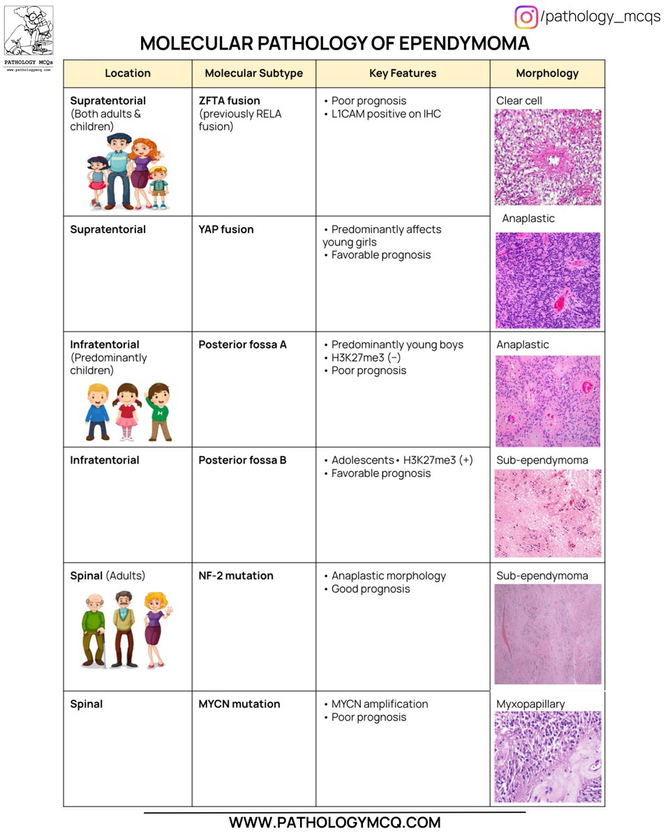
Molecular Pathology of Ependymoma — simplified & exam-ready 🧠✨
From ZFTA & YAP fusions to Posterior Fossa A/B and spinal MYCN tumors, molecular classification now drives prognosis, grading, and reporting.
#Ependymoma#Neuropathology#MolecularPathology#WHO2021#PathologyMCQs
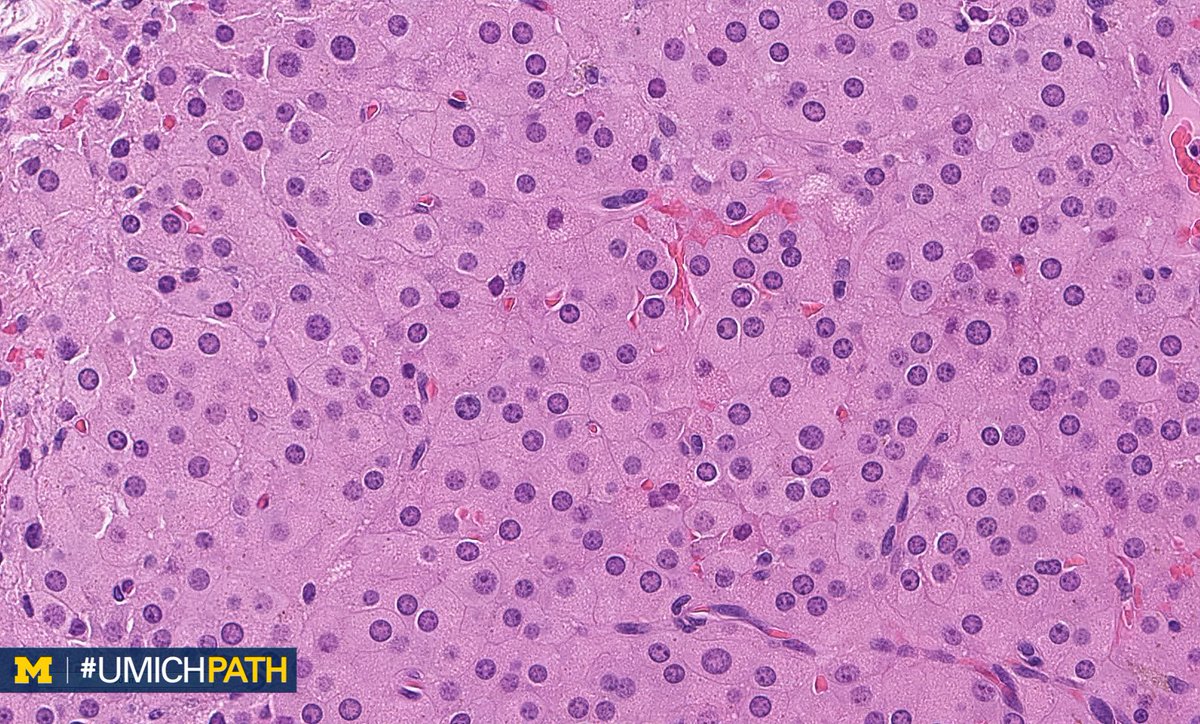
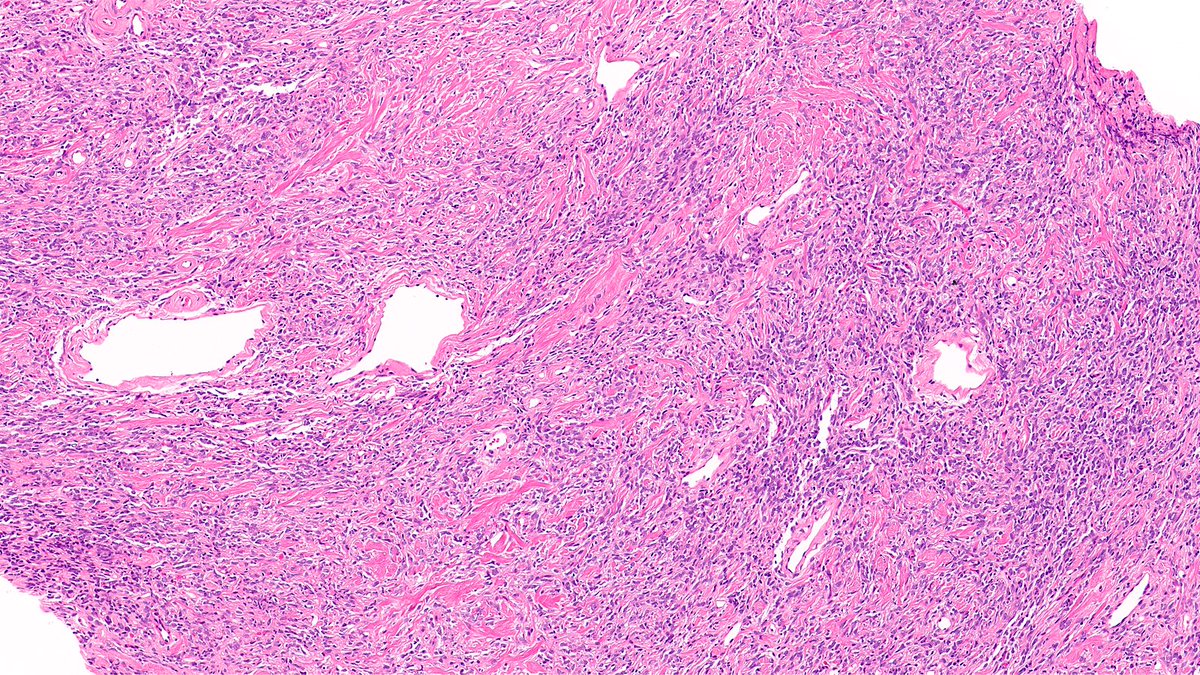
🎯 Clear cell hidradenoma
👉 64-year-old male with right arm soft tissue swelling
🔬Histology highlights:-
•Well-circumscribed dermal tumor
•Solid and cystic architecture
•Lobules of epithelial cells in hyalinized stroma
•Prominent clear cells with glycogen-rich cytoplasm
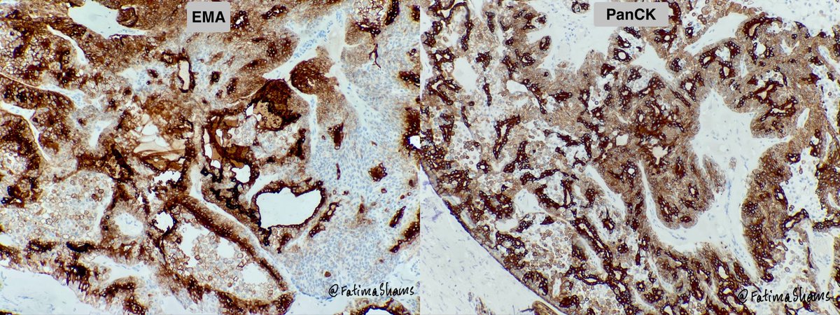
•Evidence of ductal differentiation
PanCK and EMA positivity shows epithelial and highlights ductal differentiation
🔎 Key differentials:-
•Metastatic renal cell carcinoma
•Poroma / poroid hidradenoma
•Hidradenocarcinoma
•Clear cell squamous cell carcinoma
💎 How to distinguish
•Lack of infiltrative growth, necrosis, and significant atypia
•Presence of ductal structures and biphasic cell population
•Clinicopathologic correlation is essential
⚓️ Prognosis
•Benign tumor
•Excellent prognosis after complete excision
•Local recurrence possible if incompletely excised
#PathTwitter #Dermatopathology #SkinAdnexalTumor #Hidradenoma #PathologyCase