I received this consult asking about a urothelial process given the GATA3 + in prostate tissue (NKX3.1 was+)
🔑Hx dug-up: patient had prior XRT for prostate ca
🔬Dx: benign prostatic tissue with radiation changes
(and aberrant GATA3+; see refs, oddly both 🖨️around same time!)
@GuannanTiffany You are right! Check paper below. "Pathologists should have a low threshold for performing FH, SDHB and CK20 IHC when confronted with unclassified eosinophilic RCC or ‘oncocytoma’ in young patient."
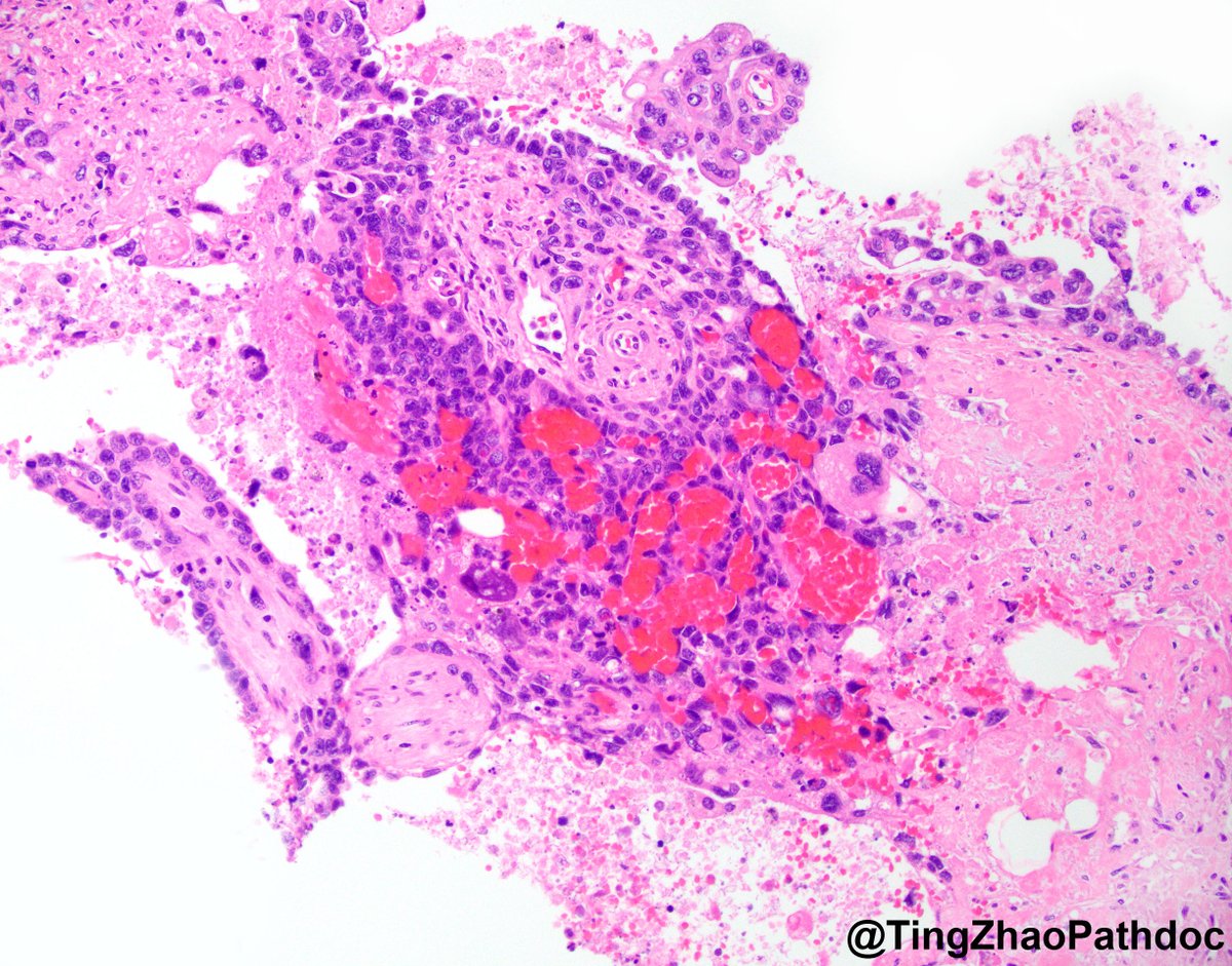
Dx: seminal vesicle stromal lipofuscinosis!
DDx could include melanocytic or mesenchymal proliferations, both benign & malignant!
4 such cases formally reported, most recently 2 by @Kiril_T_Can & another by us
Something is missing from this #renal tumor. 🤔
Diagnosis of this tumor is typically by morphology only. Missing component made this case more challenging. 🔍
What is your diagnosis on this nephrectomy #GUPath tweeps? #OnePicDx
Answer in comment👇👇👇
ALK-rearranged RCC are rare and can have variable morphologic features.
Here we describe some of these unusual tumors including an ALK-rearranged mesenchymal neoplasm:
https://t.co/SIjFXckSQt
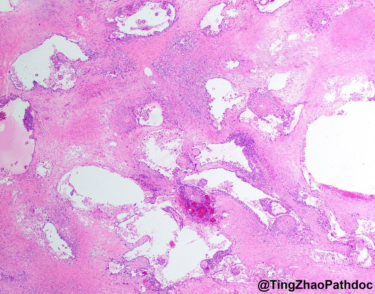
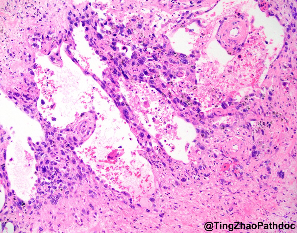
Normal vagina has no glands
Vaginal adenosis: Benign glandular epithelium involving superficial lamina propria or surface epithelium of vagina
.
Causes
Adenosis pre 1970: DES exposed children (DES daughters, now 55-60 yo). Location in upper 2/3 of vagina
Adenosis post 1970: Uncommon, due to trauma, endocrine distruption. Location is variable in the vagina
.
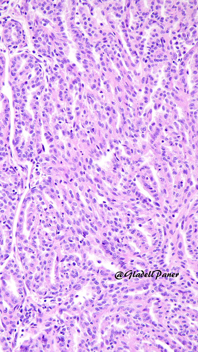
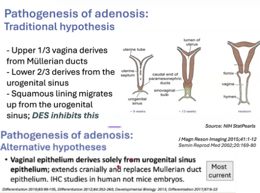
What is the source of vaginal squamous epithelium?-most current theory
Vaginal epithelium derives solely from urogenital sinus epithelium, extends cranially and replaces mullerian duct epithelium. Adenosis ocurrs due to trauma, endocrine imbalance, among others (pic 3)
Dr. Talia-Vaginal Epithelial Neoplasia: An Update-May2025 #ISGyP #gynpath #pathology
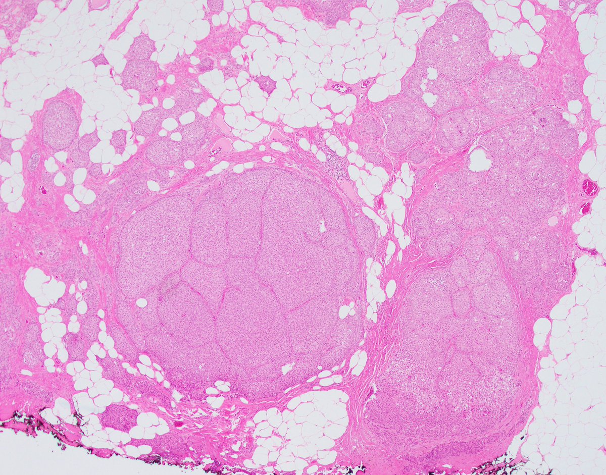
Take home messages from our study of close to 1000 prostate/bladder amyloid cases
- Often 1st diagnosis on these specimens
- systemic involvement of bladder in 7%
- SV=localized
- rare cases with different amyloid types at different sites
2 Nearly all SCLC arise in smokers and have RB1 & TP53 mutations.
Over the years, we encountered exceptional patients breaking all the rules: pathology of SCLC, but with intact RB1 & TP53, and generally never/light smokers. These are rare (3% of SCLC), but solving them was an opportunity to uncover unique biology and to inform diagnosis and treatment for when these patients are encountered in practice.
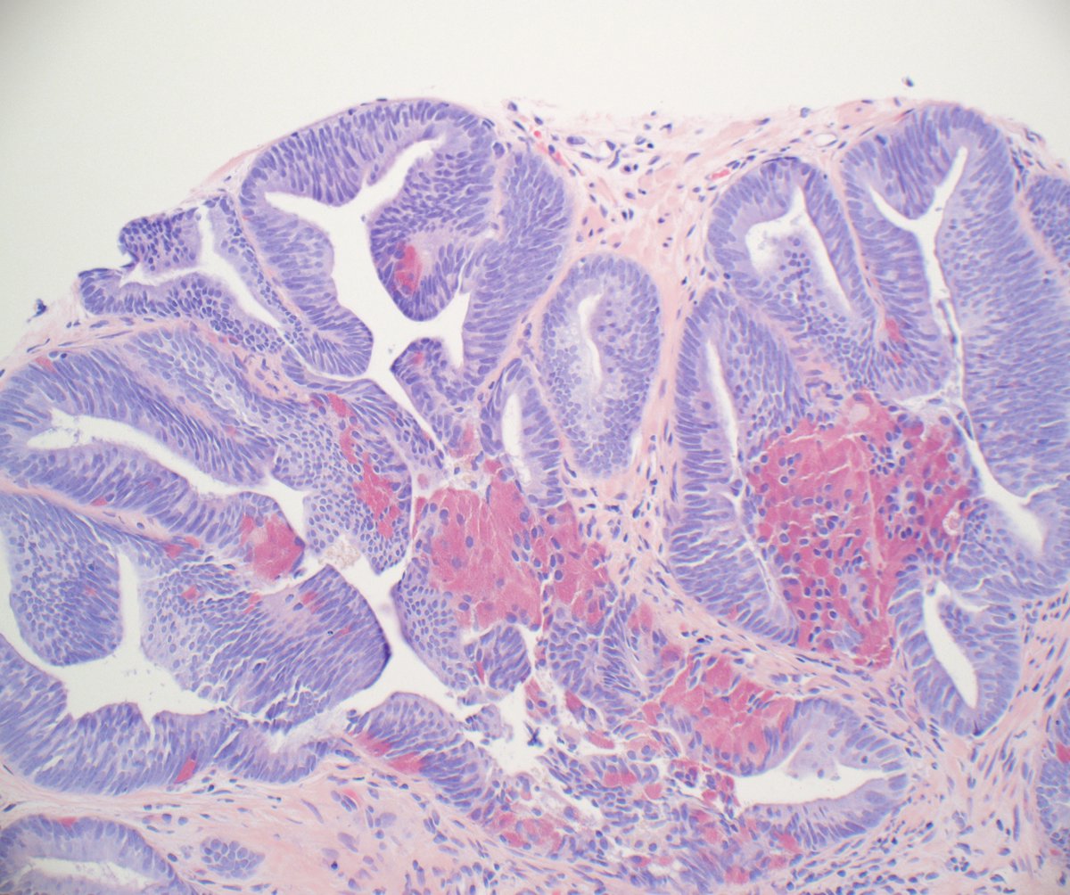
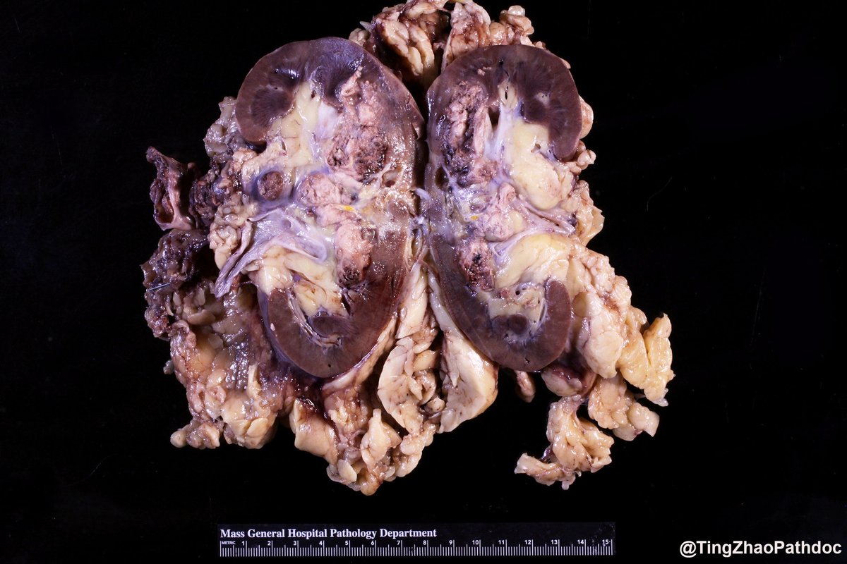
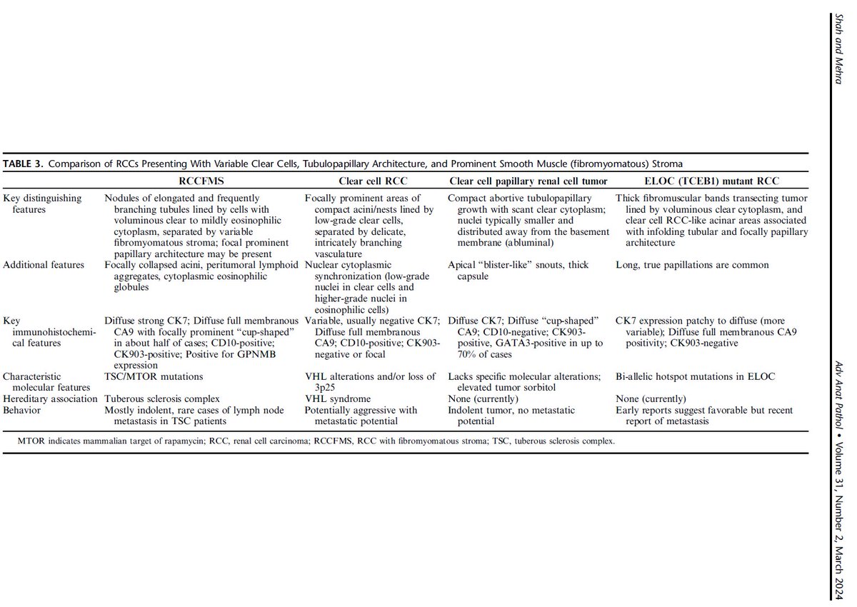
#GUpath kidney:
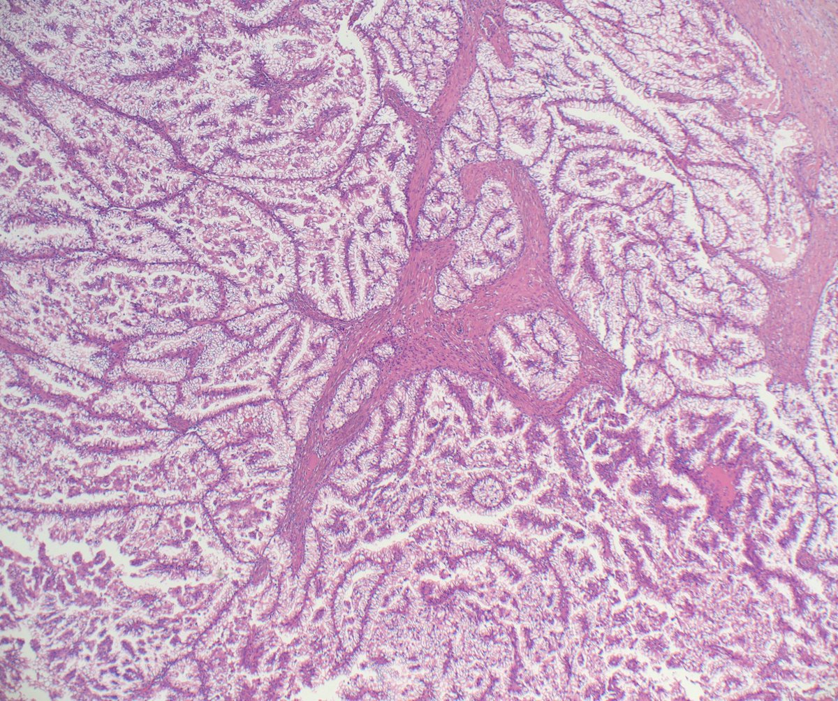
RCC with leiomyomatous (fibromyomatous) stroma (RCC-LMS/FMS)
although not currently a formal WHO RCC subtype, some still make Dx
#IHCpath: CK7+, CA9+ (often cuplike), HMWker+, GPNMB+, often CD10+
🧪often TSC/MTOR
DDx: clear cell RCC, ELOC-RCC, CCPT
✔️nice ref's
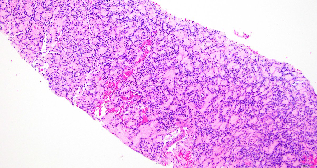
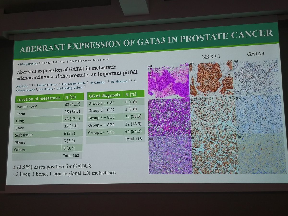
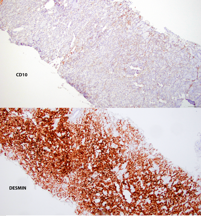
Dr. Magi-Galluzzi drawing our attention to pitfalls in work-up of PCa
GATA3 may be ➕ in HG PCa
❗Case of HG metastatic PCa to bone, initially sus MM after initial #IHCpath:
PCK- CD138+
Proved
CAM5.2+ NKX3.1+
🚨CD138+ in PCa may have adverse prognosis!
KTF meeting Mikulov, CZ
#PathTwitter #GUPath
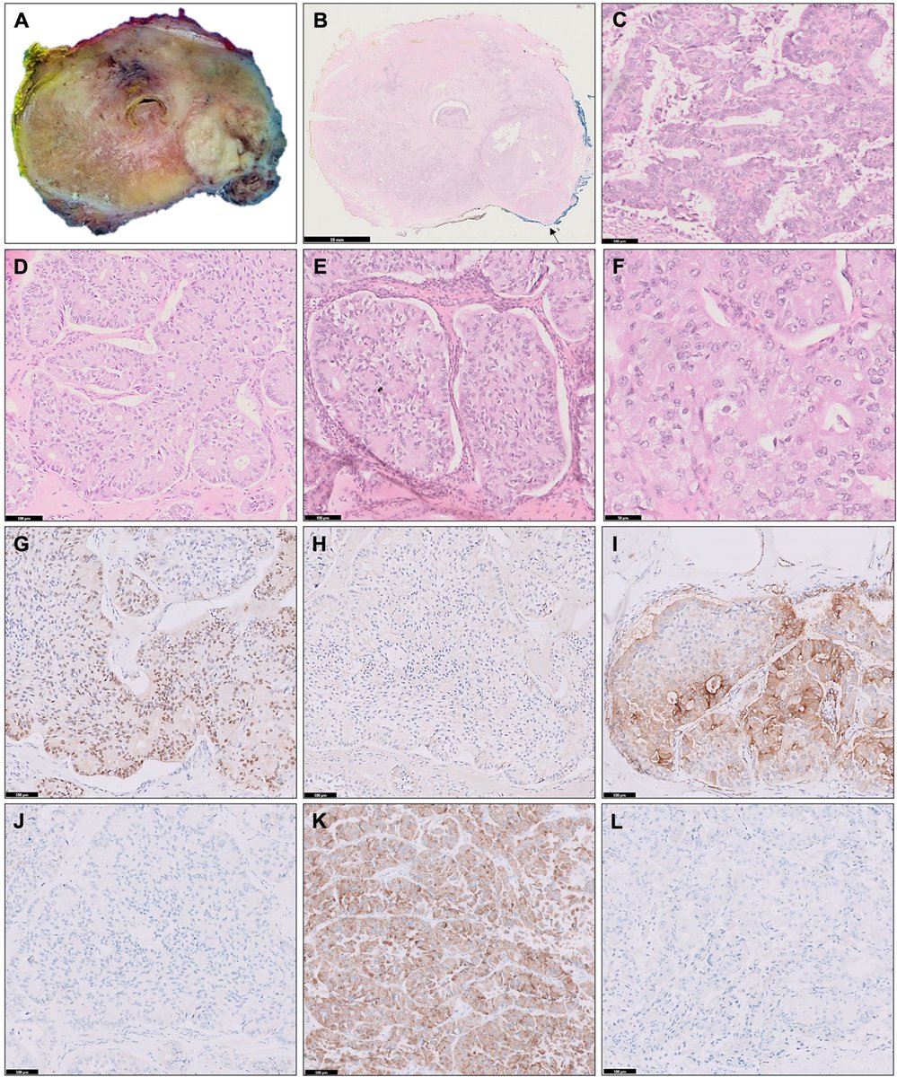
Do you recognize this rare histologic subtype of #prostatecancer? Happy to share this article just published in April issue @Pathology_RCPA. Unlike kidney tumor, pink morphology variant of #ProstateCancer is rare.
Great work by Drs. Punjabi & colleagues!
https://t.co/xaTMLFW16g
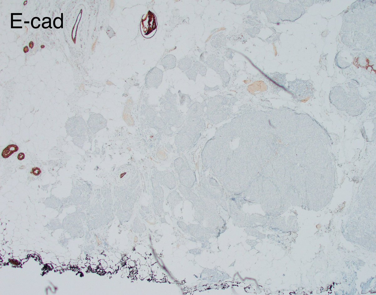
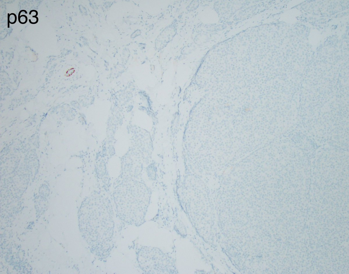
The case I posted yesterday is a resection on a biopsy that was called LCIS.
The key in this case of lobular neoplasia is to remember that E-cadherin, though often weaker than in epithelial cells, is usually retained in myoepithelial cells and its complete absence should make you pause and order some myoepithelial markers.
Here, myoepithelial cells are completely absent in all nests of lobular neoplasia, and positive internal controls are surrounded by tumor in an infiltrative pattern.
This serves as a reminder that invasive lobular carcinoma can adopt unusual morphologies like solid and alveolar. This LCIS-like pattern is highly unusual in my experience. I shall call it the large nested variant of invasive lobular carcinoma 🙂
Happy weekend everyone!
@wusm_pathology @washupathedu #PathTwitter #PathX #breastpath