Excited to share that our team's work on how pluripotent #stemcells form retinal #organoids has been accepted for publication in @stemcellreports , the official journal of the @ISSCR . Through one of the first NGS-based, mechanistic studies on "RNA Epigenetics," or “epitranscriptomics”, in the eye, we explore how chemical RNA modifications influence retinal tissue generation from stem cells.
Using cutting-edge tools like dCas13b-FTO guided RNA engineering, single nucleotide resolution mapping of RNA modifications, and transcriptome-wide RNA-protein binding, targeted protein degradation (degron), we made seminal observations in stem cell and retinal biology. Our research uncovers a novel interaction between METTL3 and RNA target Ythdf1, and shows how chromatin accessibility can be uncoupled from transcriptional dynamics in retinal organoids during acute degradation of METTL3.
Stay tuned for the publication on October 23 in @stemcellreports. Meanwhile, you can explore our #preprint on @biorxivpreprint: https://t.co/ynSEbpm4h4
Grateful for our awesome team: Jing Xu, Yuanhao Huang, Qiang Li, Ricky Han, Jie Liu, from @UMKelloggEye , and for our funders @RPB_org , @NatEyeInstitute
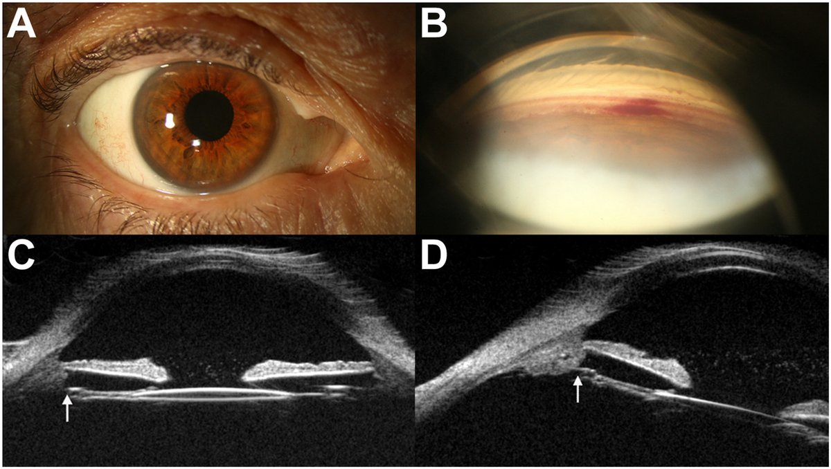
A 75-year-old physician presented with recurrent episodes of painless and “foggy” vision in the right eye (OD) lasting 3 hours. He was investigated for amaurosis and began dual anti-platelet therapy. Vision OD was 20/25, intraocular pressure was 43 mmHg, and the anterior chamber showed 2+ red blood cells without iris transillumination defects or neovascularization. (A) External photograph showed no iris abnormalities. (B) Gonioscopy demonstrated microhyphema. (C, D) Ultrasound biomicroscopy revealed an intraocular lens within the capsular bag with the nasal haptic abutting the anteriorly rotated ciliary process (arrow). Although rare with in-the-bag implants, uveitis glaucoma hyphema (UGH) syndrome may cause “white out” transient monocular vision loss.
From “In-the-Bag Uveitis Glaucoma Hyphema Syndrome Masquerading as Transient Monocular Vision Loss” by Daniel J. Espinosa, BS, Osama Ahmed, MD, Sangeeta Khanna, MD. Published by Ophthalmology online on December 8, 2025.
Read: https://t.co/YlkcnaMcdh
#AAO
Intraoperative Anti-VEGF Versus Photocoagulation During Vitrectomy for BRVO with Vitreous Hemorrhage: A Randomized Clinical Trial
https://t.co/aVA9k41TTN
#ophthalmology
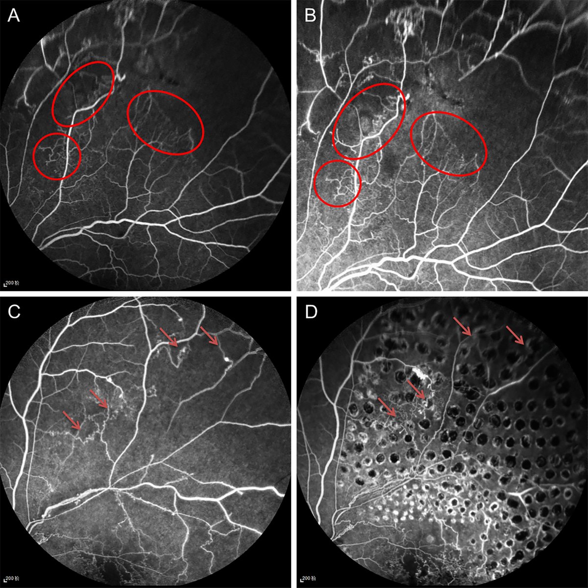
A 9-year-old girl presented with sudden painless visual loss (20/40) after a febrile illness. Fundus images show multiple discrete, round whitish retinal lesions in both eyes, more numerous in the right eye (A), involving the posterior-pole and midperiphery. The left eye shows fewer, smaller lesions predominantly in the midperiphery (B). OCT demonstrates hyperreflective inner retinal lesions with extension into the vitreous and surrounding vitritis (C). Lasiodiplodia theobromae, a plant-associated filamentous dematiaceous fungus, was identified on panfungal polymerase chain reaction sequencing of the vitreous sample. The bilateral multifocal pattern possibly reflects hematogenous spread after recent systemic infection, with significant resolution after intravitreal voriconazole, systemic amphotericin-B, and fluconazole (D).
https://t.co/1BibCKWh4E
#ophthalmology #retina
Corneal blood staining can occur after traumatic ophthalmic injuries. A temporary #keratoprosthesis can be used to improve surgical visualization. #oculartrauma#aao#ophthalmology https://t.co/JUyPO6nRe5
High-Resolution OCT of Presumed Basal Laminar Deposit and RPE Abnormalities in Extensive Macular Atrophy with Pseudodrusen-like Appearance
https://t.co/ZYIanArwJK
#ophthalmology#retina
The June issue of Ophthalmology is out now! Featuring articles on:
✅A new intraocular-lens-power calculation formula for eyes undergoing lens exchange
✅Tirzepatide and Reduced Risk of Diabetic Retinopathy and Related Complications: A Multicenter U.S. Cohort Study
✅Current Indications and Outcomes of Penetrating Keratoplasty in the United States: An IRIS® Registry (Intelligent Research in Sight) Study
✅How Well Do Ocular Trauma Scores Predict Vision After Open Globe Injury? A Systematic Review and Meta-Analysis
And much more!
https://t.co/fMBgtfGZt9
#ophthalmology
A 13-year-old boy presented with poor vision in the right eye since early childhood and esotropia noted at around 3 years of age. The right eye was not microphthalmic, with an axial length of 25.90 mm. Anterior-segment photography revealed tree-like vessels in the corneal stroma (A). Fundus examination, with limited image clarity likely due to vitreous abnormalities, showed a fibrovascular membrane anterior to the optic disc and peripapillary chorioretinal atrophy (B). Corneal fluorescein angiography demonstrated filling of these vessels beginning approximately 15 seconds after intravenous injection of fluorescein sodium, without leakage (C). Ultrasound biomicroscopy localized the vessels to the deep corneal stroma (D). These findings suggest anomalous corneal vessels in association with persistent fetal vasculature, distinct from typical corneal neovascularization.
https://t.co/KDML7Sg8HD
#ophthalmology
A 90-year-old woman presented with a scleral-fixated intraocular lens implant (IOL, CTLucia602) that was axially rotated 45° (A). Three hundred sixty-degree conjunctival peritomy was cut. A 25-gauge (5/8 inches) needle was inserted 3 mm posterior to the superior limbus immediately behind the IOL; an STC-6 needle with 10-0 Prolene was inserted 3 mm posterior to the inferior limbus into its bore and externalized. The process was repeated 5 times to form a continuous star. Visual acuity improved from counting fingers to 20/25 with IOL planar to the iris (B). Two-point IOL scleral fixation can be complicated by delayed tilt. The 5-point star technique (C), originally described by Weng for silicone oil barricade, offers a surgical solution without necessitating IOL explantation.
https://t.co/k5fCQvFKoF
#ophthalmology #retina
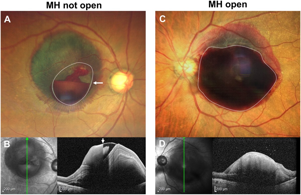
Niveau (level) as a key predictor for the absence of macular hole in sub-ILM hemorrhage secondary to retinal arterial macroaneurysm rupture
https://t.co/bbJ5wLMq8Z
#ophthalmology#retina
A 50-year-old man presented with a 7-year history of progressive bilateral exophthalmos and periorbital swelling without facial numbness. Magnetic resonance imaging demonstrated marked bilateral enlargement of the supraorbital and infraorbital nerves, accompanied by extensive involvement of multiple extraocular muscles. Histopathologic evaluation of a biopsied extraocular muscle specimen, including immunohistochemical staining, revealed numerous plasma cells with a marked predominance of IgG4-positive cells, consistent with IgG4-related ophthalmic disease. Bilateral enlargement of the infraorbital nerves without sensory changes is highly characteristic of IgG4-related orbital disease.
https://t.co/OWs9Q4ttrz
#ophthalmology
A 39-year-old man presented with left eye redness and pain. He had a history of severe gout lasting >10 years. Ocular examination revealed irregular, pale yellow crystalline deposits under the temporal conjunctiva, with well-defined borders and local hyperemia (A). No other significant abnormalities were noted on comprehensive eye examination. Systemic examination revealed swelling and deformity of the bilateral finger joints (B), along with restricted movement of both lower limbs. The conjunctival lesion was surgically excised. Histopathological analysis confirmed the presence of multicystic tophus of varying sizes within the subepithelial stroma of the conjunctiva
https://t.co/Lv1BtT76K2
#ophthalmology
A 74-year-old man presented with a pigmented tumor on the left eye suspicious for conjunctival melanoma. Visual acuity was 20/50. The anterior segment demonstrated a pigmented mass covered by the conjunctiva and Tenon’s fascia (A), with numerous episcleral sentinel vessels suggesting underlying intraocular tumor. Gonioscopy disclosed 360° angle pigmentation and fundoscopy showed a peripheral melanoma, with subtle ciliary body involvement on ultrasound biomicroscopy (B, white arrows), measuring 3.9 mm thickness. Findings represented a small ciliochoroidal melanoma with large extraocular extension (B, yellow arrow) and angle invasion. There was no primary conjunctival melanoma. Plaque radiotherapy was performed.
https://t.co/oJqoEVHpCI
#ophthalmology
Of my many amazing mentors, none greater than Prof Claes Dohlman at Harvard Med, who saved millions from blindness but never took credit for it.
Instead of naming his artificial cornea after himself, he called it “Boston KPro”.
Worked til 101, always wearing a SHORT white coat worn by medical students. Why? He would humbly say: “I am still a student. Every day, I learned from my residents, I learned from my students, and I learned from my patients.”
A man who rightfully deserved to have worn a long gold and velvet coat for all the people he selflessly helped (photo of us below - I was a lot younger then!), wearing the shortest, white coat they make, acknowledging how he continued to learn until his very last day.
I wish we had more people like Dr Dohlman in leadership and in medicine. While I will never be even one percent of the person he was, I every day remember what he taught me through his words and actions and will strive to emulate him as best I can.
A 19-year-old woman from Benin presented for myopia evaluation with no relevant personal or family history. She had undergone laser photocoagulation in the right eye at age 10 for an indeterminate lesion. Fundoscopy revealed a pearlescent, calcified lesion with adjacent retinal elevation demarcated by laser barrage in the inferior right eye (A), and 2 to 3 similar lesions in the superonasal periphery of the left eye (B). Ultra-widefield autofluorescence showed hyperautofluorescence (C). Ultra-widefield OCT revealed focal hyperreflective lesions within an undifferentiated, isoreflective retina with indiscernible layers (D). Both lesions were consistent with retinocytoma, confirmed by a germline RB1 pathogenic variant (c.2359C>T; p.Arg787∗).
https://t.co/pJtBklDa5W
#ophthalmology #retina
A 28-year-old man was referred for tumors in both eyes. He had a history of biopsy-proven pulmonary atypical carcinoid and underwent left pneumonectomy. On examination, a large mass arising in the ciliary body that extended to the contiguous choroid in the left eye (A) and multifocal yellow-white choroidal lesions in the right eye (B) were noted. Ultrasonography (A-insert) showed a medium reflective mass with “sponge-like structure” in the left eye. Local resection of the ciliary body neoplasm revealed a pink-white soft mass (C); tumor cells were uniform in appearance with finely granular nuclear chromatin (D, ×400), and stained for synaptophysin (D-insert, ×200), supporting that this neoplasm originated in the lung.
https://t.co/lD2TNV8cr6
#ophthalmology #retina
First-in-Human Clinical Evaluation of a Novel Non-Surgical Suprachoroidal Delivery Approach for Triamcinolone in Diabetic Macular Edema
https://t.co/CpEZdcLNw2
#ophthalmology
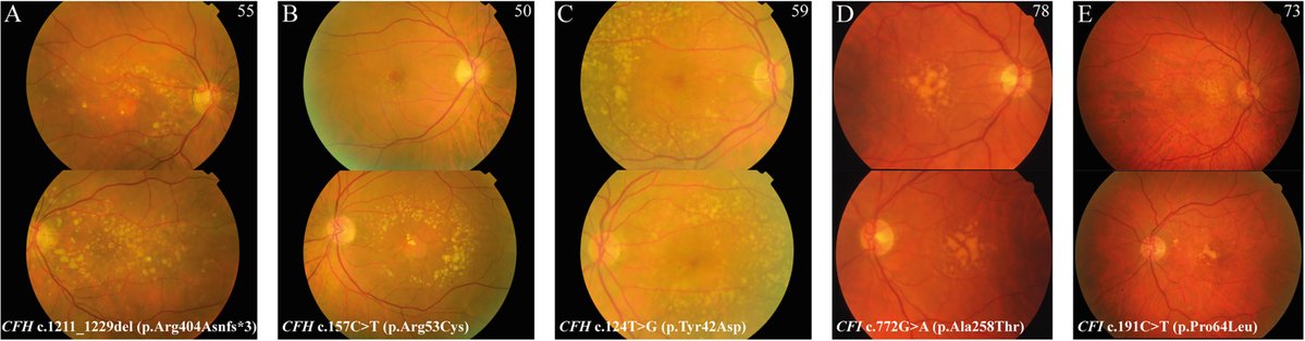
Geographic atrophy in patients with age-related macular degeneration is associated with rare variants in complement factor H and complement factor I
https://t.co/pQu5nlOman
#ophthalmology
A 56-year-old man presented with right-eye pain for 6 months. He was a known case of chorioretinal coloboma with retinal detachment, operated on 1 year earlier. Visual acuity was 1/60, and intraocular pressure was 46 mmHg. Examination revealed an inverse hypopyon (A), with emulsified silicone oil migrating into the superior angle from the pupillary area (B), and retained perfluorocarbon liquid (PFCL) bubbles inferiorly (C). The retained PFCL was likely due to intraoperative migration into the colobomatous area, leading to incomplete removal. Subsequent anterior migration of silicone oil and PFCL led to secondary glaucoma. The patient was managed with surgical removal of both silicone oil and PFCL, with improvement in intraocular pressure.
https://t.co/8qj3GJRoAL
#ophthalmology #glaucoma
An 86-year-old man with pseudoexfoliative glaucoma and asteroid hyalosis presented with elevated intraocular pressure (IOP). He underwent in-the-bag cataract surgery 10 years previously and Ahmed glaucoma valve implantation 10 months previously, with no postoperative IOP elevation until this visit, when his IOP increased from a mean of 9 to 35 mmHg. Slitlamp examination revealed intraocular lens subluxation and the vitreous humor with small glittering white particles, asteroid bodies, impacted in the tube (A), which was confirmed using anterior segment OCT (B, arrow). Pars plana vitrectomy relieved tube obstruction (C, D) and reduced the IOP to 7 mmHg
https://t.co/4CyvTMAdix
#ophthalmology #glaucoma