@JacquesCarolan Thanks! If you know any PhD candidates who has interest in building a light-sheet microscope for voltage imaging in zebrafish, please let them that I can recruiting!
SUPER excited that my first ever *real* neuroscience paper is published today in Nat Comms!🥳
We combined two-photon voltage imaging ⚡️and optogenetics 🔦to probe synaptic plasticity in the cerebellum of awake behaving animals. 🧵
Hi #Neuroscience,
Exciting news—I’m recruiting a PhD student to join my lab and study neuroscience in larval zebrafish 🧠🐟
Microscopy expertise is a big plus!
https://t.co/YjEi5VaYsz
Check out my recent paper in @CellReports, where we optically recorded action potentials in vivo from up to 300 fast-spiking interneurons at 3–4 kHz, using state-of-the-art genetically encoded voltage indicators from @StPierreLab and @mkannan3112, alongside advanced camera technology from @phantomhispeed. Voltage imaging is paving the way for addressing questions about population neural codes that were previously unreachable. https://t.co/rzVGilOXh6
The video captures 250 milliseconds of voltage imaging data from molecular layer interneurons of the cerebellum, slowed down 80-fold for visualization. Each brief flash of light corresponds to an action potential.
If you're interested in my postdoc work at @UChicago_Brains, come join my talk on ramping neuronal activity and parallel fiber–Purkinje cell LTD at the JNS 2025 (7/24, 17:30–17:45, Room 5). Grateful to @SfNtweets for the JNS Meeting Award that made this opportunity possible!
Hi #neuroscience! As I transition into my new role as an Assistant Professor at Utrecht University in the beautiful Netherlands, I’m excited to share that I’ll soon be recruiting a PhD student to study cerebellar neural plasticity and development using larval zebrafish.
Hi #neuroscience! As I transition into my new role as an Assistant Professor at Utrecht University in the beautiful Netherlands, I’m excited to share that I’ll soon be recruiting a PhD student to study cerebellar neural plasticity and development using larval zebrafish.
Our new paper is published in Current Biology! In this project, we developed a model showing how social and depth information are integrated to drive affiliative behavior in zebrafish. See: https://t.co/zhAZYFzvVy
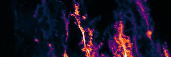
Inspired by Cajal, the Hansel lab's new work in @eLife provides a comparative histological analysis of human and mouse Purkinje cell morphology. Following our 2023 study, we detail differences of scale and kind between the neurons of the two species 🧵👇
https://t.co/8wp7g6Ds3a
Our latest research, led by @AntoineMadar, on what #SynapticPlasticity rules shape CA1 and CA3 representations in the hippocampus during familiarization has just been published in @NatureNeuro! Read here: https://t.co/eWT3j3wVSU
Our five years of work is finally out! This was truly a team effort—huge thanks to the team members in @varshneylab and to Gaurav for his great support! Proud of what we’ve accomplished together!
New preprint alert from the lab! Also you can hear an auto-generated 40 s audio summary here:
https://t.co/VhVveLXG66
More details on this exciting paper will follow soon!
@NCBS_Bangalore
Happy to announce our new preprint "A green lifetime biosensor for calcium that remains bright over its full dynamic range" which is now available on bioRxiv:
https://t.co/6p6fIaEUQK
Why yet another biosensor for Ca2+? Here's a thread 🧵
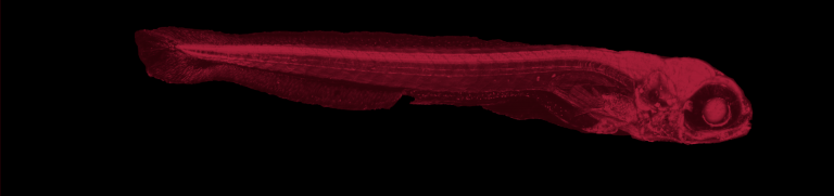
Proud to present our latest preprint by Elliot Birkett. In this paper we asked whether stimuli with different behavioural relevance evoke different adaptation in #zebrafish. https://t.co/ugccoo8Zbz
New preprint by the twitterless Tessa Herzog & colleagues: Individual cones of the vertebrate eye are exceptionally reliable and precise, but functionally heterogeneous as a population: https://t.co/aiwSwJu64S @TaYoshimatsu@neonsynapse@JosMoyaDaz1 BenJames
How do zebrafish map their environment? A new Nature paper by @rolilab found first evidence for place cells in fish. These specialized neurons play a crucial role in forming mental maps of space, social networks, and abstract relationships.
👉https://t.co/nvCp3Xu3ow
#FluorescenceFriday#zebrafish#bioart To see cells as they really are, it's necessary to see them in the native multicellular environment in which they evolved. However, the deeper we peer into living tissue, the more our view is obscured by optical aberrations. Here, take a trip as we dive 200 um down from the optic tectum to the hindbrain in a living zebrafish, turning on adaptive optics to correct these aberrations as we go, to see oligodendrocytes (orange) and neural nuclei (green). https://t.co/VtRNfzY6np
We're recruiting a lab technician at the University of Tübingen for our neurobio research group. It is an E9b position (TV-L, 100%) supporting our zebrafish animal facility and our mol-bio lab. https://t.co/arCTdDx6OA