The classic answer is vitamin B5 (pantothenic acid) deficiency.
It causes burning feet syndrome, also called Grierson–Gopalan syndrome.
It was first described in malnourished prisoners of war in WWII, and volunteers fed a pantothenate-deficient diet developed burning paraesthesia of the feet.
Medication-related osteonecrosis of the jaw (MRONJ), most likely from antiresorptive therapy for osteoporosis (bisphosphonates or denosumab).
Key features: exposed necrotic bone in the mouth with surrounding inflammation, an extraoral draining sinus on the chin/jaw, and a radiograph showing a mottled, moth-eaten lytic area in the mandible with sequestrum.
Management: stop or hold the antiresorptive after discussion with the prescriber, chlorhexidine rinses, antibiotics for infection, and conservative debridement. Surgical resection is reserved for advanced or refractory disease. Refer to oral and maxillofacial surgery.
Prevention: a dental check-up before starting therapy and avoiding invasive dental procedures during treatment.
Rhinophyma, a severe form of phymatous rosacea (glandular type).
Key features: bulbous, erythematous nose with thickened skin, prominent dilated follicular pores, and nodular contour irregularity from sebaceous gland hyperplasia and fibrosis.
Next steps: confirm clinically and biopsy if the course is atypical to exclude BCC, which can coexist. Start medical therapy for the underlying rosacea (topical ivermectin or metronidazole, oral doxycycline), and consider isotretinoin in early or moderate disease. Established tissue overgrowth needs surgical debulking (scalpel, CO₂ laser, or electrosurgery). Advise trigger avoidance and sun protection.
Note: it's not caused by alcohol, a common myth.
@TrackYourHeart A. Iron-deficiency anemia
Why? Spoon-shaped nails (koilonychia) combined with fatigue and pallor are the classic clinical hallmark of chronic iron-deficiency anemia.
@simplify_drugs Heparin (including Unfractionated Heparin) and Low-Molecular-Weight Heparin (such as enoxaparin) act by indirectly activating antithrombin III (also known as antithrombin).
@Madihealth2026 Vitamin C (Ascorbic acid) should be given with iron supplements for better absorption.
✒Why? Vitamin C creates an acidic environment in the stomach and acts as a reducing agent, converting insoluble ferric iron (Fe³) into the much more easily absorbable ferrous iron (Fe²).
16-year-old girl presents with:
- Low-grade fever for 6 months
- Progressive weight loss, anorexia, short stature, and thin build
- Previous history of abdominal tuberculosis, treated with anti-tuberculous therapy (ATT) for 6 months; treatment records are unavailable.
Investigations:
- Chest X-ray: Bilateral hilar lymphadenopathy with patchy areas of consolidation.
- Abdominal ultrasonography: Necrotic and matted lymph nodes.
- Skin examination: Extensive, bilateral, reticulated brown hyperpigmented patches over the abdomen and flanks.
What is your differential diagnosis❓
@DrAyubaD The most common content of an inguinal hernia is small intestine (specifically the ileum) or preperitoneal fat / omentum, with the small bowel being the most frequently encountered intra-abdominal viscus.
☑Diagnosis: May-Thurner Syndrome
⚪Responsible Artery: The right common iliac artery.
✒Clinical Pearl: May-Thurner syndrome occurs when the right common iliac artery compresses the left common iliac vein against the lumbar spine, resulting in venous outflow obstruction, stasis, and a significantly increased risk of extensive left-sided deep vein thrombosis (DVT).
@md1talk "الـ Lactulose يتم تكسيره بواسطة بكتيريا القولون إلى أحماض، مما يقلل الـ pH ويحول الـNH₃ القابل للامتصاص إلى NH₄ غير قابل للامتصاص (Ion Trapping)، ثم يطرده خارج الجسم عن طريق الإسهال الأسموزي."
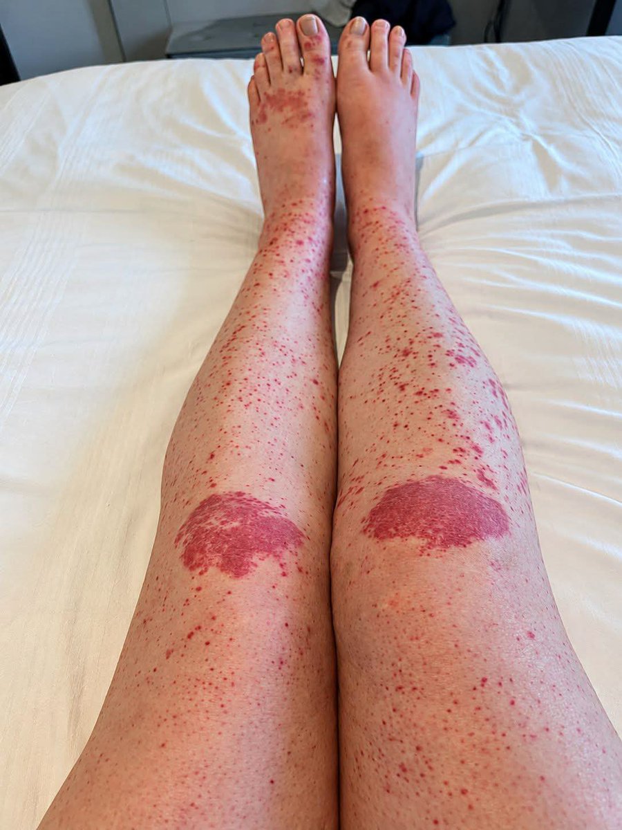
patient with a history of Crohn���s disease presented with a rapidly progressive skin rash affecting both lower limbs.
A few days later, both ankles and knees became markedly swollen and painful, leaving the patient unable to walk for 2 days.
The patient was admitted through the Emergency Department , and further evaluation established the diagnosis.
❓ Questions:
1. What is the most likely diagnosis?
2. What is the characteristic type of skin lesion?
3. Which organ involvement must be actively screened for?
4. What is the possible association between this condition and Crohn’s disease?