Anyone can see the bright spot on diffusion—what sets you apart is if you can tell them why it’s there!
Can you tell a stroke’s etiology from its appearance on MRI?
Main stroke types:
Artery to artery embolism
—Vulnerable plaque ruptures & causes clot formation
—Occludes the artery & distal blood flow
—Makes a wedge shape
Distal hypoperfusion:
—At the border zones
—Remember borderzones almost look like a fancy letter H for Hypoperfusion = two vertical lines (ant, internal watershed) that have curls at each end (ant & post external watersheds)
Vasculitis
—Inflammation ofvessel wall
—Idiopathic, autoimmune, or infectious
—Remember: Vascu-LIGHT-us = tons of little regions LIGHT up on DWI
—Remember Vascu-LITE-us = usually in LITE vessels or small vessel territories
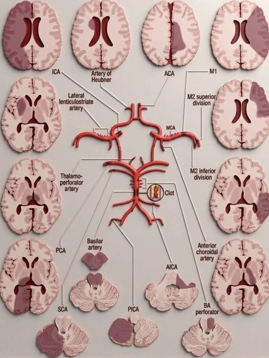
Impingement on perforators:
—Large vessel plaque covers opening of small perforator in its wall
—Perforators affect the Ps = pons, putamen (lenticulostriate)
Small vessel dz
—Many different pathologies that cause occlusion of small, unnamed vessels
—Remember: Small & subcortical both start w/—tend to be subcortical
Cardioembolic
—Emboli from heart stasis or vegetations
—Remember: Emboli & everywhere both start w/E = emboli go everywhere
So now you know how different etiologies have different distributions on MRI.
Remember, catching a stroke on DWI isn’t the end of your job—it’s the beginning!
This one's for me!!
I was tired of seeing small MCA infarcts that were clearly from a branch occlusion& not knowing the name of the branch that was occluded!
I decided I would find a way to remember these territories myself-and then share it w/ you!!
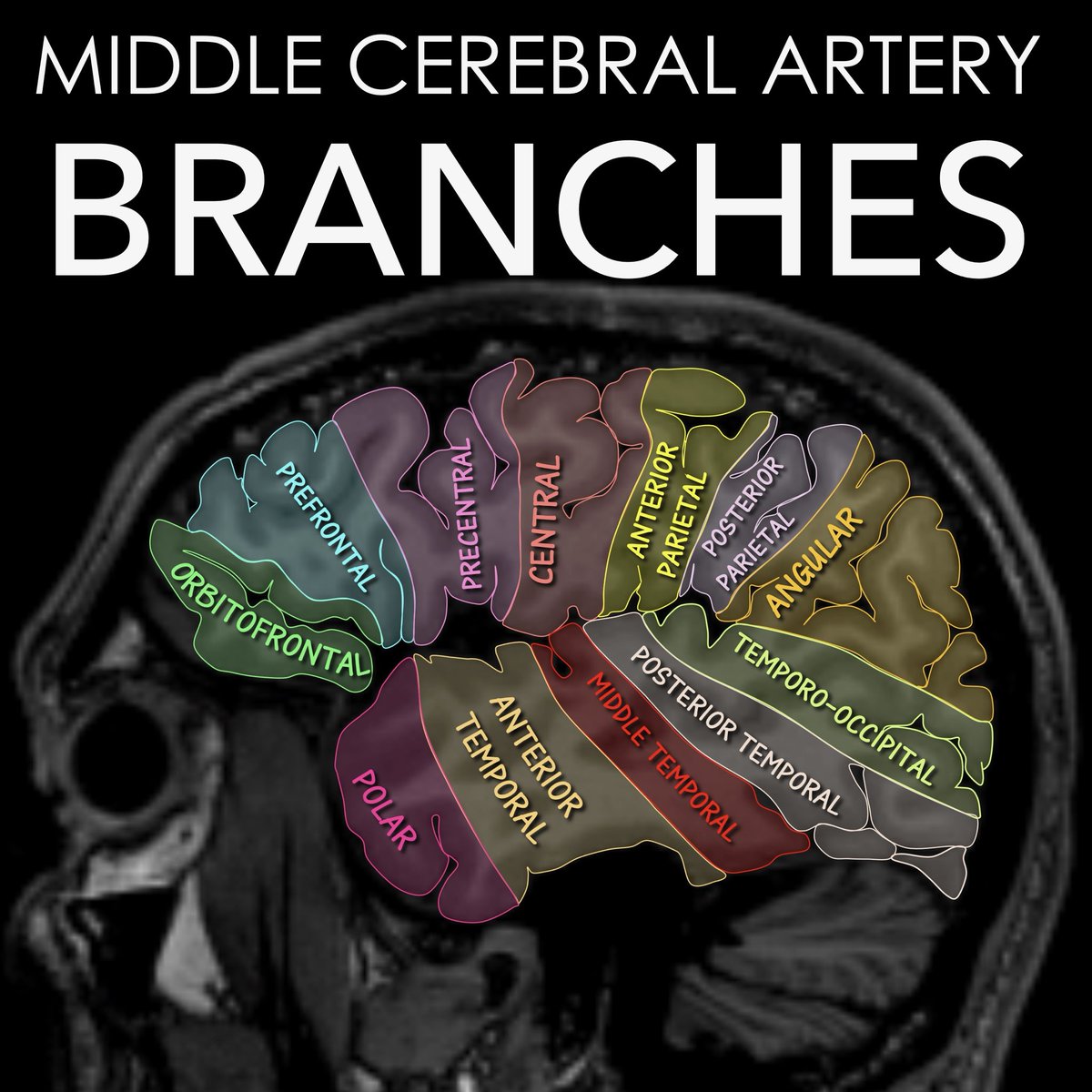
Here's how you can remember the different territories
Start w/4 cardinal territories
-These are the skeleton from which you can remember the rest
Orbitofrontal: Easy, it overlies orbit in the frontal region, like its name
Central: East, it's the territory surrounding central sulcus
Angular: Easy, it literally looks like an angle between parietal & temporal region
Polar: Easy, it's the temporal pole!
Bc it's the MCA, there is an M or two humps between each of these cardinal branch territories & their names reflect the anatomy
Finally, the temporal territories look like fingers on a hand:
Polar is the thumb of the hand: remember polar sounds pollex which means thumb
Middle temporal is literally the middle finger!!
Now you know the middle cerebral branch territories— so your reports will never be middling!
Here’s a head start on head CT anatomy!!
Cisterns: think “CT loves clear space.”
If it’s dark space around the brainstem, that’s a cistern.
Suprasellar = the starfish, S is for star and Suprasellar!
Ambient = hugging the midbrain. Remember Amb-brace cistern, it embraces the midbrain!
Quadrigeminal = the smile behind it. Remember, you are GEM if you smile!!
Lobes made easy:
Frontal lobe = Get in FRONT of things = planning, personality, decisions.
Parietal lobe = “pair-ietal” = pair of hands to feel things = sensation and spatial awareness.
Temporal lobe = Remember conTEMPlation =memory. And remember tempo = hearing
Occipital lobe = “optic-pital” in the back = vision.
Hopefully this will help you wrap your head around CT head anatomy!!
🔔New pub🔔We conducted a twin study on sexual behaviors & found a #genetic link:
"Pilot evidence of #genetic and environmental contributions to problematic #pornography use, pornography use frequency, & moral disapproval to pornography" @sexmedjournals
https://t.co/QimPLwGyAO
🔔And to share a final paper we put out this year:
"Far Out: #Psychedelics are Becoming a Current Treatment Option in #Psychiatry"
My close friend (Nicholas Borgogna) & I opine on pros & cons of #psychedelic assisted therapy for #mentalhealth disorders.
https://t.co/jvWQD2ql7b
2025 had its highs and lows. Rounding off the year with some new pubs!
"The #Disparities in #MyastheniaGravis#ClinicalTrial Enrollment in the #UnitedStates and #Canada
We evaluate trends in demographic representation in myasthenia gravis pharma trials.
https://t.co/i8gHrod5pc