One of the most practical discussions at the 36th Annual Aspen Movement Course centered on a terrific new JAMA review of restless legs syndrome (RLS). What really resonated with our faculty was the simplicity of the diagnostic and treatment flow charts, which translate a complex disorder into a stepwise approach clinicians can use immediately. The review reinforces starting with the essential diagnostic criteria while carefully considering common mimics, risk factors, and associated conditions, then emphasizes checking serum ferritin and transferrin saturation early because iron deficiency remains one of the most treatable contributors to RLS. We especially liked the updated treatment algorithm: replenish iron first when stores are low, transition to gabapentinoids (gabapentin, gabapentin enacarbil, or pregabalin) as preferred first-line therapy for patients with bothersome, frequent symptoms, reserve as-needed levodopa or dopamine agonists for those with only infrequent symptoms, and thoughtfully escalate to combination strategies, low-dose opioids, peroneal nerve stimulation, or carefully monitored dopamine agonists for refractory disease. If you use an agonist be ready for augmentation. This paper provides an outstanding example of how a well-designed clinical flow chart can help clinicians deliver evidence-based, individualized care while minimizing complications such as augmentation.
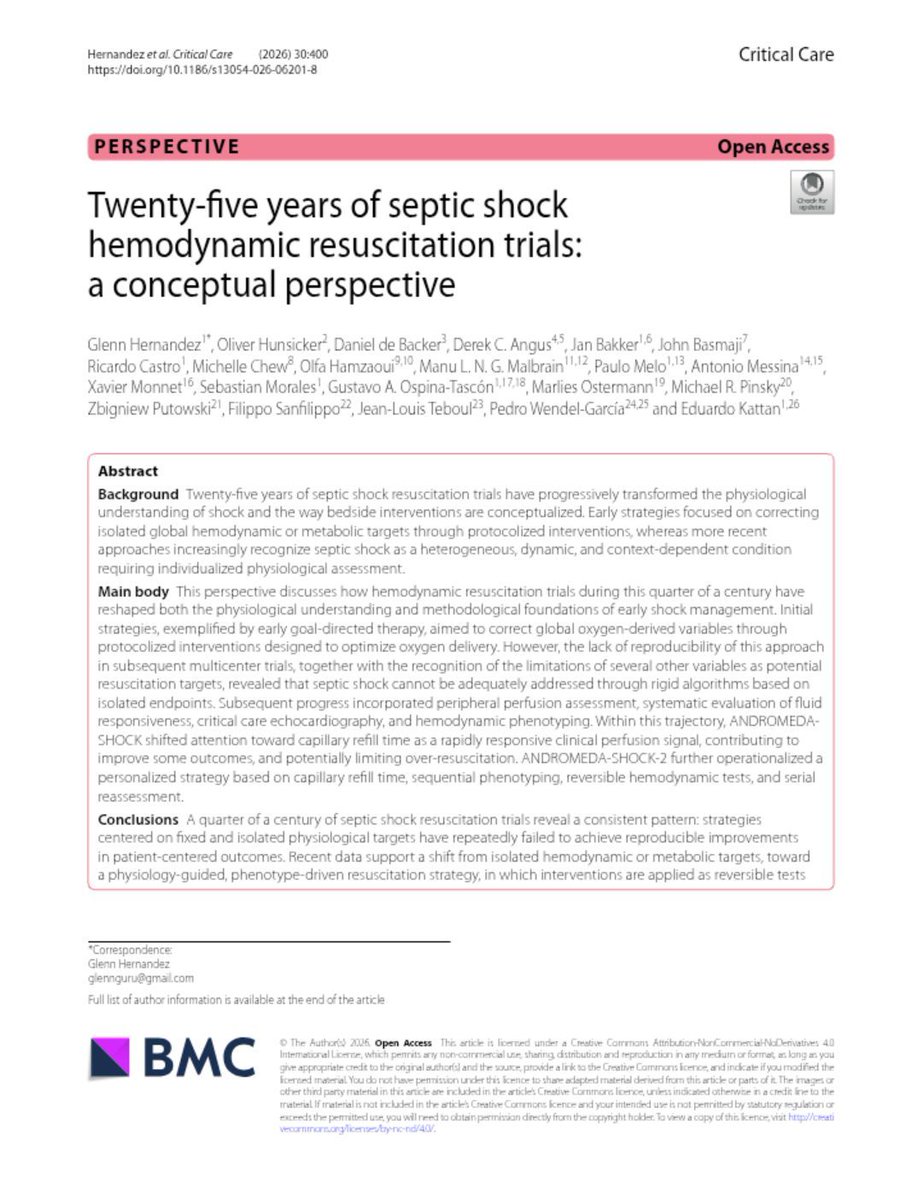
🫀Septic shock resuscitation is entering a new era. Physiology is replacing protocols.
For more than two decades, septic shock research has challenged one assumption after another. We targeted central venous oxygen saturation. We targeted lactate. We targeted mean arterial pressure. We restricted fluids. Yet despite these advances, improvements in patient-centered outcomes remained inconsistent.
This remarkable perspective from many of the world's leading experts argues that the problem was never the individual variables. The problem was expecting a single physiological target to represent an extraordinarily heterogeneous disease.
The evolution of septic shock management tells an important story. Early Goal Directed Therapy demonstrated that protocolized resuscitation could improve outcomes, but subsequent multicenter trials showed that fixed algorithms cannot account for the diversity of septic shock phenotypes. Lactate proved invaluable for risk stratification but increasingly unreliable as an isolated treatment target beyond the initial hours of resuscitation. Mean arterial pressure also evolved from a universal threshold to an individualized physiological variable whose optimal value depends on the patient's underlying cardiovascular state.
The most important conceptual shift has been moving from treating numbers to treating perfusion. Capillary refill time emerged as a rapidly responsive bedside marker that reflects the adequacy of tissue reperfusion. The ANDROMEDA-SHOCK program demonstrated that combining capillary refill time with systematic assessment of fluid responsiveness, focused critical care echocardiography, and sequential hemodynamic phenotyping allows clinicians to individualize therapy while reducing unnecessary interventions.
Another major lesson is that fluids are no longer considered inherently beneficial. Every fluid bolus should answer a physiological question. Is the patient fluid responsive? Will the patient tolerate additional volume? Is venous congestion already limiting organ perfusion? Fluid administration has evolved from a routine intervention into a reversible physiological test.
Perhaps the strongest message of this review is that hemodynamic resuscitation is no longer protocol driven but physiology guided. Septic shock should be understood as a dynamic process requiring repeated cycles of observation, bedside testing, intervention, and reassessment.
The future of resuscitation will not be defined by new numbers, but by a deeper understanding of physiology.
Reference 📚
Hernandez, G., Hunsicker, O., de Backer, D., Monnet, X., Pinsky, M. R., Teboul, J. L., et al. (2026). Twenty-five years of septic shock hemodynamic resuscitation trials: a conceptual perspective. Critical Care, 30, 400. https://t.co/57pYcSZ5de
🫀Congestion is the disease. Diuresis is only the treatment.
For decades, the success of acute heart failure therapy has been measured by urine output, weight loss, or symptom relief. However, these surrogate endpoints do not necessarily indicate that congestion has resolved. Residual congestion at discharge remains one of the strongest predictors of rehospitalization and mortality.
This review proposes a more standardized and physiology-based strategy for decongestion. The objective is not simply to increase urine output, but to achieve effective natriuresis, restore euvolemia, and eliminate congestion before discharge.
The first message is urgency. Intravenous loop diuretics should be administered within 60 minutes of hospital arrival whenever acute heart failure with congestion is diagnosed. Delays may reduce the effectiveness of treatment and worsen outcomes.
The second message is even more important. Early treatment response should be measured objectively. Rather than waiting for body weight to change over several days, clinicians should evaluate:
• Spot urinary sodium after 2 hours, targeting ≥50to70 mmol/L
• Urine output after 6 hours, targeting ≥100to150 mL/hour
Failure to achieve these targets should prompt early intensification of therapy instead of continuing an ineffective regimen.
The review also supports the growing concept of early sequential nephron blockade. Randomized trials such as ADVOR and CLOROTIC demonstrated that adding acetazolamide or thiazide therapy to loop diuretics can improve decongestion in selected patients, particularly when diuretic resistance develops. Acetazolamide appears especially useful in patients with elevated serum bicarbonate, a marker of increased proximal sodium reabsorption and neurohormonal activation.
Another clinically relevant finding is that continuous loop diuretic infusion offers no clear advantage over intermittent bolus administration, consistent with the DOSE trial. Success depends more on achieving an adequate natriuretic response than on the method of administration.
Perhaps the most important lesson is that patients should not leave the hospital while still congested. Successful decongestion must be followed by rapid optimization of guideline directed medical therapy and early outpatient follow up, as demonstrated by the STRONG HF strategy.
Modern heart failure management is evolving from simply prescribing diuretics toward measuring the physiological response to therapy and adapting treatment early. Congestion should no longer be treated empirically. It should be monitored, quantified, and resolved before discharge.
Reference 📚
Bilgeri, V., Spitaler, P., Puelacher, C., et al. (2024). Decongestion in Acute Heart Failure: Time to Rethink and Standardize Current Clinical Practice? Journal of Clinical Medicine, 13(2), 311. https://t.co/fic1FPNt7x
📌 MENSAJE PARA RECORDAR
No toda pericarditis es igual.
👉 El futuro consiste en tratar el mecanismo, no solo el síntoma.
🔥 Inflamación → bloquear IL-1.
🧬 Autoinmunidad/no inflamatoria → inmunosupresión dirigida.
Ese es el cambio que introduce el ACC 2025.
Most people mess up steroid conversions.
Here’s the quick bedside hack I actually use:
4 × 5 = 20 × 1
Methylprednisolone 4 mg
≈ Prednisolone 5 mg
≈ Hydrocortisone 20 mg
≈ Dexamethasone 1 mg ( actual is 0.75 mg)
Not perfectly accurate… but gets you close enough in emergencies.
A normal LDL does not rule out an atherosclerotic stroke.
A familiar stroke-clinic pattern:
A patient in his late 40s develops an MCA infarct. No diabetes. Never smoked. LDL 88 mg/dL.
Yet vascular imaging shows significant ipsilateral carotid atherosclerosis.
The lipid profile looks reassuring.
The artery does not.
This is when lipoprotein(a), or Lp(a), becomes clinically relevant.
Lp(a) is often described as sticky cholesterol. That explanation is incomplete.
Lp(a) is an LDL-like apoB particle with an additional protein, apolipoprotein(a), attached to it. Like LDL, it carries cholesterol into the arterial wall.
But it also carries oxidised phospholipids, which promote vascular inflammation, plaque progression and plaque vulnerability.
The usual sequence is:
Lp(a) enters the arterial wall
Plaque develops and becomes inflamed
The plaque ruptures or erodes
A thrombus forms
Ischaemic stroke follows
The clot is generally the final event. High Lp(a) does not mean that the patient’s entire blood is systemically hypercoagulable.
Apolipoprotein(a) resembles plasminogen and may interfere with fibrinolysis, but the clinical importance of this effect remains uncertain. The strongest human evidence supports an atherogenic and pro-inflammatory mechanism.
This mechanism also predicts the stroke phenotype.
Recent Mendelian-randomisation data found that a genetically predicted 100 nmol/L increase in Lp(a) was associated with:
Large-artery atherosclerotic stroke: OR 1.23
Early-onset large-artery stroke: OR 1.37
Small-vessel stroke: OR 0.98
The main signal is therefore for large-artery atherosclerotic stroke, not lacunar stroke.
Lp(a) should not be used alone to decide whether an acute stroke is cardioembolic, lacunar or atherosclerotic.
Stroke mechanism still depends on the clinical syndrome, vascular imaging, cardiac evaluation and the complete diagnostic work-up.
Lp(a) testing is especially useful when:
• Stroke or TIA occurs before 60
• Carotid or intracranial atherosclerosis appears greater than expected
• Significant plaque is present despite a relatively bland lipid profile
• Large-artery events recur despite good LDL control
• There is a family history of premature stroke or myocardial infarction
Lp(a) is largely genetically determined and relatively stable throughout life.
Current lipid guidance recommends measuring it at least once in every adult.
Reporting in nmol/L is preferred when available.
1. Below 75 nmol/L is considered lower risk.
2. 75–124 nmol/L is intermediate.
3. 125 nmol/L or more is considered high risk.
A fixed formula should not be used to convert mg/dL into nmol/L. Apo(a) particle size varies, making direct conversion unreliable.
What changes when Lp(a) is high?
Not the acute stroke treatment.
Not the antiplatelet strategy automatically.
Not an indication for anticoagulation.
And definitely not the statin.
In patients with ischaemic stroke or TIA and atherosclerotic disease, intensive LDL lowering remains essential, with an LDL-C target below 70 mg/dL.
Ezetimibe should be added when required, and a PCSK9 inhibitor may be considered in an appropriate very-high-risk patient who remains above target despite maximally tolerated therapy.
Blood pressure, diabetes, smoking, exercise and weight require equally rigorous attention.
First-degree relatives should also be tested.
A high Lp(a) result often identifies an at-risk family, not merely one patient.
As of July 2026, no randomised outcomes trial has yet proved that selectively lowering Lp(a) prevents stroke. The phase 3 pelacarsen Lp(a)HORIZON trial is still awaiting definitive outcome results.
Until those data arrive, Lp(a) should not be treated as an isolated number.
The atherosclerotic risk associated with it should be treated aggressively.
LDL builds the plaque. Lp(a) accelerates it.
When the atherosclerosis does not match the conventional risk factors, do not stop at the routine lipid profile.
Sometimes the missing answer is Lp(a).
#Stroke #LipoproteinA #Atherosclerosis #Neurology #NeuroTwitter #MedX
Before calling it ICU-acquired weakness... look at the face.
Before writing CIP/CIM in the notes, do one examination first.
Look at the face.
Why?
Because critical illness polyneuropathy (CIP) and critical illness myopathy (CIM) usually follow a predictable pattern:
✓ Symmetrical limb weakness
✓ Proximal > distal weakness
✓ Difficulty weaning from the ventilator
✓ Facial and extraocular muscles are usually spared.
So if you find:
• Ptosis
• Diplopia
• Facial weakness
• Dysarthria
• Dysphagia
• Ophthalmoplegia
Pause. Reopen the diagnosis.
Now think of:
→ Myasthenia gravis
→ Guillain-Barré syndrome
→ Brainstem stroke
→ Botulism
→ Intubation-related cranial nerve injury
My bedside rule
Legs tell you there is weakness.
The face tells you what the weakness is.
Never diagnose ICU-acquired weakness by the duration of ICU stay.
Diagnose it by the pattern of weakness.
That one-minute facial examination may save the patient from the wrong diagnosis and the wrong treatment.
Reference: Neurologic Complications of Critical Illness (Continuum, 2026): “The limbs are affected symmetrically and proximally more than distally... Facial and extraocular muscles are typically spared.”
#NeuroX #NeuroICU #CriticalCare #MedicalEducation #MedX
A hidden pearl about colonoscopy is that adding Coke Zero to bowel preparation makes it much easier to drink and improves quality of views for the endoscopist.