🎤 Tune in to our new series of #podcasts: 𝗧𝗵𝗲 𝗟𝗶𝗴𝗵𝘁-𝗦𝗵𝗲𝗲𝘁 𝗖𝗵𝗿𝗼𝗻𝗶𝗰𝗹𝗲𝘀!
▶️ https://t.co/fgRfFc5kqf
Listen to Dr. Malte Wachsmuth (Bruker), Dr. Johanna Perens (Gubra), and Dr. Jacob Hecksher-Sørensen (Gubra) share the advantages and advancements of #LSFM!
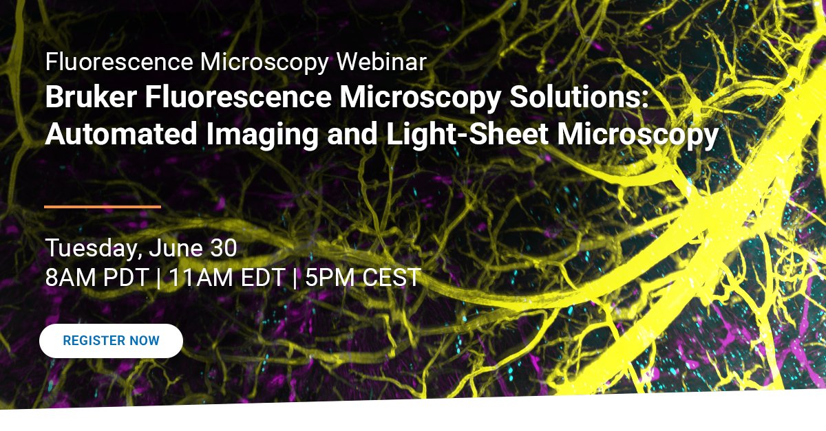
🖥️ Automated Imaging and Light Sheet Microscopy
▶️ https://t.co/CupoUjYBCc
This webinar presents the Bruker Luxendo imaging ecosystem, which connects automated #microscopy, light-sheet #imaging, and high-performance #data infrastructure into a cohesive, scalable workflow.
🖥️ Automated Imaging and Light Sheet Microscopy
▶️ https://t.co/xhgB5NqN24
This webinar presents the Bruker Luxendo imaging ecosystem, which connects automated #microscopy, light-sheet #imaging, and high-performance #data infrastructure into a cohesive, scalable workflow.
❓ Can you efficiently view data immediately following your experiments?
▶️ https://t.co/Tm4wIZGpUZ
🎙️This segment was taken from Episode 4 of our 𝘓𝘪𝘨𝘩𝘵-𝘚𝘩𝘦𝘦𝘵 𝘊𝘩𝘳𝘰𝘯𝘪𝘤𝘭𝘦𝘴 𝘗𝘰𝘥𝘤𝘢𝘴𝘵
#lifesciences#microscopy#MicroscopyMonday
🎤 Tune in to our newest episode of 𝗧𝗵𝗲 𝗟𝗶𝗴𝗵𝘁-𝗦𝗵𝗲𝗲𝘁 𝗖𝗵𝗿𝗼𝗻𝗶𝗰𝗹𝗲𝘀!
▶️ https://t.co/VUmEPtQsbU
Learn about computational, algorithmic, and workflow strategies necessary for #biomedical image analysis of light-sheet data.
#lifescience#microscopy#podcasts
🔬 𝗙𝘂𝘀𝗶𝗼𝗻 𝗼𝗳 𝗵𝗲𝗮𝗹𝘁𝗵𝘆 𝗮𝗻𝗱 𝗱𝗶𝘀𝗲𝗮𝘀𝗲𝗱 𝗜𝗣𝗦𝗖 𝗻𝗲𝘂𝗿𝗼-𝘀𝗽𝗵𝗲𝗿𝗼𝗶𝗱 𝗹𝗮𝗯𝗲𝗹𝗲𝗱 ����𝗶𝘁𝗵 𝗝𝗙𝟱𝟱𝟮.
Imaged with Ultima 2Pplus and the NEW Inverter Module.
Courtesy of Trish Hoang, Promega.
#MicroscopyMonday #neuroscience #organoids
🔬 Acquisition in CA1 and ACC regions with over 200 cells in CA1 identified across multiple days, and ~80% of cells registered across multiple sessions.
Acquired with Quartet.
Courtesy of Gal Elyasaf, Ph.D., Weizmann Institute.
#MicroscopyMonday#Neuroscience#Microscopy