Can you guess the diagnosis?
.
.
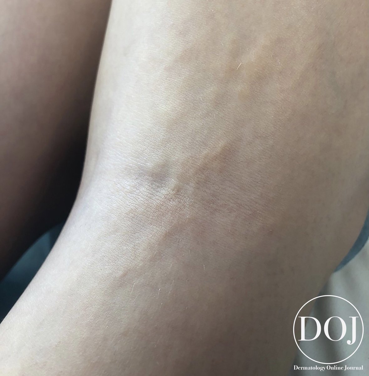
An 8-year-old girl presented with asymptomatic sclerotic flesh-yellowish colored papules and nodules in a linear distribution over her right popliteal space, not following a specific dermatome.
#dermtwitter#dermatology#diagnosisoftheday
H&E stain showed a normal epidermis, increased thick collagen bundles in the dermis, and dermal thickening. Trichrome stain showed haphazardly arranged thick collagen bundles in the dermis and orcein stain showed dense collagen fibers with diminished elastic fibers.
Can you guess the diagnosis?
.
.
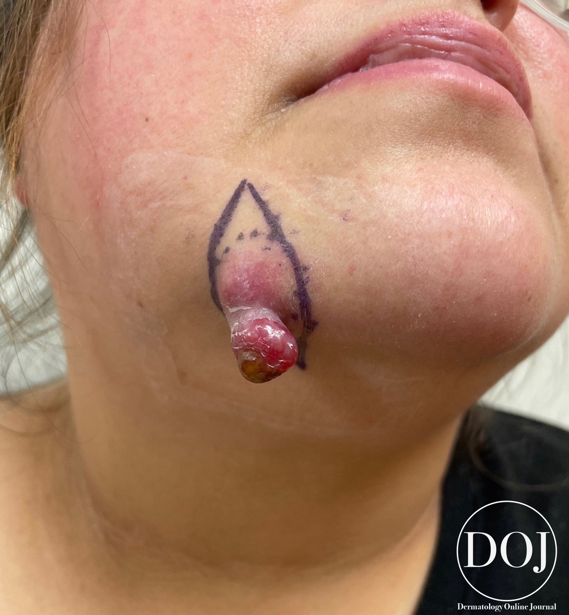
A 45-year-old woman presented with a four-month history of a painful, bleeding chin nodule previously treated with warm compresses, antibiotics, and incision and drainage.
Of note, the patient had a history of RCC that was treated 7 years prior with a nephrectomy and inferior vena cava thrombectomy with no evidence of recurrence.
Can you guess the diagnosis?
.
.
A 45-year-old woman presented with a four-month history of a painful, bleeding chin nodule previously treated with warm compresses, antibiotics, and incision and drainage. Because this nodule continued to grow, she presented to dermatology.
Can you guess the diagnosis?
.
.
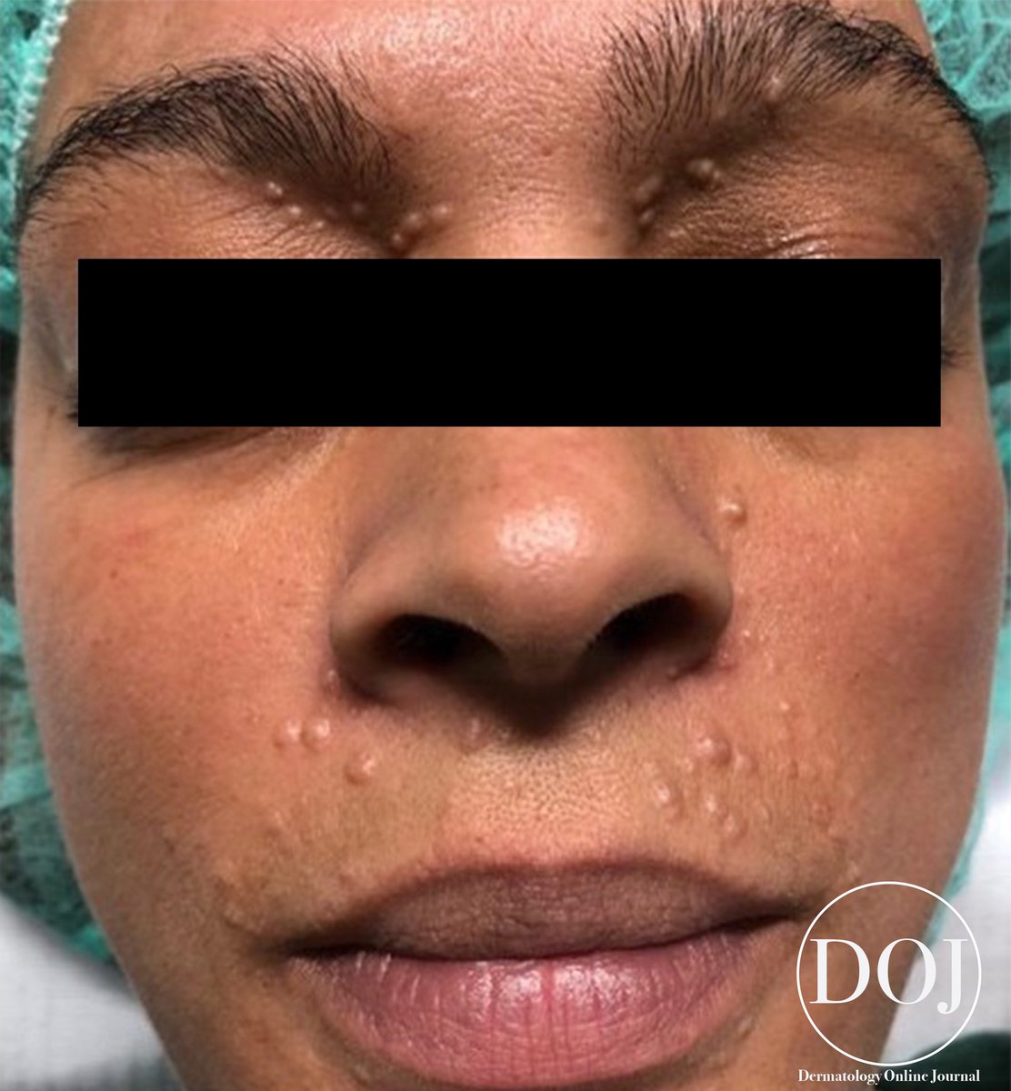
A 38-year-old woman presented with a 5-year history of multiple small, asymptomatic, follicular, skin-colored papules over the face, particularly in the periorbital region and the upper cutaneous lip.
Histopathology of an upper lip lesion showed an irregular proliferation of basaloid cells in anastomosing strands and cords with peripheral palisading and cysts with trichilemmal keratinization. Immunohistochemistry was positive for BerEP4.
Can you guess the diagnosis?
.
.
A 60-year-old man presented for evaluation of a recurrent asymptomatic rash consisting of variably-sized, arcuate-to-annular, erythematous papules and small plaques of the upper back, as shown in the image.
Biopsy was notable for a dense infiltrate consisting of monomorphic lymphocytes oriented around superficial and deep vascular and adnexal structures. Epidermal change, plasma cells, and dermal mucin were absent. IHC confirmed the presence of a predominantly T cell population.