Paper I’m reading today — September 21, 2026
Can MRI help approximate the disease-relevant information clinicians seek from FDG-PET—without treating the cortex like an ordinary volume?
In this arXiv paper, Yitong Li et al. introduce DB-SUiT, a surface-based diffusion bridge for translating MRI to PET on the cortical manifold. The authors focus on cortical hypometabolism as a dementia biomarker, while addressing the cost, radiation exposure, and accessibility constraints of PET. Rather than generating volumetric images alone, their approach models the folded cortical geometry directly and conditions synthesis on demographic and subcortical information.
The model combines:
• Spherical convolutional encoders for multi-scale surface features
• Bottleneck Transformers for long-range spatial dependencies
• A conditional spherical U-shaped vision Transformer (SUiT)
• A diffusion bridge for MRI-to-PET translation
• Cortical surface topology with demographic and subcortical conditions
What I find most interesting is the representation choice: the paper treats cortical geometry as part of the problem, not just as a post-processing detail. The abstract reports evaluation across two datasets, including different dementia types, as well as an external cohort without retraining. It also reports gains in automated dementia classification and a blinded reader study comparing synthetic PET with MRI and real PET. These are encouraging findings, but synthetic PET should be evaluated carefully as a complementary research tool rather than assumed to replace measured PET in every setting.
The broader direction matters for AI × Medicine: modality translation could make informative imaging signals more accessible, provided that anatomical fidelity, cross-cohort robustness, clinical validity, and failure modes are rigorously characterized. I’m looking forward to seeing how surface-native generation develops beyond this study.
Reading note—not my work.
Paper: “Bridging Modalities on the Cortex: Surface-based MRI to PET Translation with a Diffusion Bridge,” Yitong Li et al., arXiv:2609.20147v1 (2026). https://t.co/aLF57SzfcT
Not medical advice.
Figures from Yitong Li et al.: Figure page 4, Figure page 7. Original images/PDF page screenshots. https://t.co/DSBbw5ATwU
Paper I’m reading today — September 20, 2026
Can non-contrast CT support reliable infarct segmentation and net water uptake (NWU) quantification across hospitals—without relying on advanced imaging?
In this arXiv study, Linus Britt et al. investigate a domain-aware deep learning framework for ischemic stroke lesion segmentation on NCCT. Using data from 801 patients across four datasets, the authors train an nnU-Net-based model and fine-tune it with small target-domain subsets before evaluating it on held-out external cases. The goal is practical: automate lesion delineation and extract NWU, a quantitative biomarker, from baseline NCCT. This is my reading note and a recommendation to explore the authors’ work—not my own research.
The model combines:
• NCCT input
• nnU-Net-based lesion segmentation
• supervised target-domain adaptation
• external evaluation across heterogeneous cohorts
• automated NWU quantification against expert references
What I find most interesting is the paper’s focus on domain shift as a central clinical deployment problem, rather than treating segmentation performance on one dataset as sufficient. The reported results vary across cohorts and lesion sizes, which makes the adaptation strategy—and the remaining limitations—particularly important. The abstract reports median Dice values of 0.68 for lesions ≥30 mL in Boston and 0.56 in ISLES, while automated NWU quantification in Boston had a mean absolute error of 1.37 percentage points.
The direction is promising, but the authors appropriately frame it as supporting further prospective clinical evaluation. For AI × Medicine, this is a useful example of how domain-aware adaptation may help translate imaging models from curated datasets toward more heterogeneous real-world settings—while keeping validation and generalization in view.
Paper: Linus Britt et al., “Ischemic Stroke Segmentation and Net Water Uptake Quantification on Multicenter Non-Contrast CT Using Supervised Target-Domain Adaptation,” arXiv:2609.20151v1 (2026).
https://t.co/M7mniAAddx
Not medical advice.
Figures from Linus Britt et al.: Figure page 8, Figure page 15, Figure page 16. Original images/PDF page screenshots. https://t.co/DSBbw5ATwU
Paper I’m reading today — September 19, 2026
Can a medical LLM do more than answer exam questions—can it take a focused history, assess urgency, and avoid dangerous next steps in Japanese?
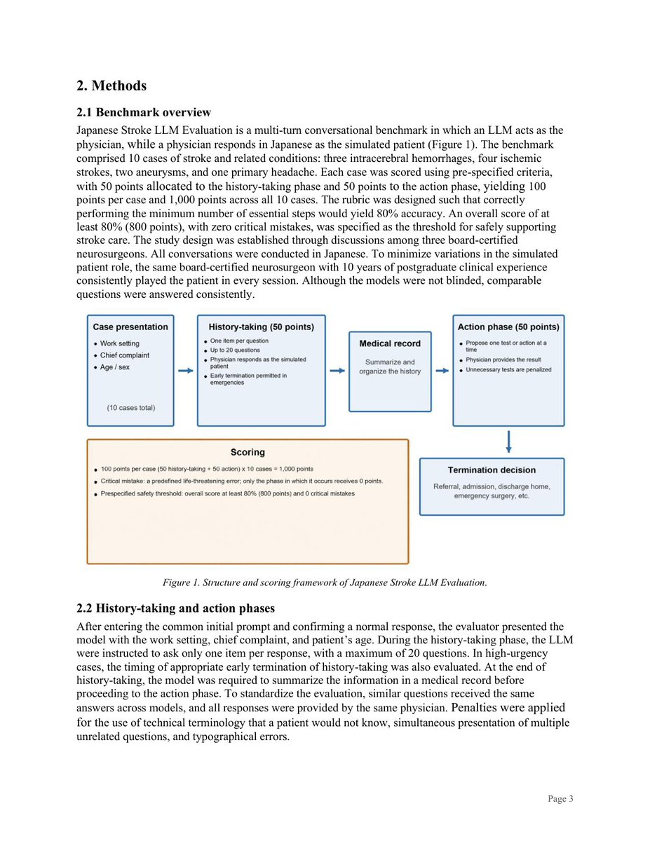
In this arXiv paper, Keisuke Masuda et al. introduce Japanese Stroke LLM Evaluation, a multi-turn conversational benchmark for stroke and related-condition care. The authors evaluated 18 models in October 2025 and June 2026, with the LLM acting as physician and a board-certified neurosurgeon serving as simulated patient and evaluator. Cases were scored across history-taking and action phases, with critical mistakes defined as errors that could directly threaten life.
The benchmark combines:
• 10 practice-oriented Japanese cases
• Multi-turn history-taking with a cap on questions
• Separate history and action scoring
• Specialist-created cases and evaluation
• A safety threshold of ≥80% overall with zero critical mistakes
The reported results are a useful reminder that overall scores alone are not enough. Claude Fable 5 achieved 87.4% with zero critical mistakes, followed by Claude Opus 4.7 at 80.3% and GLM-5.2 at 75.6%; two models met the stated safety threshold. Eleven models made 17 critical mistakes, including failures related to confirming laboratory results or blood glucose before t-PA, airway stabilization before surgery, cervical vascular evaluation, and t-PA indication. The paper also reports that question count correlated with history-taking score (r = 0.648, p = 0.007).
What I find most interesting... is the evaluation design. It tests conversational process and safety—not just recall—and avoids an LLM-as-judge setup by using neurosurgical specialists. That makes the benchmark feel closer to the kinds of constraints clinical systems face, while still leaving the authors’ own caveat central: further evaluation with real-world cases is needed.
This direction matters for AI x Medicine because deployment-quality evaluation has to measure uncertainty, questioning, urgency, and critical failure modes in the language and workflow where a system would actually be used. These results are promising for some models, but they are not a substitute for clinical validation or safe oversight.
Reading note / promotion of someone else’s work.
Paper: “Japanese Stroke LLM Evaluation: A Conversational Benchmark for Safe Stroke Care in Japanese Using Large Language Models” — Keisuke Masuda, Kazutaka Yatsushiro, Hirohumi Iwamoto, Hirofumi Hirano, and Ryosuke Hanaya. arXiv:2609.16739v1 (2026). https://t.co/P5ccznMCcY
Not medical advice.
Figures from Keisuke Masuda et al.: Figure page 3, Figure page 6. Original images/PDF page screenshots. https://t.co/DSBbw5ATwU
Paper I’m reading today — September 18, 2026
If clinicians know where and how to find a concept in a patient note, why is it still so difficult to make an LLM do the same?
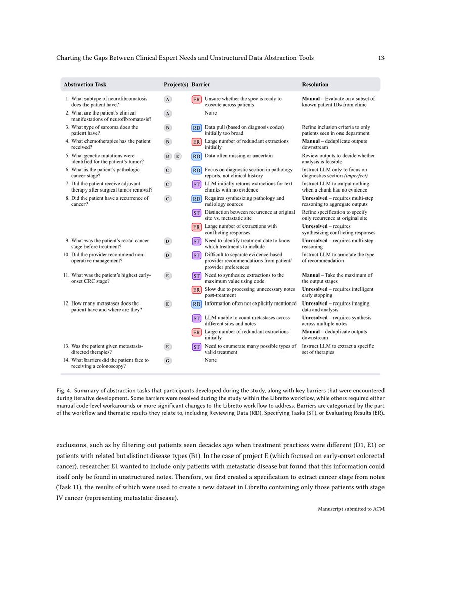
In this arXiv paper, Venkatesh Sivaraman et al. study that gap through Libretto, an interactive LLM-based clinical data abstraction system co-designed with seven cancer research teams. The authors evaluate whether the system helps researchers answer real-world questions from patient records—without assuming deep AI expertise. Their findings suggest that clinical intuition does not automatically translate into effective LLM workflows: across 12 of 14 tasks, teams encountered barriers involving contextual judgments about note reliability, steering “vibe-coded” prompts, and inflexible evaluation strategies.
The workflow combines:
• Clinical researchers’ domain knowledge
• Unstructured patient notes
• An interactive LLM-based abstraction system, Libretto
• Annotation, prompting, and evaluation for research questions
What I find most interesting... is the distinction between knowing where to look and being able to operationalize that knowledge with current AI tools. The challenge is not simply extracting more text or writing better prompts. It is designing systems that respect how clinical researchers judge context, ambiguity, and evidence while making those judgments usable in an information-extraction workflow.
This points toward an important direction for AI x Medicine: tools should be built around clinical research practices, not just adapted from generic LLM interfaces. The open problems here seem substantial, but studying them with real users and real abstraction tasks is a useful step toward more reliable and usable systems.
Reading note / paper promotion—not my work.
Citation: Venkatesh Sivaraman et al., “I Know Where to Look,” But Does the LLM? Charting the Gaps Between Clinical Expert Needs and Unstructured Data Abstraction Tools, arXiv:2609.19318v1. https://t.co/IUV95TYnMD
Not medical advice.
Figures from Venkatesh Sivaraman et al.: Figure page 13. Original images/PDF page screenshots. https://t.co/DSBbw5ATwU
Paper I’m reading today — September 17, 2026
Can multimodal AI make sarcopenia screening more scalable for patients with gastric cancer?
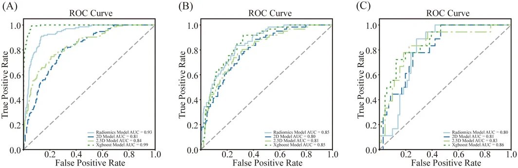
In this study, Huaiqing Zhi et al. explore that question using data from 1,067 patients across two medical centres. The authors develop and evaluate models for identifying sarcopenia and severe sarcopenia, combining information from clinical variables, CT-based radiomics, and deep learning features. The paper is indexed on PubMed; this is my reading note on work by the authors, not my own research.
The model combines:
Clinical variables
+ CT radiomics from the third lumbar vertebral level
+ 2.5D deep learning features from a ResNet50-based model
→ transformer-based multimodal sarcopenia model
An XGBoost fusion model using the same three modalities serves as a comparator. The abstract reports external-test AUCs of 0.89 for sarcopenia and 0.84 for severe sarcopenia for the multimodal model, with performance assessed across training, validation, and external test sets.
What I find most interesting... is the explicit comparison between single-modality and multimodal approaches. Radiomics and deep learning each provide useful imaging signals, but the paper frames clinical variables, engineered image features, and learned image features as complementary rather than interchangeable. That is a practical design choice for AI x Medicine, where the value of a model depends not only on predictive performance but also on whether it can support a clinically meaningful screening and risk-stratification workflow.
The direction is promising, while prospective evaluation, workflow integration, and assessment across broader patient populations will remain important before models like this can be considered for routine use.
Huaiqing Zhi et al., “Integrated Assessment of Sarcopenia in Patients with Gastric Cancer Using Deep Learning and Radiomics.” DOI: 10.1002/jcsm.70377 | PubMed: https://t.co/jd5Tg2VJej
Not medical advice.
Figures from Huaiqing Zhi et al.: FIGURE 2, FIGURE 4, FIGURE 5. Original images/PDF page screenshots. https://t.co/DSBbw5ATwU
Paper I’m reading today — September 12, 2026
What if dental AI could help public health teams see gaps in care—not just support decisions in a clinic?
In this review, Panagiotis Douris and George Kodovazenitis examine how aggregated, de-identified outputs from AI diagnostic systems might become real-time signals for dental public health surveillance. They discuss opportunities alongside methodological, privacy, governance, and ethical constraints, including bias, interpretability, and accountability. The article does not present a new model; it proposes a framework for thinking about how existing AI outputs could be connected to population-level monitoring.
The framework combines:
1. Data ingestion
2. Spatiotemporal aggregation
3. Socioeconomic enrichment
4. Predictive modeling
5. Dashboard deployment
What I find most interesting is the shift in scale: diagnostic systems are framed not only as tools for individual workflows, but also as possible sources of information about structural barriers to care when their outputs are combined with geospatial and socioeconomic data. That promise also makes safeguards essential—especially around privacy, bias, and who is accountable for acting on the resulting intelligence.
The next step, as I read it, is not simply building more capable systems. It is testing whether these signals can be governed and interpreted responsibly enough to support equitable public health action.
Panagiotis Douris et al., “AI-Driven Dentistry and Public Health Surveillance: Opportunities and Challenges.” DOI: 10.1002/puh2.70278
https://t.co/Xp5DBAB3hH
Not medical advice.
Figures from Panagiotis Douris et al.: FIGURE 2. Original figure; WebP converted to PNG. https://t.co/DSBbw5ATwU
Paper I’m reading today — September 8, 2026
How much spatial transcriptomic information can histology images recover when direct ST data are expensive and time-consuming to generate?
In “Spatially guided translation from histology images to transcriptomic profiles using foundation model-driven contrastive learning,” Zi Huai Huang, Ziyang Xu, and Pingzhao Hu introduce FOCST, a framework for predicting and imputing spatial transcriptomic profiles from tissue images. The approach uses UNI, a histopathology foundation model, to extract visual features; contrastive learning to align image and expression information in a shared embedding space; and a graph neural network to incorporate positional relationships and improve spatial awareness.
The model combines:
• Histology-image features from UNI
• Cross-modal contrastive learning between images and expression data
• A graph neural network for positional and regional context
• Predicted transcriptomic profiles for downstream analyses
What I find most interesting is the attempt to treat morphology and spatial organization as complementary signals rather than solving image-to-expression translation with visual features alone. The abstract reports benchmarking against existing methods and alternative vision encoders, with statistically significant differences across six images, and describes downstream patient stratification by treatment response with ROC AUC = 0.79. Those results are encouraging, but they also make the evaluation setting and generalization question especially important.
A promising direction for reducing barriers to ST analysis—but I’d want to see broader validation before treating image-derived profiles as a substitute for measured spatial transcriptomics.
Citation: Huang ZH, Xu Z, Hu P. “Spatially guided translation from histology images to transcriptomic profiles using foundation model-driven contrastive learning.” DOI: 10.1371/journal.pcbi.1014762
https://t.co/DCSJHaMo9h
Not medical advice.
Figures from Zi Huai Huang et al.: Fig 1. Original figure; WebP converted to PNG. https://t.co/DSBbw5ATwU
Paper I’m reading today — September 16, 2026
How should a survival-prediction system reason when the patient data it needs is multimodal—and some modalities may be missing or vary across cases?
In this arXiv work, H Mathavan and H Liu explore multi-modal tumor survival prediction using clinical data, cell slides, and genomics. Rather than training one foundation model end to end, the authors propose a graph-guided Mixture of Experts (MoE) framework that coordinates multiple existing models and manages their use as tools. The abstract reports experiments on the TCGA-LUAD dataset, with improved performance over individual-modality models and a vanilla ensemble.
The model combines:
• Clinical data + cell-slide information + genomics
• A graph structure to guide prediction
• Multiple expert models in an MoE-style ensemble
• Automatic model calling to accommodate complex, heterogeneous inputs
What I find most interesting is the shift from “one model must absorb everything” toward structured coordination among specialized models. In real-world biomedical settings, the challenge is not only learning from more modalities; it is also handling incomplete inputs, modality variation, and the practical question of which model should contribute when.
This is still a reading note about someone else’s work, not my own research. The direction seems promising for AI × Medicine, but the broader value will depend on how robustly these systems handle missing data, generalize beyond the reported setting, and support careful evaluation in clinically meaningful workflows.
Paper: H Mathavan and H Liu, “Multi-Modal Tumor Survival Prediction via Graph-Guided Mixture of Experts,” arXiv:2609.14072v1, 2026.
https://t.co/e588kU43Fl
Not medical advice.
Figures from H Mathavan et al.: Figure page 28. Original images/PDF page screenshots. https://t.co/DSBbw5ATwU
Paper I’m reading today — September 13, 2026
What would it take to move AI in cancer theranostics from promising tools to routine, clinically validated workflows?
In this review, Zahra Mansouri et al. critically examine AI across the cancer theranostic workflow, focusing on the technical and clinical barriers that limit personalized radiopharmaceutical therapy—including complex dosimetry, demanding imaging protocols, and quantitative image analysis. The paper considers both current maturity and readiness for clinical translation.
The model combines:
• Automated image interpretation
• Quantitative imaging and accelerated acquisition
• Single-time-point dosimetry
• Radiomics and multi-omics analyses
• Emerging digital twins, physics- and biology-informed neural networks, and explainable AI
What I find most interesting is the workflow perspective. The promise is not limited to one prediction task: AI could potentially connect imaging, dosimetry, treatment planning, and biological information. But the review is appropriately cautious—data quality, interpretability, ethics, privacy, standardization, and clinical validation remain central obstacles.
The next step is not simply building more capable models. It is establishing trustworthy, reproducible, clinically evaluated systems that can support personalized radiopharmaceutical therapy without obscuring uncertainty. Whether that leads to routine dosimetry-guided treatment will depend on evidence gathered across real clinical workflows.
Citation: Mansouri Z, Salimi Y, Akhavanallaf A, Zaidi H. “Artificial Intelligence Across the Cancer Theranostics Workflow: Critical Appraisal of Current Evidence and Future Clinical Translation.” DOI: 10.1016/j.neunet.2020.07.010
https://t.co/2r0o4A9ZJO
Not medical advice.
Paper I’m reading today — September 13, 2026
What could AI-assisted analysis add to breast cancer screening and diagnosis without replacing clinical expertise?
Rana et al. present a systematic review of artificial intelligence and machine learning applications in breast cancer pathology, covering screening, diagnosis, classification, and tumor-marker scoring. Their search focused on PubMed and Google Scholar and included articles involving breast cancer pathology, with attention to cytology, histology, medical imaging, and biomarker expression.
The model family combines:
• Artificial neural networks (ANNs)
• Support vector machines (SVMs)
• Convolutional neural networks (CNNs)
• Faster R-CNN
• Imaging, pathology, and tumor-biomarker analysis
What I find most interesting is the breadth of the proposed assistive role: AI is discussed not only for image interpretation, but also for connecting imaging with pathology and supporting more consistent, rapid, and potentially cost-effective workflows. The review also points to the growing translation of these tools into approved software products, while emphasizing their relevance to diagnosis, research, precision medicine, and patient care.
The cautious takeaway is that AI may help address workload and consistency challenges in pathology, but its practical value will depend on how reliably these systems are evaluated and integrated into clinical workflows. I’ll be watching for evidence that clarifies where assistance is most useful—and where expert judgment remains essential.
Paper: Manjit Kaur Rana et al., “Artificial Intelligence as an Assistive Tool in Breast Cancer Screening and Diagnosis in the Digital Era.” DOI: 10.1155/ijbc/5575969
https://t.co/QShI6Z8mMd
Not medical advice.
Paper I’m reading today — September 12, 2026
Can modeling the meaning of biomedical entities—and the type of side effect being predicted—improve polypharmacy risk prediction?
In “Beyond Structural Representations: A Semantic-Relational Graph Neural Network for Polypharmacy Side Effect Prediction,” Fatma Zahra Abdeldjouad and Menaouer Brahami introduce SIREN, a graph neural network designed to address limitations of relation-agnostic prediction in polypharmacy side-effect modeling. The paper evaluates the approach on the TWOSIDES dataset in transductive and inductive settings.
The model combines:
• Semantic embeddings from ChemBERTa and BioBERT
• A multirelational biomedical graph structure
• A relation-specific decoder that adapts drug-pair scores to the target side-effect type
What I find most interesting is the modeling choice to make the decoder explicitly sensitive to the predicted relation. In this setting, the same drug pair may need to be interpreted differently depending on which side effect is being considered. The reported ablations also point to the importance of both relation-specific decoding and pretrained node embeddings, while the evaluation reports strong performance in transductive settings and competitiveness in inductive ones.
A useful direction to watch is whether combining semantic representations with graph reasoning continues to generalize across settings and datasets. As always, model performance is one part of the broader validation needed before such systems can support real-world pharmacovigilance workflows.
Paper: Fatma Zahra Abdeldjouad and Menaouer Brahami, “Beyond Structural Representations: A Semantic-Relational Graph Neural Network for Polypharmacy Side Effect Prediction.” DOI: 10.3390/ijms20020386 | PubMed: https://t.co/g90x1zlWUQ
Not medical advice.
Paper I’m reading today — September 11, 2026
How is AI research in out-of-hospital cardiac arrest (OHCA) developing—and where is the field still concentrated?
Fee et al. present a bibliometric analysis of the AI–OHCA literature using the Web of Science Core Collection. They identified 290 publications spanning 1996–2026 and examined publication and citation trends, countries, institutions, collaboration patterns, journals, subject categories, and keyword networks using descriptive analyses and VOSviewer. The analysis describes a sharp increase in research output since 2019, with contributions concentrated among a relatively small number of countries, institutions, and collaborative groups.
The research landscape spans:
• Cardiac arrest recognition
• Rhythm analysis
• Prognostication and survival prediction
• Clinical decision support
• Resuscitation and post-arrest outcomes
What I find most interesting is the field-level perspective: the literature appears clinically oriented, but its growth does not automatically establish readiness for real-world EMS use. Mapping who is publishing and which themes are emerging can help clarify where evidence is accumulating—and where broader validation, implementation work, and attention to diverse patient populations and EMS systems are still needed.
A useful next step will be seeing whether future studies move beyond promising applications toward prospective, externally validated evaluation in real-world settings. This paper provides a snapshot of that evolving research landscape, not a clinical recommendation.
Paper: Fee S, Nichols E, Ganti L, Banerjee P. “Artificial intelligence applications in out-of-hospital cardiac arrest: an analysis of emerging research trends.” Resuscitation Plus. DOI: 10.1016/j.resplu.2025.100973
https://t.co/grqPmRVBFi
Not medical advice.
Paper I’m reading today — September 11, 2026
How much does segmentation performance depend on the organ—and on the imaging modality—rather than on the model name alone?
Nguyen and Bui present a scoping review of deep-learning approaches for segmenting the liver, kidneys, and pancreas across ultrasound, MRI, and CT. Following scoping-review reporting guidance, they searched five databases for studies published from 2020–2026 and charted 72 studies by organ, modality, method family, dataset, and performance.
The review’s schematic:
• Organ tractability: liver > kidney >> pancreas
• Modality maturity: CT > MRI > US
• Method waves: CNNs → transformers → segmentation foundation models → LLM-based agents
• Persistent gap: tumor segmentation trails organ segmentation
• Emerging pattern: agents augment rather than replace specialist segmentation
What I find most interesting is the review’s emphasis on evaluation design, not just headline scores. Foundation models matched or exceeded equally box-prompted specialists on individual unseen tasks, but the cited comparison used the Segment Anything Model and gave every comparator the same ground-truth-derived box. Across the two benchmarks in the review pool with a fully automatic comparator, that comparator led on one and came within about one Dice point on the other. No benchmark evaluated specialists, foundation models, and agents under one common protocol.
The picture is therefore promising but uneven: performance degrades sharply for the pancreas and ultrasound, while LLM/agentic methods have not surpassed specialists on overlap metrics. Their potential contribution currently appears to be generality, autonomy, and annotation efficiency. More standardized, fully automatic, modality-stratified comparisons may clarify how far these approaches can go—especially in the areas the review identifies as underserved.
Paper: “Deep Learning Segmentation of Abdominal Organs Across Ultrasound, MRI, and CT: A Scoping Review of Modality-Specific Approaches and the Emerging Role of Foundation Models and LLM-Based Agents.” Thi My Tien Nguyen and Ngoc Thang Bui. DOI: 10.2196/72109
https://t.co/FiIT4Zpyon
Not medical advice.
Paper I’m reading today — September 10, 2026
Can heterogeneous diabetes data be combined without sacrificing privacy, personalization, or interpretability?
I’m reading “FedMediFormer-XAI” by Netra Patil, Naveenkumar Jayakumar, and Sudan Jha. The paper describes a framework for diabetes intelligence that brings together clinical records, population indicators, continuous glucose monitoring, retinal fundus images, wearable measurements, and pharmacological information. Its reported representative implementation includes 94.2% accuracy and an AUC-ROC of 0.96, while the authors note that prospective multicenter clinical evaluation is still needed.
The model combines:
• Federated learning for collaborative training without sharing raw patient data
• Multimodal transformers for learning across different data types
• Diffusion-based augmentation for synthetic samples and class imbalance
• Graph neural networks for patient–drug and drug–drug relationships
• XAI methods including SHAP, attention visualization, Integrated Gradients, and counterfactual reasoning
What I find most interesting is the attempt to treat privacy, multimodality, recommendation, and explainability as parts of one system rather than isolated technical problems. The abstract also reports graph-based recommendation results—precision 0.89, recall 0.84, and NDCG 0.91—but these figures should be interpreted in the context of the reported implementation, not as evidence of clinical utility.
The framework suggests a promising direction for decentralized healthcare AI, but its generalizability, robustness, and real-world usefulness will depend on external validation and prospective evaluation. In particular, combining synthetic data, multimodal inputs, and recommendations makes careful assessment of bias, calibration, and explanation quality important next steps.
Citation: Patil N, Jayakumar N, Jha S. “FedMediFormer-XAI: Federated Multimodal Transformers with Diffusion Augmentation and Graph-Based Drug Recommendation for Diabetes.” DOI: 10.3791/73113. PubMed: https://t.co/ui8K1lKP3L
Not medical advice.
Paper I’m reading today — September 10, 2026
Can routinely collected ultrasound and clinical data help estimate breast cancer recurrence risk over five years without relying on costly genomic assays?
Chang et al. present a feasibility study of a deep learning framework for five-year breast cancer recurrence prediction. The approach is designed around real-world clinical data and combines ultrasound, radiomics, clinical variables, and report-derived features rather than relying on a single information source. The abstract reports a final-model AUC of 0.7540, accuracy of 74.61%, sensitivity of 70.41%, and specificity of 76.44%.
The model combines:
• Automated tumor segmentation from ultrasound images using MedSAM
• Radiomics extracted from the segmented tumor regions
• Report features generated with a MedCPT-based approach and clinically informed queries
• Image representation using the Ultrasound Foundation Model (USFM)
• Structured clinical data processed with FT-Transformer
• Multimodal feature fusion through a mixture-of-experts classifier
• Progressive fusion training
What I find most interesting is the emphasis on integrating complementary clinical signals. The study does not treat imaging, reports, and structured data as interchangeable; instead, it gives each modality its own processing pathway before fusion. The reported additive value of multimodal integration—and the feasibility of using tumor-mask information with ultrasound—makes this a useful example of how accessible data sources might be combined for risk assessment.
This is still a feasibility study, so the findings should be read as an encouraging step rather than evidence of ready clinical deployment. Further evaluation will be important before judging how robustly such a framework performs across settings and populations.
Paper: Chang RF, Huang HW, Kuo SY, Lee YW, Wang MY, Huang CS. “Breast Cancer Recurrence Status Assessment in 5 Years Using Multimodal Integrated Learning: A Feasibility Study.” DOI: 10.1007/s10278-026-02252-z
https://t.co/VRuyHF2I1D
Not medical advice.
Paper I’m reading today — September 9, 2026
What if a medical imaging model learned the radiographic state behind both a diagnosis and the evidence used to support it?
In this arXiv paper, Suyang Xi et al. introduce MedDream, a radiographic world model trained on paired chest radiograph–text observations. Rather than treating diagnostic prediction and image generation as separate tasks, the authors explore a shared continuous latent representation that can support clinical reasoning, report-conditioned radiograph generation, and targeted evidence construction. The paper reports evaluation across eight clinical datasets and two independent reader cohorts, including tests of disease recognition, label-scarce adaptation, severity assessment, localization, and synthetic-data augmentation.
The model combines:
Chest radiograph + clinical text
↓
Shared continuous latent radiographic state
↙ ↘
Diagnostic reasoning Report-conditioned evidence generation
↓ ↓
Clinical readout Targeted synthetic augmentation
What I find most interesting... is the move from “predict a label” or “generate an image” toward modeling an underlying state that can support both interpretation and simulation. The abstract also describes conditioning evidence generation on prespecified subgroup performance gaps, rather than relying only on unguided augmentation. That points toward a more deliberate use of generative models: not just making more data, but constructing data around a measurable limitation.
This is a reading note and recommendation of someone else’s paper, not my own work. The direction is promising for AI x Medicine, while still inviting careful scrutiny of generalization, synthetic-data validity, and how these systems perform in real clinical workflows. World models may matter if their internal representations are clinically useful—not merely visually convincing.
Paper: “A radiographic world model for clinical reasoning and evidence generation” — Suyang Xi et al., arXiv:2609.07719v1 (2026). https://t.co/X0G9EF0MSE
Not medical advice.
Paper I’m reading today — Aug 29, 2026
Can a world model help plan cancer treatment?
CLARITY (Ding et al., ECCV 2026) explores this idea in glioma by modeling how a patient’s disease state may evolve under different therapies.
The model combines:
MRI + clinical context + treatment + time
→ predicted post-treatment latent state
Instead of only predicting a single outcome, CLARITY learns treatment-conditioned disease trajectories in latent space. Candidate therapies can then be evaluated through the predicted future states and associated survival risk.
What I find most interesting is the shift from using AI only to predict what happens next, toward asking a counterfactual-style question:
“What might happen to this patient under different treatments?”
That is much closer to how I think medical world models could eventually become useful for treatment planning and clinical decision support.
Interesting direction, especially as world models start moving from robotics and general AI into longitudinal patient modeling.
📄 CLARITY: arXiv:2512.08029
Figure from Ding et al., CLARITY (ECCV 2026).
Hi everyone👋, I'm Dijkstra Liu, and I just started my PhD in Computational Precision Health at UCSF & UC Berkeley.
I work on AI × Medicine and Health. I’ll use this account to share 1–3 papers I find worth reading each day.