Classic scenario for diagnosis of chronic eosinophilic pneumonia CEP in HRCT. Here is a male patient 31 years old non smoker presented with cough and SOB for 3 months duration and blood eosinophilia. HRCT shows upper lobar predominant peripheral and bronchocentric consolidation and ground glass opacities. What is further characteristic finding of CEP could be noted in HRCT and shown in this case?
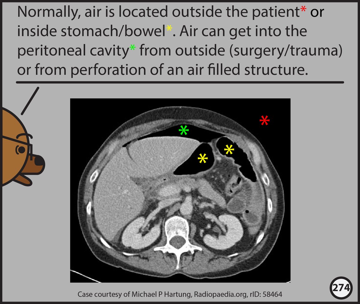
1/5 Interested in learning about abnormal abdominal air collections? This thread, from chapter 7 of our (@MP_Hartung ) still gestating Abdominal CT comic will help! In the meantime, if you want to learn how to read Head CTs, our free comic is available at: https://t.co/wPeVKtgGBe