A new study in the European Heart Journal – Imaging Methods and Practice evaluated whether measuring diastolic flow reversal in the distal abdominal aorta could improve the echocardiographic assessment of aortic regurgitation.
Abdominal aortic holodiastolic flow reversal is an established marker of significant aortic regurgitation, but guidelines do not clearly define the optimal measurement location.
The study evaluated 103 patients with moderate or severe chronic aortic regurgitation.
Key findings:
✅ Distal flow-reversal assessment was feasible in 94% of patients
✅ Proximal assessment was feasible in only 54%
✅ Diagnostic yield was higher with distal measurements: 82% vs 50%
✅ In paired analyses, distal assessment showed 92% sensitivity, 86% specificity, and 88% accuracy for severe aortic regurgitation
✅ Reproducibility between and within observers was excellent
✅ In a small exploratory subgroup, distal echocardiographic findings showed 92% agreement with cardiac MRI
🔗 Read the full study:
https://t.co/IRiqyMHDHd
@EHJIMPEiC@VikashJaiswalMD@JGrapsa
#AorticRegurgitation #AorticValveDisease #Echocardiography #CardiacImaging #CardiacMRI #ValvularHeartDisease #HeartValveDisease #Cardiology #ImagingInnovation #MedicalResearch #StructuralHeartDisease #CardioX #CardioEd
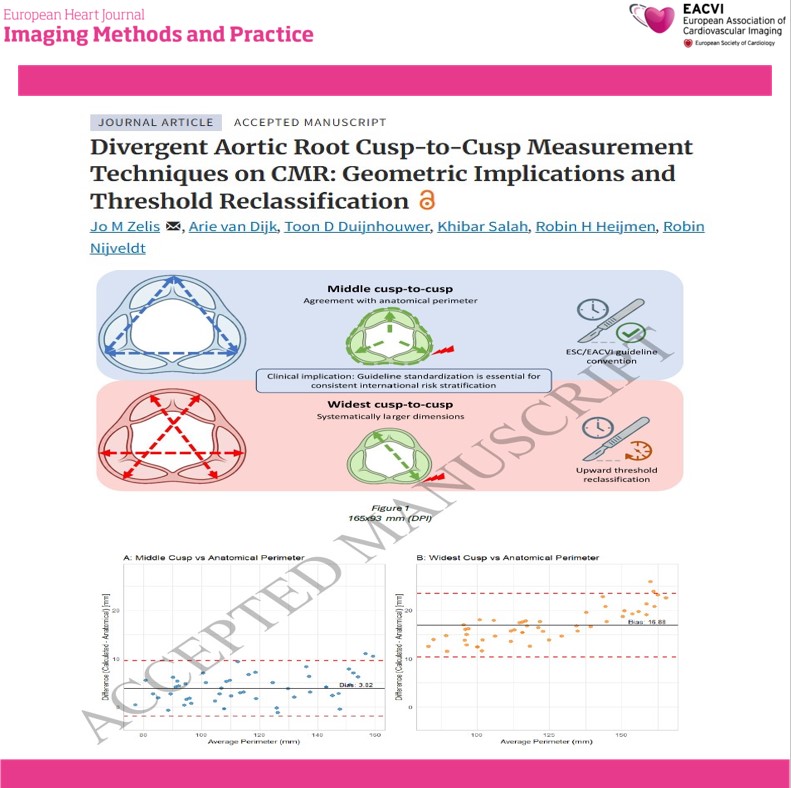
A new study in the #EHJIMP examined differences between ESC and EACTS cusp-to-cusp measurement techniques using 3D cardiovascular magnetic resonance imaging.
The study included 50 patients with a mean age of 42 years and compared:
🔹 Middle cusp-to-cusp measurements
🔹 Widest cusp-to-cusp measurements
🔹 Traced anatomical aortic root perimeter
Key findings:
✅ Widest measurements were, on average, 3.60 mm larger than middle measurements
✅ Middle measurements showed better agreement with the traced aortic root perimeter
✅ Lin’s concordance correlation coefficient: 0.98 vs 0.80
✅ Mean absolute error: 3.96 mm vs 16.88 mm
⚠️ Use of the widest measurement reclassified 10% of patients at the 40-mm threshold and 16% at the 45-mm threshold
These findings highlight an important challenge in aortic surveillance: different guideline-recommended measurement conventions can place the same patient in different clinical risk categories.
The study emphasizes the need for:
📌 Standardized aortic root measurement techniques
📌 Improved comparability across imaging studies
📌 Consistent international risk stratification
📌 Prospective validation against clinical outcomes
🔗 Read the full study:
https://t.co/OjoEZOHQZX
@EHJIMPEiC@VikashJaiswalMD@saramoscatelli7@JGrapsa
#AorticDisease #AorticRoot #CardiacMRI #CMR #CardiovascularImaging #AorticAneurysm #AorticSurgery #Cardiology #CardioTwitter #CardioX #CardioED
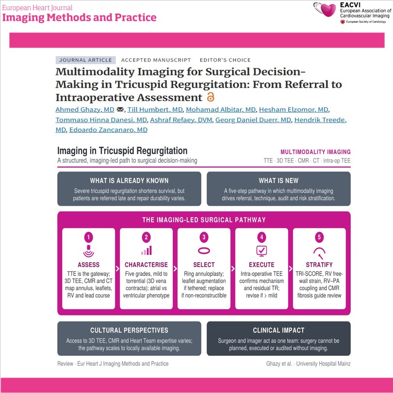
A new state-of-the-art review in the #EHJ-IMP highlights how multimodality imaging can guide the complete management pathway for tricuspid regurgitation—from referral to surgery, transcatheter intervention, and follow-up.
The review outlines the complementary role of:
🔹 Transthoracic echocardiography: initial assessment and disease severity
🔹 3D transesophageal echocardiography: detailed leaflet and annular anatomy
🔹 Cardiac magnetic resonance: reproducible assessment of right ventricular volumes and function
🔹 Cardiac CT: annular geometry, adjacent structures, device leads, and vascular access
The authors emphasize that treatment decisions should integrate:
✅ Mechanism of tricuspid regurgitation
✅ Annular dilatation and leaflet tethering
✅ Right ventricular function and reserve
✅ Pulmonary hemodynamics
✅ End-organ involvement
✅ Surgical, transcatheter, or surveillance options
🔗 Read the full review:
https://t.co/iBJIwVQ9wV
@EHJIMPEiC@VikashJaiswalMD
#TricuspidRegurgitation #HeartValveDisease #StructuralHeartDisease #Echocardiography #CardiacMRI #CardiacCT #MultimodalityImaging #CardiacSurgery #TranscatheterTherapy #RightHeartFailure #HeartTeam #Cardiology #CardiovascularImaging #MedicalResearch #PatientCare
A new state-of-the-art review in the #EHJ-IMP highlights how multimodality imaging can guide the complete management pathway for tricuspid regurgitation—from referral to surgery, transcatheter intervention, and follow-up.
The review outlines the complementary role of:
🔹 Transthoracic echocardiography: initial assessment and disease severity
🔹 3D transesophageal echocardiography: detailed leaflet and annular anatomy
🔹 Cardiac magnetic resonance: reproducible assessment of right ventricular volumes and function
🔹 Cardiac CT: annular geometry, adjacent structures, device leads, and vascular access
The authors emphasize that treatment decisions should integrate:
✅ Mechanism of tricuspid regurgitation
✅ Annular dilatation and leaflet tethering
✅ Right ventricular function and reserve
✅ Pulmonary hemodynamics
✅ End-organ involvement
✅ Surgical, transcatheter, or surveillance options
🔗 Read the full review:
https://t.co/iBJIwVQ9wV
@EHJIMPEiC@VikashJaiswalMD
#TricuspidRegurgitation #HeartValveDisease #StructuralHeartDisease #Echocardiography #CardiacMRI #CardiacCT #MultimodalityImaging #CardiacSurgery #TranscatheterTherapy #RightHeartFailure #HeartTeam #Cardiology #CardiovascularImaging #MedicalResearch #PatientCare
A new short report in the #EHJ-IMP highlights a rare but clinically important complication following mitral transcatheter edge-to-edge repair (M-TEER).
Acute oxygen desaturation after M-TEER should prompt immediate assessment of:
🔍 Right ventricular function
🔍 Right atrial and pulmonary pressures
🔍 Residual or iatrogenic atrial septal defect
🔍 Direction of interatrial blood flow
🔍 New right-to-left shunting
🔍 Changes in left atrial pressure after repair
Although an iatrogenic atrial septal defect is common after transseptal procedures and is often hemodynamically tolerated, elevated right-sided pressures or acute right ventricular dysfunction can reverse the direction of flow—resulting in a clinically significant right-to-left shunt and sudden hypoxemia.
This report reinforces the importance of:
✅ Continuous hemodynamic monitoring
✅ Prompt transesophageal echocardiographic assessment
✅ Careful evaluation of right-sided heart function
✅ Identifying the mechanism of hypoxemia before considering device closure
✅ Multidisciplinary decision-making during structural heart procedures
🔗 Read the full report:
https://t.co/U1YiunyiqM
@EHJIMPEiC@VikashJaiswalMD@saramoscatelli7@JGrapsa
#MTEER #MitralRegurgitation #StructuralHeartDisease #InterventionalCardiology #Echocardiography #TransesophagealEcho #RightHeartFailure #CardiacImaging #StructuralHeartInterventions #Cardiology #CardioX #CardioEd
A recent study from the Yorkshire Heart Centre demonstrates how targeted CMR protocol redesign can reduce scan times and expand imaging capacity.
The study evaluated a “Short Aorta” CMR protocol for patients referred primarily for thoracic aortic assessment. Unlike the standard protocol, the shorter approach focused only on the thoracic aorta when additional assessment of the aortic valve and left ventricular function was not clinically required.
Key findings:
✅ 137 referrals were reviewed
✅ 69% were clinically suitable for the shorter protocol
✅ 46% of scans used the “Short Aorta” protocol after implementation
✅ Median scan time decreased from 37 to 18 minutes
✅ Approximately 1.2 hours of scanner time per week was saved
✅ With optimized scheduling, potential savings increased to 1.9 hours per week
These findings highlight how thoughtful protocol redesign—not only new technology—can improve CMR workflow, reduce patient waiting times, and increase imaging capacity.
The shorter protocol should be used selectively and with appropriate clinical triage. Patients requiring assessment of the aortic valve, left ventricular function, or other cardiac structures should continue to undergo the full CMR protocol.
🔗 Read the full study:
https://t.co/NsghIpehMK
@EHJIMPEiC@VikashJaiswalMD@saramoscatelli7
#CardiacMRI #CMR #CardiovascularImaging #AorticDisease #MedicalImaging #HealthcareInnovation #QualityImprovement #Radiology #Cardiology #ImagingEfficiency #PatientCare #CardioX #CardioEd
A new study in the #EHJ-IMP assessed cardiac image quality from single-probe handheld ultrasound devices.
Authors evaluated 518 cine-loops from 206 patients using Butterfly IQ+, Butterfly IQ3, and Vave devices.
Key findings:
✅ 88% of cine-loops were interpretable
✅ 91% of patients had at least one interpretable image
⚠️ Only 12% achieved good-quality imaging
📉 Image quality declined with increasing BMI
⚖️ No significant difference was observed by sex
🫀 Parasternal long-axis views provided the best image quality
The findings support the potential of handheld ultrasound to assist with early detection of structural heart disease and point-of-care cardiac assessment.
However, image quality assessment remains essential. Handheld ultrasound should complement not replace comprehensive echocardiography, particularly when evaluating complex valvular disease, cardiomyopathies, or Doppler-dependent conditions.
The next step: integrating standardized image-quality assessment and artificial intelligence to identify inadequate studies and guide referral for formal echocardiography.
🔗 Read the full study:
https://t.co/Tamqu7oF7C
@VikashJaiswalMD@saramoscatelli7@EHJIMPEiC
#Cardiology #Echocardiography #POCUS #HandheldUltrasound #CardiacImaging #StructuralHeartDisease #MedicalResearch #CardiovascularMedicine
🫀 Could a traumatic partial pericardial defect be hiding in plain sight?
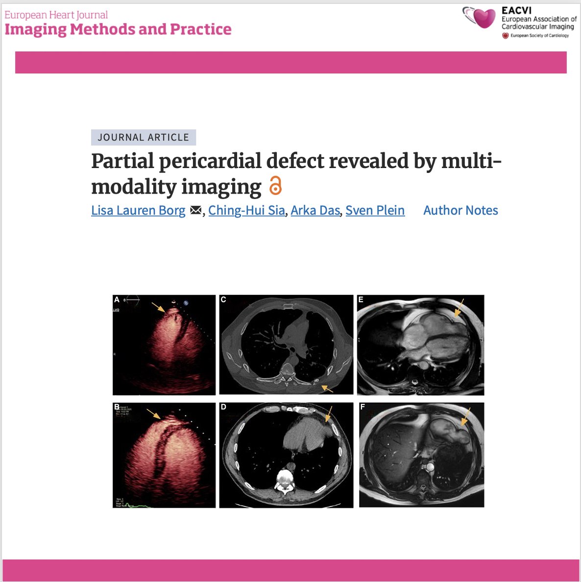
A fascinating multimodality imaging case highlights how echocardiography, CT and cardiac MRI uncovered an acquired partial pericardial defect with right ventricular apical herniation in a patient presenting with intermittent chest pain years after major thoracic trauma.
🔍 Key findings:
✅ Stress echocardiography showed an apparently dilated right ventricle with an aneurysmal-appearing apex, raising suspicion for structural heart disease.
✅ Chest CT demonstrated loss of the pericardial contour at the RV apex in the region of previous left-sided rib fractures.
✅ Cardiac MRI confirmed a partial pericardial defect with herniation of the RV apex, while showing preserved RV size, systolic function and normal myocardial tissue characteristics.
✅ As the patient remained clinically stable without evidence of progressive herniation or ventricular dysfunction, conservative management with serial imaging was chosen instead of surgery.
💡 Clinical takeaway:
Not every "RV aneurysm" is myocardial disease. Multimodality imaging is essential to distinguish pericardial defects from cardiomyopathies and can reveal rare acquired complications of previous chest trauma, preventing unnecessary interventions.
📖 https://t.co/8tQ6IsfafZ
#CardioX #MultimodalityImaging #CardiacMRI #CardiacCT #Echocardiography #PericardialDisease #RightVentricle #CardiacImaging #Cardiology #OpenScience
🫀 When an incidental cardiac mass reveals a complication from the past…
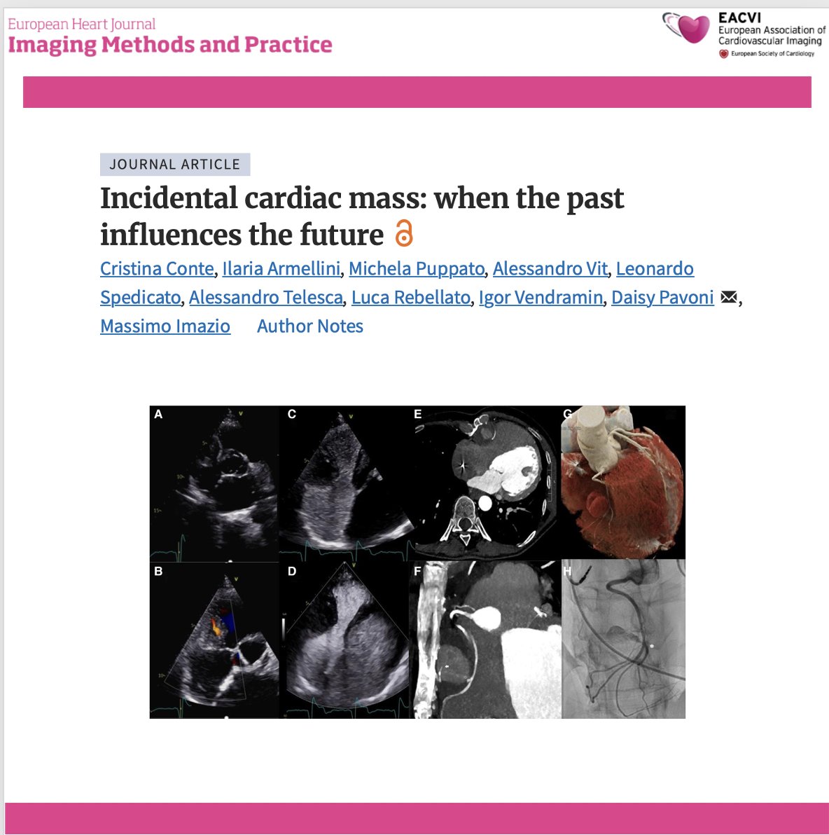
This striking multimodality imaging case describes a 53-year-old woman with previous surgical atrial septal defect closure in whom an incidental mediastinal mass was ultimately identified as a right coronary artery pseudoaneurysm with peripheral thrombosis—likely related to chronic erosion from nearby sternal wires.
🔍 Key findings:
✅ Transthoracic echocardiography detected a mass adjacent to the right atrioventricular groove, with systolic–diastolic flow and suspected circumferential thrombosis.
✅ Agitated saline excluded communication with the right heart, while contrast-enhanced ultrasound demonstrated delayed enhancement, suggesting a systemic arterial supply.
✅ Coronary CT angiography revealed marked sternal malalignment, close contact with the sternal wires, and kinking of the mid-to-distal right coronary artery.
✅ Invasive angiography confirmed active RCA perfusion, supporting the diagnosis of a coronary pseudoaneurysm. Because both percutaneous and surgical treatment were considered high risk, conservative management was chosen.
💡 Clinical takeaway:
Incidental cardiac masses require more than anatomical description. Integrating echocardiography, contrast ultrasound, CT and invasive angiography can clarify vascular origin, define relationships with previous surgical material and guide complex management decisions.
📖 https://t.co/OJWa9tP76E
#CardioX #MultimodalityImaging #Echocardiography #CardiacCT #CoronaryPseudoaneurysm #CardiacMass #AdultCongenitalHeartDisease #CardiacSurgery #CardiacImaging #OpenScience
🫀 Can routine cardiac PET provide prognostic information beyond myocardial perfusion?
A study of 783 patients referred for rest–stress ¹⁵O-water PET shows that left ventricular hypertrophy can be quantified directly from routine, non-ECG-gated PET images—and may identify patients at increased cardiovascular risk.
🔍 Key findings:
✅ PET-derived LV mass index (LVMi) and interventricular septal wall thickness were significantly higher in patients with a previous diagnosis of heart failure.
✅ Among patients without known heart failure, both LVMi and wall thickness independently predicted major adverse cardiac events, defined as cardiovascular death or acute heart failure.
✅ The prognostic associations remained significant after adjustment for age, sex, and myocardial flow reserve.
✅ Measurements showed high test–retest repeatability, with no significant systematic bias in patients undergoing repeated PET examinations.
💡 Clinical takeaway:
Routine ¹⁵O-water PET may offer more than perfusion assessment. Extracting LV hypertrophy metrics from the same examination could provide additional, reproducible prognostic information without further imaging or radiation exposure.
📖 https://t.co/UQFaSVSegW
#CardioX #NuclearCardiology #CardiacPET #PETImaging #LeftVentricularHypertrophy #HeartFailure #CoronaryArteryDisease #CardiacImaging #Cardiology #OpenScience
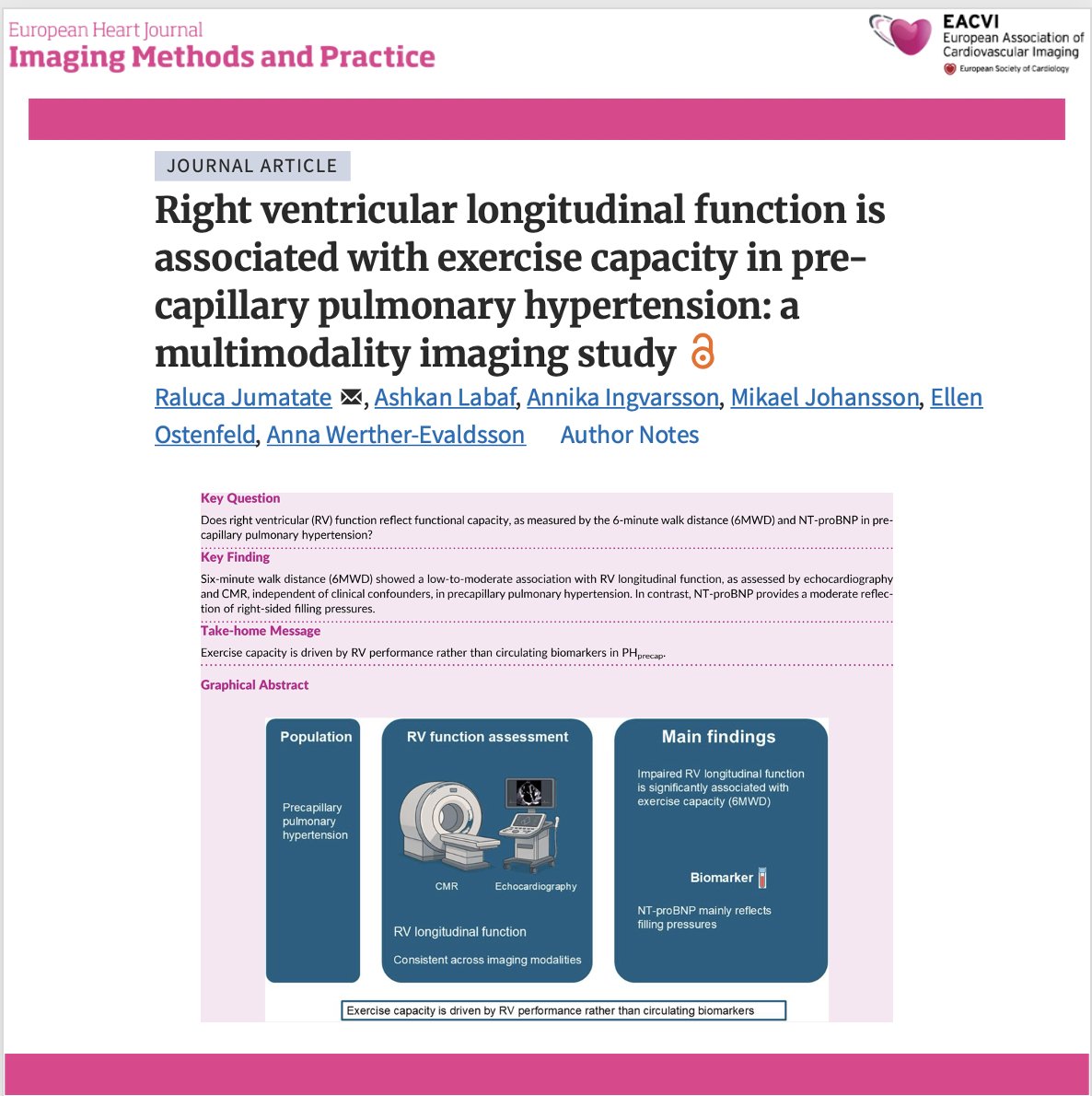
🫀 What is the best imaging marker of exercise capacity in precapillary pulmonary hypertension?
In precapillary pulmonary hypertension (PH), right ventricular (RV) adaptation determines prognosis. This multimodality imaging study shows that RV longitudinal function is more closely associated with exercise capacity than conventional biomarkers, highlighting its value for patient assessment.
🔍 Key findings:
✅ Impaired RV longitudinal function was independently associated with reduced exercise capacity, regardless of whether it was assessed by echocardiography or cardiac magnetic resonance.
✅ These associations remained significant after adjustment for clinical confounders, supporting RV longitudinal mechanics as a robust functional marker.
✅ In contrast, NT-proBNP was more strongly related to right-sided filling pressures than to functional capacity, suggesting it reflects a different aspect of disease severity.
✅ The findings reinforce the complementary role of multimodality imaging in evaluating RV performance beyond resting haemodynamics alone.
💡 Clinical takeaway: Resting haemodynamics and biomarkers tell only part of the story. Assessing RV longitudinal function may provide a more clinically meaningful estimate of functional limitation in patients with precapillary pulmonary hypertension and could improve risk stratification.
📖 Read the full article: https://t.co/AoT0pLhzke
#CardioX #PulmonaryHypertension #RightVentricle #Echocardiography #CardiacMRI #CardiacImaging #HeartFailure #Cardiology #MedTwitter #OpenScience
🫀 What happens to the heart after transcatheter mitral valve repair with the PASCAL® system?
Chronic mitral regurgitation leads to progressive cardiac remodelling, but how much reverse remodelling occurs after PASCAL® transcatheter edge-to-edge repair (TEER)? This study evaluates structural cardiac changes and their relationship with clinical outcomes following the procedure.
🔍 Key findings:
✅ Successful PASCAL® TEER was associated with favourable reverse cardiac remodelling, reflecting the haemodynamic benefit of reducing mitral regurgitation.
✅ Improvements in cardiac structure were accompanied by better clinical outcomes and functional status during follow-up.
✅ The study highlights the importance of serial imaging after TEER to monitor ventricular and atrial remodelling beyond procedural success.
✅ These findings further support the role of multimodality imaging in evaluating response to transcatheter mitral valve interventions and guiding long-term patient management.
💡 Clinical takeaway: The success of mitral TEER should not be judged solely by immediate MR reduction. Reverse cardiac remodelling is an important marker of sustained benefit, and follow-up imaging is essential to identify patients who derive the greatest long-term advantage.
📖 Read the full article: https://t.co/tuRBZuev3x
#CardioX #MitralRegurgitation #TEER #PASCAL #StructuralHeart #ValveDisease #CardiacImaging #Echocardiography #Cardiology #MedTwitter
🫀 Is left ventricular untwisting really just passive recoil?
A fascinating new speckle-tracking echocardiography study challenges a long-held concept in cardiac physiology, suggesting that papillary muscles actively contribute to left ventricular untwisting during diastole, rather than simply following ventricular motion.
🔍 Key findings:
✅ In 75 individuals with normal echocardiograms, papillary muscle mechanics were analysed using 2D speckle-tracking alongside LV torsion measurements.
✅ Both the anterolateral and posteromedial papillary muscles reached peak contraction later than peak LV torsion and GLS, indicating an active role during diastole rather than systole alone.
✅ Greater papillary muscle contraction was significantly associated with enhanced LV untwisting during early and mid-diastole through advanced time-dependent functional modelling.
✅ These findings suggest that papillary muscles may facilitate diastolic suction, providing a previously underappreciated contributor to normal ventricular filling.
💡 Clinical takeaway: This study reshapes our understanding of diastolic mechanics, proposing that papillary muscles are active participants in ventricular relaxation. The findings may open new avenues for studying diastolic dysfunction and diseases affecting papillary muscle mechanics.
📖 Read the full article: https://t.co/GGMB4lpgFt
#CardioX #Echocardiography #SpeckleTracking #DiastolicFunction #CardiacMechanics #PapillaryMuscles #CardiacImaging #Cardiology #MedTwitter #OpenScience
🫀 Can serial ultrasound congestion scores predict outcomes better than a single assessment in acute heart failure?
A new prospective study introduces ΔVExPLUs, combining serial changes in Venous Excess Ultrasound (VExUS) and lung ultrasound (LUS) to improve risk stratification in patients hospitalized with acute decompensated heart failure (ADHF).
🔍 Key findings:
✅ 104 patients with ADHF underwent serial assessment of VExUS and lung ultrasound during hospitalization to evaluate changes in systemic and pulmonary congestion.
✅ The combined change (ΔVExPLUs) provided stronger prognostic information than either VExUS or lung ultrasound alone, highlighting the value of integrating venous and pulmonary congestion assessment.
✅ Patients with persistent or worsening ultrasound congestion experienced significantly worse clinical outcomes, emphasizing that successful decongestion should be assessed with imaging rather than symptoms alone.
✅ The study supports multiorgan ultrasound as a practical bedside tool to guide treatment response and identify patients at higher risk before discharge.
💡 Clinical takeaway: Heart failure congestion extends beyond the lungs. Integrating VExUS and lung ultrasound may provide a more complete picture of decongestion and improve risk stratification, paving the way for more personalized management of ADHF.
📖 Read the full article: https://t.co/eAUK9cZ2sa
#CardioX #HeartFailure #POCUS #LungUltrasound #VExUS #Echocardiography #CardiacImaging #AcuteHeartFailure #Cardiology #MedTwitter
🩺 Can AI enable high-quality cardiac ultrasound in places without expert sonographers?
A new study explores whether artificial intelligence-guided point-of-care ultrasound (AI-POCUS) can help non-expert operators acquire diagnostic cardiac ultrasound images in rural and remote communities—bringing echocardiography closer to the patients who need it most.
🔍 Key findings:
✅ AI-guided POCUS enabled non-expert users to acquire diagnostic cardiac ultrasound images, supporting task-shifting in settings with limited access to expert sonographers.
✅ The study evaluated patient characteristics, image quality, and operator learning curves, providing real-world evidence on how AI-assisted scanning performs outside specialist centres.
✅ Image quality improved as operators gained experience, suggesting that AI guidance can accelerate skill acquisition while maintaining clinically useful examinations.
✅ AI-guided POCUS has the potential to expand access to cardiac imaging in underserved and remote communities by supporting frontline healthcare professionals rather than replacing expert echocardiographers.
💡 Clinical takeaway: AI is not just transforming image interpretation—it is also improving image acquisition. AI-guided POCUS could help bridge inequalities in cardiovascular care by making high-quality echocardiography accessible in resource-limited settings while keeping experts connected for interpretation and oversight.
📖 Read the full article: https://t.co/KFym4CZUM7
#CardioX #ArtificialIntelligence #POCUS #Echocardiography #GlobalHealth #DigitalHealth #CardiacImaging #Cardiology #MedTwitter #OpenScience
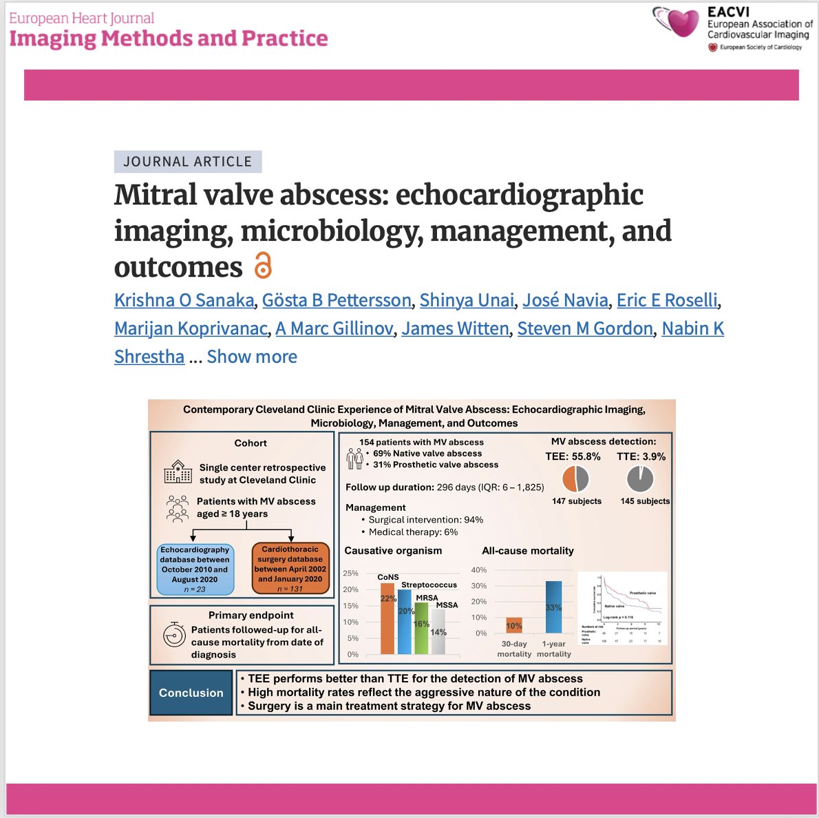
🫀 Can echocardiography reliably detect mitral valve abscess before surgery?
Mitral valve abscess is a rare but life-threatening complication of infective endocarditis. This large contemporary study of 154 patients highlights the pivotal role of transoesophageal echocardiography (TEE) while revealing that advanced disease often requires complex surgical reconstruction despite modern imaging.
🔍 Key findings:
✅ TEE vastly outperformed transthoracic echocardiography (TTE) for detecting mitral valve abscess (55.8% vs. 3.9%), confirming its central role when periannular extension is suspected.
✅ 94% of patients underwent surgery, and more than one-quarter required highly complex procedures (including Commando-type reconstructions), reflecting the severity of this condition.
✅ Mitral valve abscess frequently involved extensive tissue destruction, including leaflet perforation, fistulae, pseudoaneurysms, and multivalvular disease, particularly in prosthetic valve endocarditis.
✅ Independent predictors of mortality included chronic kidney disease, Staphylococcus aureus infection, native valve involvement, and female sex, emphasizing the need for early diagnosis and multidisciplinary management.
💡 Clinical takeaway: When infective endocarditis is complicated by suspected periannular extension, early TEE—especially with 3D imaging—should be performed promptly. However, this study also reminds us that echocardiography may miss earlier stages of mitral valve abscess, making close collaboration between imaging specialists and cardiac surgeons essential.
📖 Read the full article: https://t.co/XWowinU3Jz
#CardioX #Echocardiography #TEE #InfectiveEndocarditis #MitralValve #CardiacImaging #ValveDisease #CardiacSurgery #Cardiology #MedTwitter
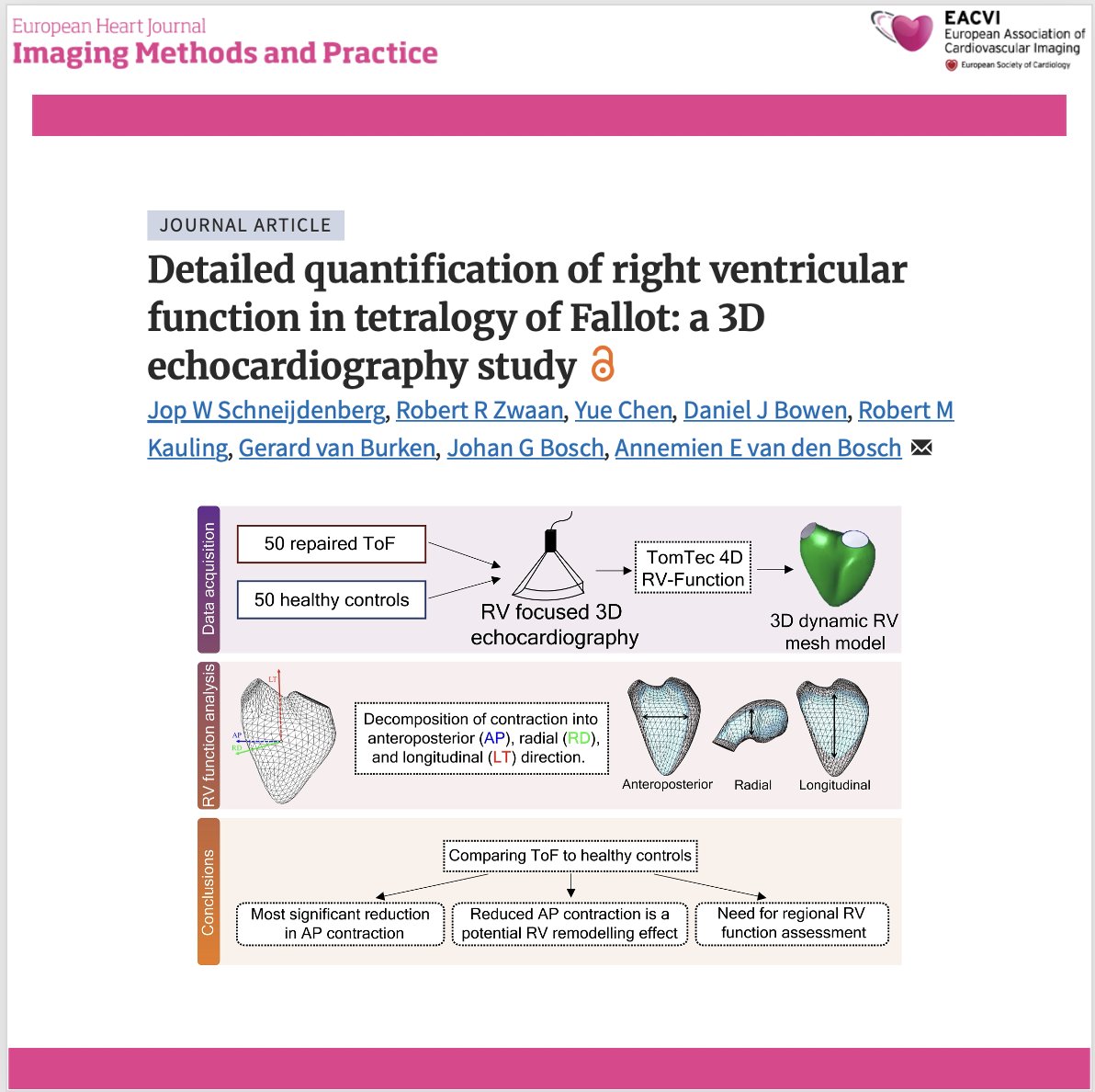
🫀 Are we missing the most important component of right ventricular dysfunction in repaired Tetralogy of Fallot?
A novel 3D echocardiography approach provides a more detailed assessment of right ventricular (RV) mechanics by separating RV contraction into longitudinal, radial, and anteroposterior (AP) components—revealing previously unrecognized patterns of RV remodelling in patients with repaired Tetralogy of Fallot (ToF).
🔍 Key findings:
✅ Anteroposterior (AP) contraction was the main contributor to RV dysfunction, showing the greatest reduction in ToF patients compared with healthy controls.
✅ Despite overall RV impairment, the relative contribution of longitudinal contraction increased, suggesting a compensatory mechanism rather than uniform deterioration of RV function.
✅ The anterior free wall mirrored the global remodelling pattern, highlighting regional heterogeneity and the importance of moving beyond conventional global RV indices.
✅ This novel software enables automated directional decomposition of RV ejection fraction, offering a more comprehensive assessment of RV mechanics that could improve future risk stratification in congenital heart disease.
💡 By looking beyond conventional measures such as TAPSE or global RVEF, 3D echocardiography may provide new insights into right ventricular remodelling and help identify earlier markers of disease progression in adults with repaired Tetralogy of Fallot.
📖 Read the full article: https://t.co/LuRRozAIRR
#CardioX #TetralogyOfFallot #CongenitalHeartDisease #Echocardiography #3DE #RightVentricle #CardiacImaging #ACHD #Cardiology #MedTwitter #OpenScience
🫀 Is the failing heart really "running out of fuel"?
A comprehensive meta-analysis of 176 studies and 7,843 cardiac MR spectroscopy examinations provides the largest reference dataset to date on myocardial energetics, confirming that impaired cardiac energy metabolism is a common feature across multiple cardiovascular diseases.
v
🔍 Key findings:
✅ The normal phosphocreatine-to-ATP (PCr/ATP) ratio in healthy hearts is 1.96, providing an important benchmark for future clinical and research studies.
✅ Cardiac energetics were significantly impaired in HFrEF (−21%) and HFpEF (−18%), with similar reductions also observed in hypertrophic cardiomyopathy, valvular heart disease, and diabetes.
✅ Lower PCr/ATP ratios were associated with higher BMI and natriuretic peptide levels, while better left ventricular ejection fraction correlated with improved myocardial energetics.
✅ Although acquisition methods (field strength, sequence, voxel location) influenced individual measurements, they had minimal impact on pooled estimates, supporting meaningful comparisons across studies.
💡 This work establishes robust reference values for cardiac energetics and reinforces the concept that metabolic dysfunction is a shared pathway across cardiovascular diseases, highlighting myocardial metabolism as an important therapeutic target.
📖 Read the full article: https://t.co/OKX3X7RwVW
#CardioX #CMR #CardiacMRI #MRSpectroscopy #HeartFailure #CardiacMetabolism #CardiacImaging #Cardiology #MedTwitter #OpenScience
🫀 Is the failing heart really "running out of fuel"?
A comprehensive meta-analysis of 176 studies and 7,843 cardiac MR spectroscopy examinations provides the largest reference dataset to date on myocardial energetics, confirming that impaired cardiac energy metabolism is a common feature across multiple cardiovascular diseases.
v
🔍 Key findings:
✅ The normal phosphocreatine-to-ATP (PCr/ATP) ratio in healthy hearts is 1.96, providing an important benchmark for future clinical and research studies.
✅ Cardiac energetics were significantly impaired in HFrEF (−21%) and HFpEF (−18%), with similar reductions also observed in hypertrophic cardiomyopathy, valvular heart disease, and diabetes.
✅ Lower PCr/ATP ratios were associated with higher BMI and natriuretic peptide levels, while better left ventricular ejection fraction correlated with improved myocardial energetics.
✅ Although acquisition methods (field strength, sequence, voxel location) influenced individual measurements, they had minimal impact on pooled estimates, supporting meaningful comparisons across studies.
💡 This work establishes robust reference values for cardiac energetics and reinforces the concept that metabolic dysfunction is a shared pathway across cardiovascular diseases, highlighting myocardial metabolism as an important therapeutic target.
📖 Read the full article: https://t.co/OKX3X7RwVW
#CardioX #CMR #CardiacMRI #MRSpectroscopy #HeartFailure #CardiacMetabolism #CardiacImaging #Cardiology #MedTwitter #OpenScience
🫀 Can we use much less regadenoson in paediatric stress perfusion CMR without compromising image quality?
A new study introduces a heart rate-targeted regadenoson titration strategy for paediatric stress perfusion CMR, demonstrating that individualized dosing can achieve reliable stress imaging while substantially reducing drug exposure.
🔍 Key findings:
✅ Heart rate-targeted titration reduced the median regadenoson dose by 60% compared with conventional weight-based protocols (3.2 vs. 8 mcg/kg), while achieving the target heart rate response in 100% of successful studies.
✅ All examinations produced diagnostic-quality images, with no major adverse events, despite the lower drug exposure.
✅ In children weighing >50 kg, 83% achieved adequate stress with ≤160 μg, less than half the conventional adult dose of 400 μg.
✅ Stress-induced perfusion defects were detected in 24% of this high-risk cohort, suggesting that individualized titration maintained adequate diagnostic performance while avoiding unnecessary pharmacological stress.
💡 This study suggests that physiology-guided regadenoson dosing may represent a safer and more patient-centred approach to paediatric stress perfusion CMR, although prospective validation with quantitative myocardial perfusion is still needed.
📖 Read the full article: https://t.co/Wb4kG9b8xj
#CardioX #CMR #StressCMR #PaediatricCardiology #CardiacMRI #CongenitalHeartDisease #CardiacImaging #PerfusionCMR #Cardiology #MedTwitter #OpenScience