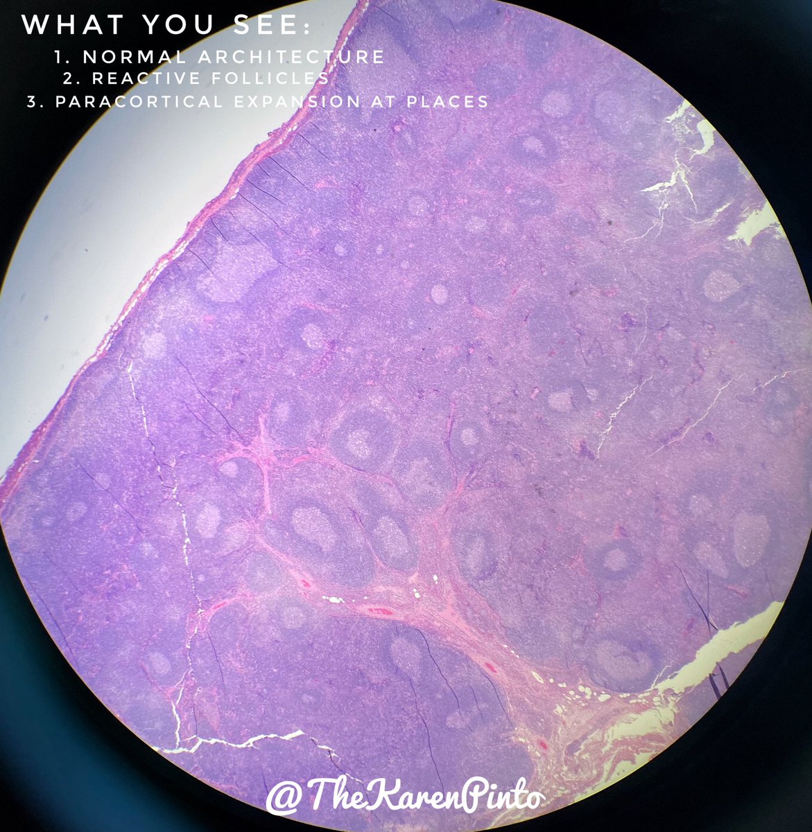
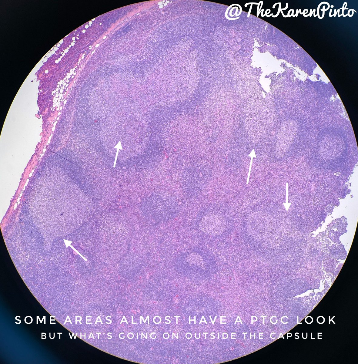
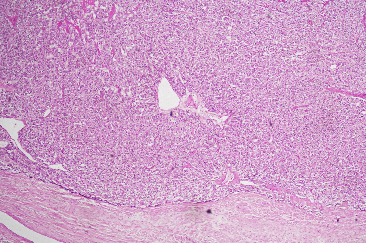
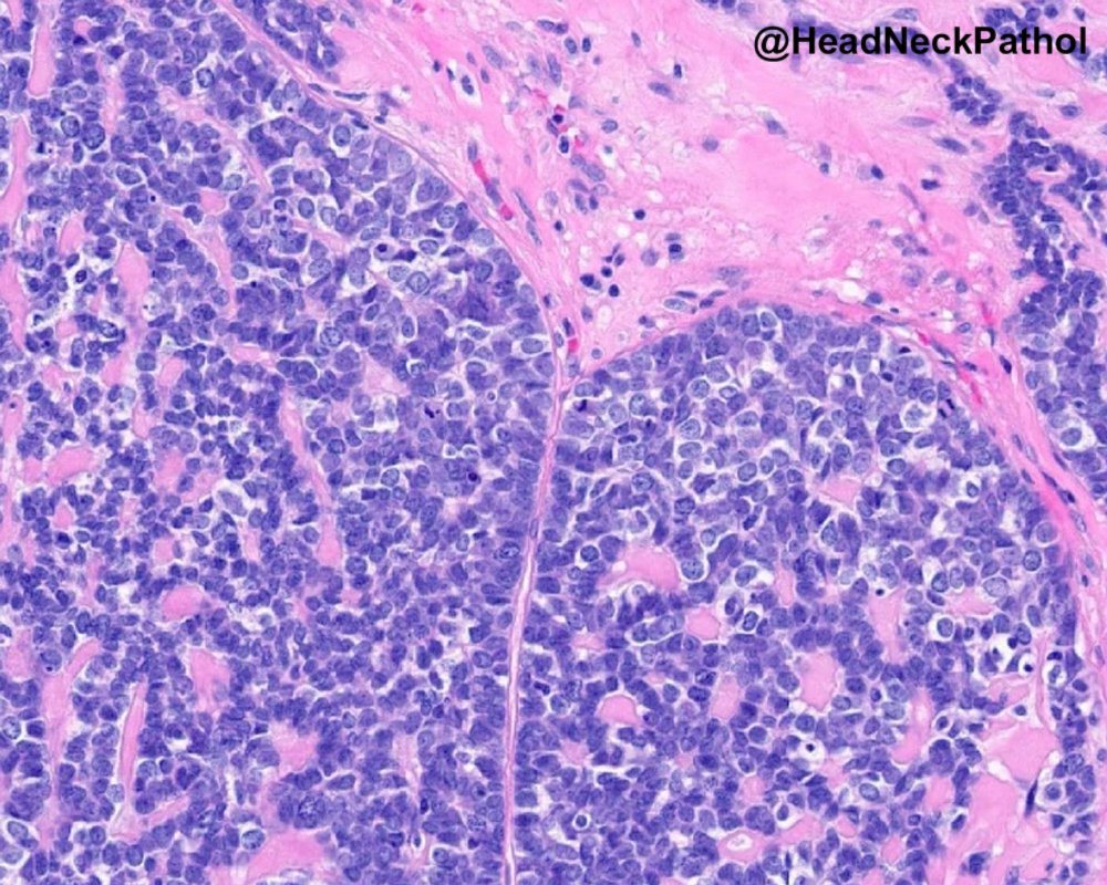
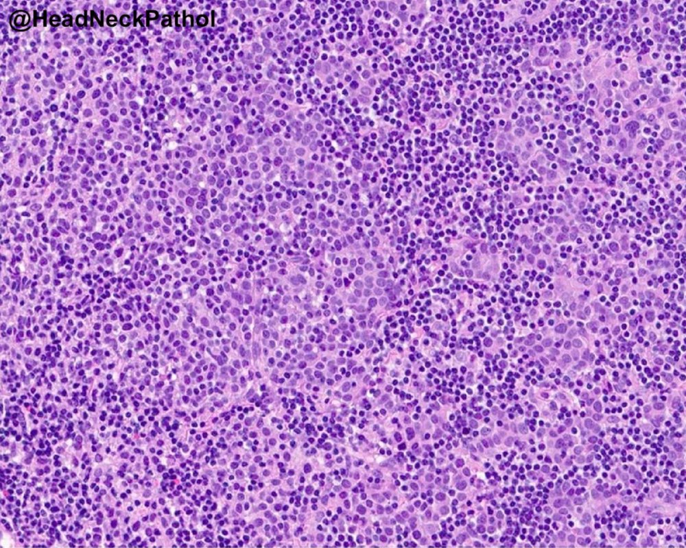
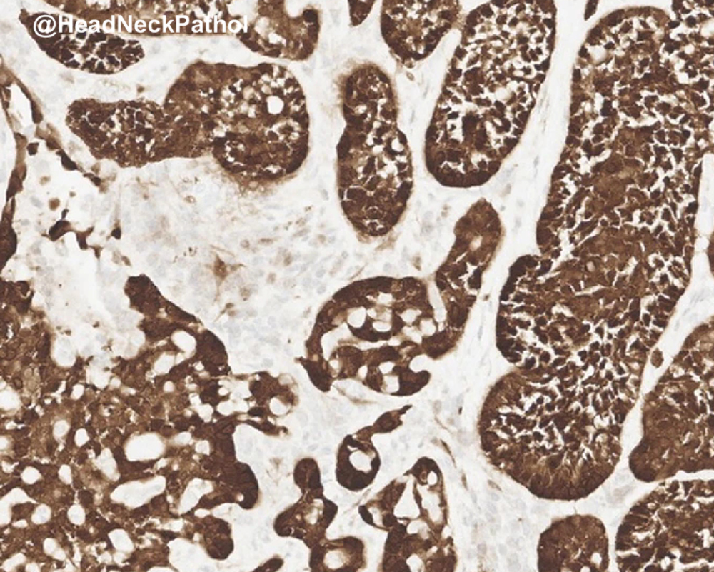
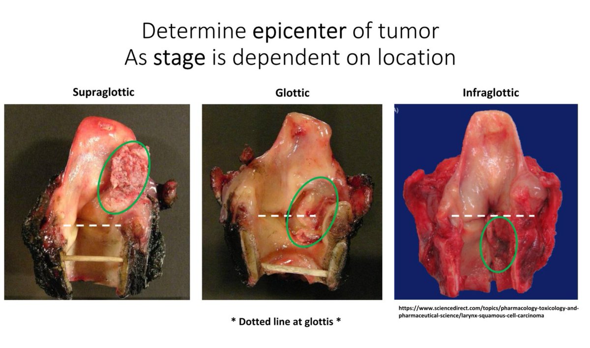
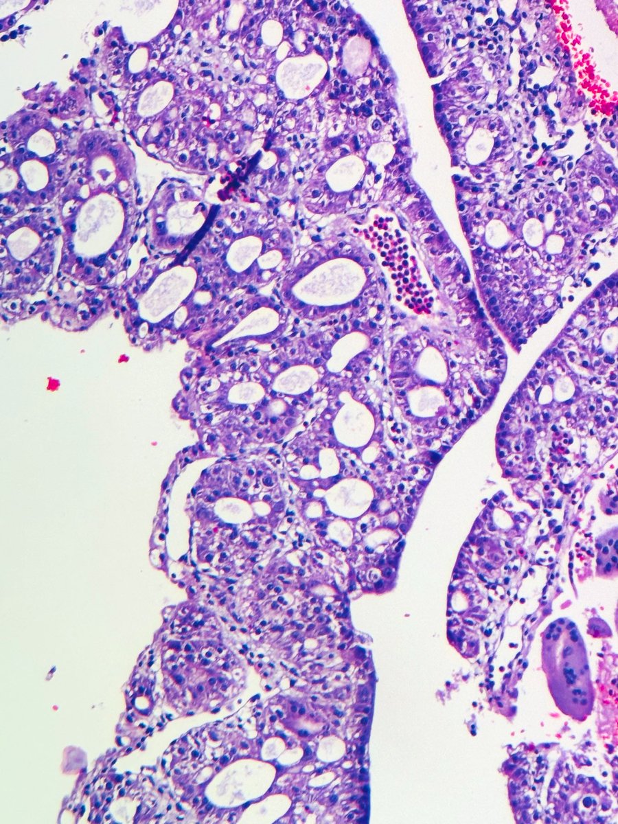
Staging scan for sigmoid adenocarcinoma picks up this inguinal nodal mass which was biopsied, what is your diagnosis and what IHC would you perform if any? https://t.co/vFtioedigU
Crowded endometrial glands that fall short of EIN criteria are often reported as “atypical glandular crowding” or “glandular crowding.”
What’s the clinical significance?
A large retrospective study (79,998 specimens) identified 167 such cases. Within 1 year, 28% were diagnosed with EIN, most within the first 4 months.
Roughly 1 in 4 patients will be found to have EIN or carcinoma on follow-up.
Pic, Left side: minute ( < 1 mm) detached fragment of crowded, atypical endometrial glands—a focus too small to meet the diagnostic size threshold for EIN
Pic, right side: Follow-up endometrial curettage from the same patient (20×) demonstrating a FIGO grade 1 endometrioid adenocarcinoma
Dr. Zeidel et al. Glandular Crowding of the Endometrium: Clinical Outcomes of a Diagnostic Gray Zone
DOI: 0.1097/PGP.0000000000001165
#Gynpath #pathology #PathX #PathTwitter
Variability in diagnosis of invasive lobular carcinoma (ILC)
Sources of discordance:
• E-cadherin-positive ILCs harboring CDH1 missense mutations (Pic 2; this case showed retained β-catenin staining and interestingly, E-cadherin-negative LCIS)
• E-cadherin-negative ILCs with tubular elements (Pic 3)
• ILCs with trabecular pattern (Pic 4; most experts in this study classified it as non-lobular breast carcinoma based only on H&E morphology). Check this interesting study: DOI: 10.1002/cjp2.253
Dr. Calhoun #BreastPath #PathX #PathTwitter #USCAP
Breast - Infiltrating epitheliosis
Key histologic features (Eusebi & Millis)
• Predominantly florid usual ductal hyperplasia (UDH) with focal squamoid areas
• Scleroelastotic stroma distributed throughout the lesion (not limited to a central nidus like radial scar (RS)/CSL)
• Frequent desmoplastic reaction + keloid-like fibrous bands
Unlike classic UDH, ducts show jagged/irregular edges with epithelium “flowing out” into stroma → more infiltrative appearance
IE is possibly related to radial scar/complex sclerosing lesion family. Lacks clear zonal architecture; myoepithelial cells are frequently discontinuous or absent
Dr. Lerwill #USCAP2026 #pathology #PathX #PathTwitter
Expanding the Histologic Spectrum of HPV-Associated Oropharyngeal Carcinoma: A Study of 379 Cases Focusing on Classification, Histologic Features, & their Prognostic Significance
https://t.co/53B8fWkS2c
#HeadNeckPathol#HNPJ#HeadandNeckPath#Pathology#ENTPath#PathologyTweet
Post treatment: rare case of residual carcinoma only within lymphovascular spaces.
Not accounted for in the AJCC TNM staging system. Some have advocated for the use of ypTX or ypT-LVI
See CD31 IHC!
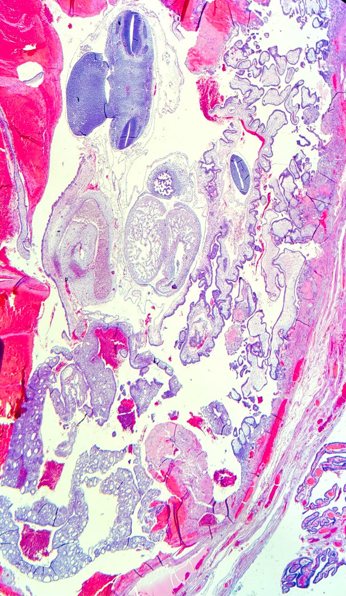
Ectopic pregnancy with fetal parts and yolk sac remnant (or simply yolk sac), Pic 1
It is not common to see a yolk sac in a POC specimen or even in ectopic pregnancy. It looks concerning, but once you see one, you'll recognize it faster next time.
Yolk sac, high power (Pic 2)
For the best educational thread on yolk sac, check @leon_metlay's post: https://t.co/wNu2SXpYqe
Another yolk sac post from Dr. Kaur's ISGYP presentation: https://t.co/x8H67pzXBZ
#everydayGYN #PathX #pathtwitter
Breast biopsy with the following histology. Classical 3 questions 😀
1. What is the phenotype of the cells?
2. What is your understanding about the GATA3 expression of the cells?
3. If you see the same lesion in cervical biopsy what would you call it?
#pathology#breastpath
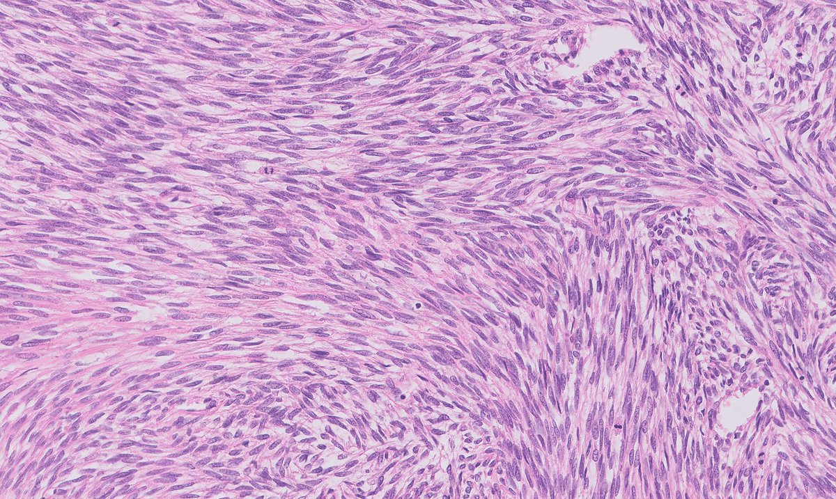
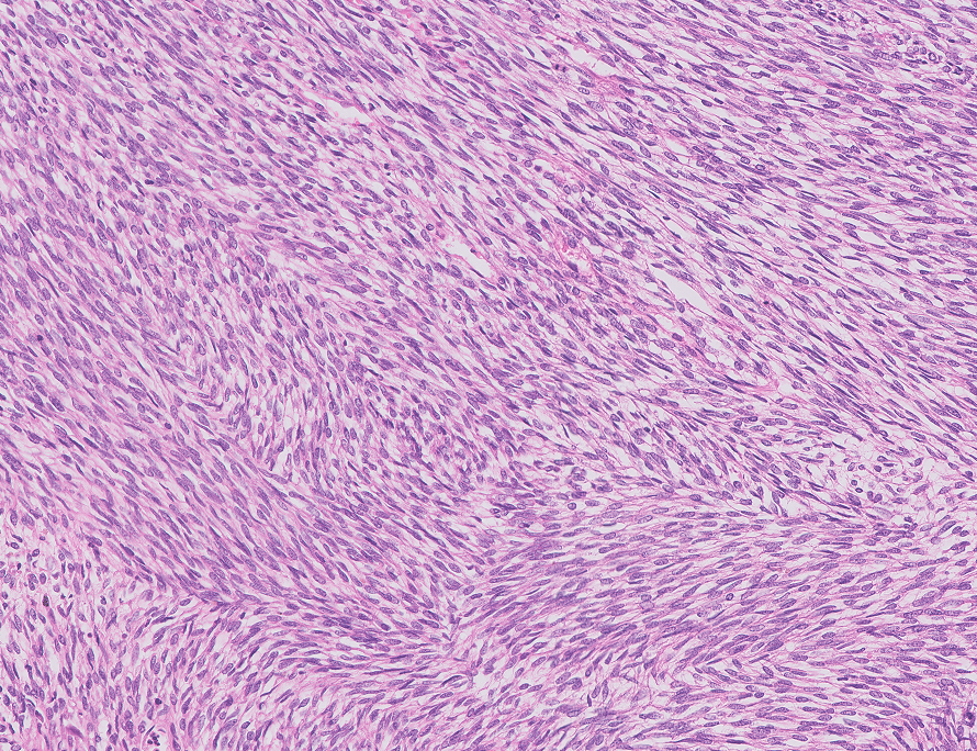
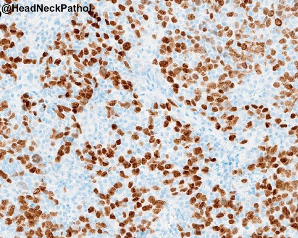
A classic monophasic spindle cell Synovial Sarcoma and its next generation immunohistochemical marker, the antibody SSX::SS18, which identifies the fusion protein of this fusion driven sarcoma.
Baranov E, et al. PMID: 32141887