

Top Tweets for #SnapshotsInNeuroscience

#SnapshotsinNeuroscience | Featured on the #eNeuro blog from DeWitt et al., an image of the zebrafish olfactory rosette showcasing olfactory sensory neurons and leukocytes following hypoxic insult. Image credit: Kate DeMann.
https://t.co/Uzoft9ICPd
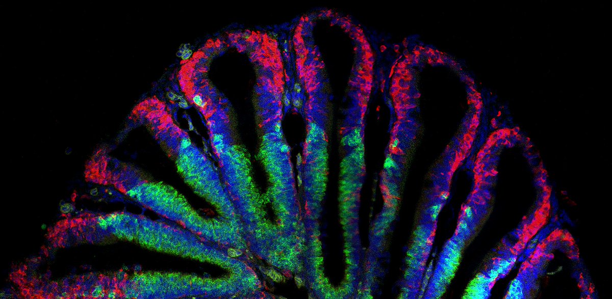
#SnapshotsinNeuroscience | Featured on the #eNeuro blog from Jeong et al., a confocal image showing Drd2-expressing hilar neurons in the mouse dorsal hippocampus. Image credit: Yong-Seok Oh.
https://t.co/XKnA1I1mJA
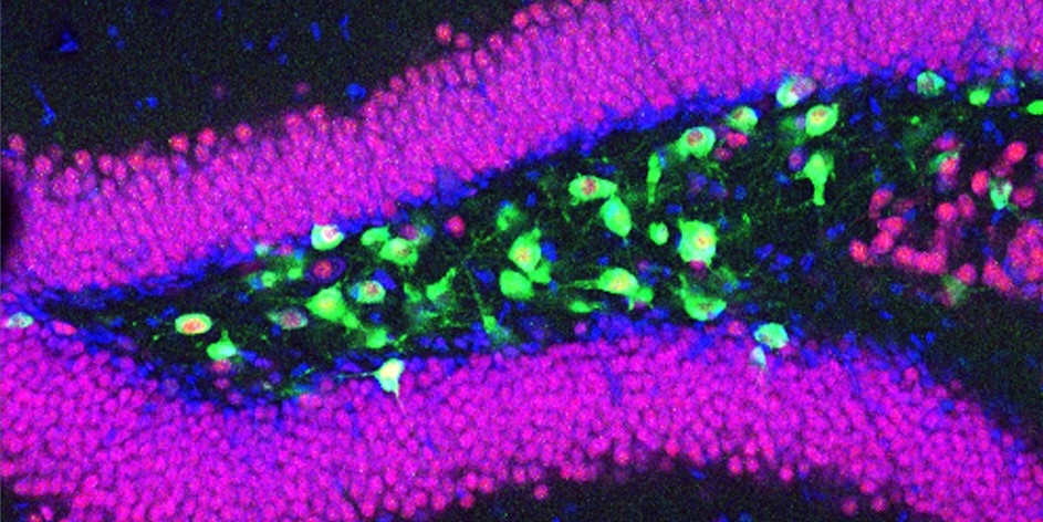
#SnapshotsinNeuroscience | Featured on the #eNeuro blog from Luo. et al., neurons in the male mouse striatum that are activated by social vocalizations during courtship. Image credit: Shi-Xiang Luo.
https://t.co/0QM29mQ66t

#SnapshotsinNeuroscience | Featured on the #eNeuro blog from Quainoo et al., bipolar cells and cone photoreceptor cells in the inner and outer nuclear layers of the adult mouse retina. Image credit: Ebenezer Quainoo.
https://t.co/3qI42tqCgj

#SnapshotsinNeuroscience | Featured on the #eNeuro blog from Meftah et al., neurotoxic plaques and a CA1 pyramidal neuron in the hippocampus of an Alzheimer’s disease mouse model. Photo Credit: Jamie Elliott
https://t.co/ki0Ld3gRzE

#SnapshotsinNeuroscience | Featured on the #eNeuro blog from Soma et al. vivid vertical tracks targeting the Cornu Ammonis 1 region of the dorsal hippocampus of a transgenic rat. Photo Credit: Shogo Soma
https://t.co/d6LQMLW75C
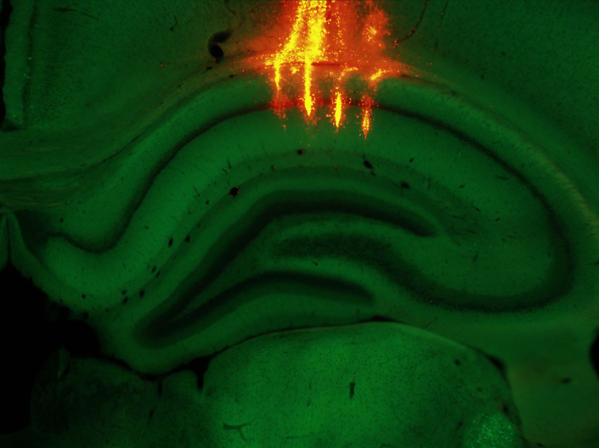
#SnapshotsinNeuroscience | Featured on the #eNeuro blog from Pofahl et al., granule cells in the dentate gyrus of a mouse, with visible extensions of their dendrites reaching out into the molecular layer. Photo Credit: Martin Pofahl
https://t.co/plE5Py7o1w

#SnapshotsinNeuroscience | Featured on the #eNeuro blog from Tarannum et al., expression of four neuropil-localized mRNA in hippocampal subregions of a young mouse. Photo Credit: Shannon Farris
https://t.co/uUcDeh1w86
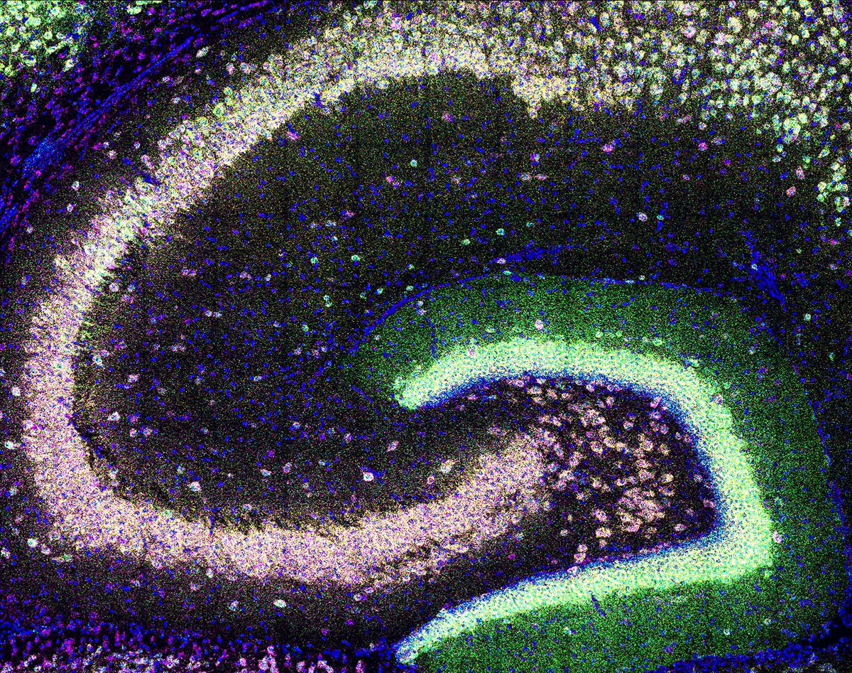
#SnapshotsinNeuroscience | Featured on the #eNeuro blog from Orozco Ramirez et al., an adult male fruit fly brain with expression of membrane-tethered GFP in neurons that express the neuropeptide F receptor and detected with anti-GFP antibodies.
https://t.co/5yIaLogtSa

#SnapshotsinNeuroscience | Featured on the #eNeuro blog from Lustig et al., a whole mount view of a young mouse brain with all cortical layer 5 neurons expressing dystonia-related gene Klhl14, highlighted in green.
Photo Credit: Alexander Lammers
https://t.co/g879RVjCIO
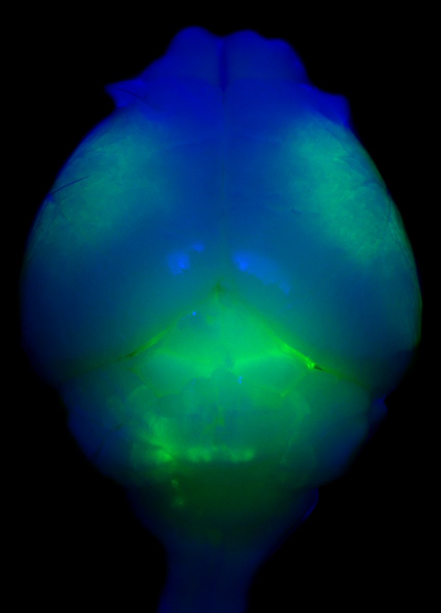
#SnapshotsinNeuroscience | This image from Smith et al. shows a mouse cerebellar section with a single layer of Purkinje cells extending their elaborate dendritic branches upwards into the dense molecular layer. Image credit: Kathleen McCoy
https://t.co/VV5K1nFvtw

#SnapshotsinNeuroscience | Featured on the #eNeuro blog, an image from Sandoval, Rychlik, and Choe showing real-time calcium dynamics of astrocytes and oxytocin neurons in the mouse paraventricular nucleus of the hypothalamus.
Image credit: Katrina Choe
https://t.co/IaBP4YnAPH

#SnapshotsinNeuroscience: Mitral and Tufted Cells
This image from Gadiwalla et al. shows principal neuron dendrites in the adult mouse olfactory bulb extending up towards the glomerular layer where they receive sensory input. Image credit: Huang & Galliano
https://t.co/yoQyyT7HI1
#SnapshotsinNeuroscience | Featured on the #eNeuro blog, an image from Fyrberg et al. showing neurons of the spinal cord dorsal horn within a newly developed mouse model for inducing targeted gene expression.
Image credit: Eric Fyrberg
https://t.co/tLKDO1sQob

#SnapshotsinNeuroscience | Featured on the #eNeuro blog, an image showing immunostaining of cell bodies and nerve fibers of neurons in an adult mouse small intestinal myenteric ganglion from Gorecki, Kulkarni, et al.
Image credit: Subhash Kulkarni
https://t.co/o5BrqyDrOo

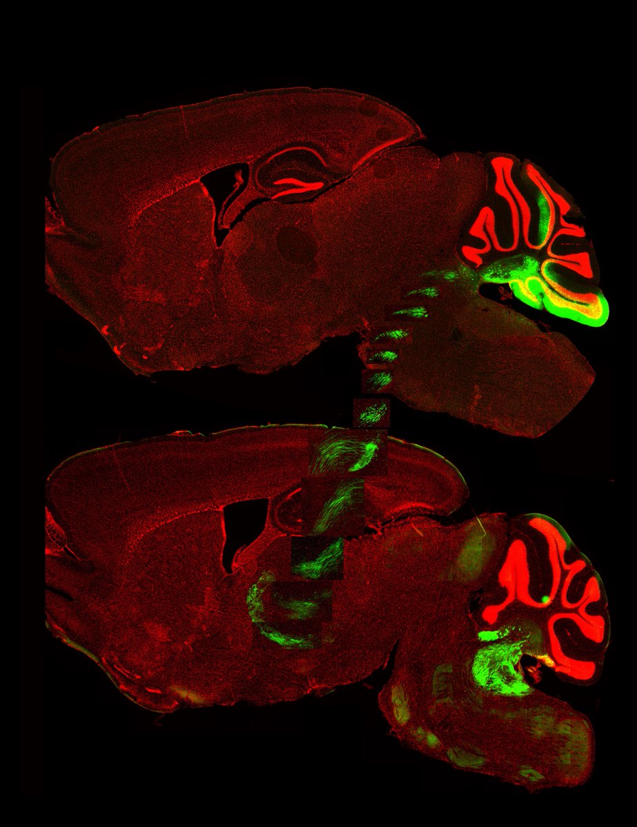
#SnapshotsinNeuroscience | Featured on the #eNeuro blog, sagittal mouse brain sections showing cerebellar nuclei sending afferents to the ventral posterolateral nucleus of the thalamus from Witteveen and Balmer.
Image credit: Isabelle Witteveen.
https://t.co/P2uJGYbRGE
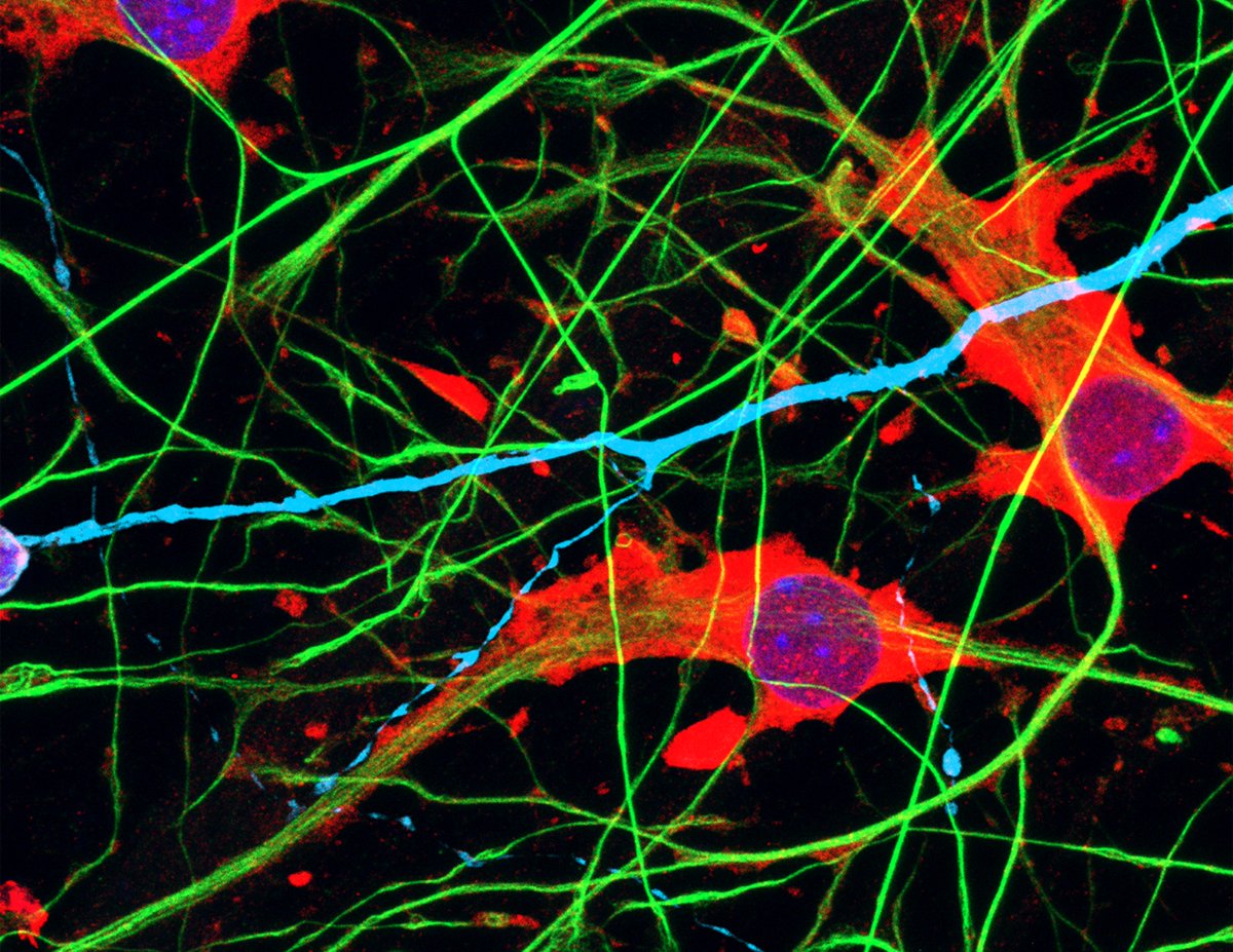
#SnapshotsinNeuroscience | Featured on the #eNeuro blog, human induced pluripotent stem cell-derived astrocytes and neurons grown in vitro as a coculture from Lendemeijer et al.
Image credit: Bas Lendemeijer
https://t.co/vFIAZhgeAQ
#SnapshotsinNeuroscience | Featured on the #eNeuro blog, an image of a mouse brain containing norepinephrine-synthesizing neurons in the locus coeruleus with a calcium indicator (green) & red fluorescent protein (magenta) from Sciolino, Fan, & Engborg.
https://t.co/z9UHtQ6zwu

#SnapshotsinNeuroscience | Featured on the #eNeuro blog, an image of proprioceptive sensory afferents and Atoh1-lineage neurons in the lower thoracic mouse spinal cord from Espinosa, Pop, and Lai.
Image credit: Iliodora Pop and Helen Lai.
https://t.co/zZPCIUXYUL

#SnapshotsinNeuroscience | Featured on the #eNeuro blog, an image of the cellular layers of an adult mouse retina, stained for markers of amacrine cells and type 3b bipolar cells from @SihRong_Wu and Huda Y. Zoghbi.
Image credit: Sih-rong Wu.
https://t.co/sujeSK8Pdc

Last Seen Hashtags on Sotwe
Most Popular Users

Elon Musk 
@elonmusk
241.5M followers

Barack Obama
@barackobama
118.8M followers

Cristiano Ronaldo
@cristiano
114.5M followers

Donald J. Trump
@realdonaldtrump
111.8M followers

Narendra Modi
@narendramodi
107.1M followers

Rihanna
@rihanna
98.7M followers

NASA
@nasa
92.3M followers

Justin Bieber
@justinbieber
91.8M followers

KATY PERRY
@katyperry
90M followers

Taylor Swift
@taylorswift13
84M followers

Lady Gaga
@ladygaga
75.5M followers

Virat Kohli
@imvkohli
73.5M followers

Kim Kardashian
@kimkardashian
70.9M followers

YouTube
@youtube
68.8M followers

Neymar Jr
@neymarjr
66.5M followers

Bill Gates
@billgates
65.3M followers

Selena Gomez
@selenagomez
63.1M followers

The Ellen Show
@theellenshow
62.2M followers

CNN
@cnn
61.7M followers

X
@x
60.7M followers