A physics-informed deep learning model enabled acquisition-free correction of severe prostate DWI distortions, improving diagnostic confidence while identifying all histologically confirmed lesions.
@RSNA@OxfordJournals@SusannaLeeRad
https://t.co/cIXdsptDbQ
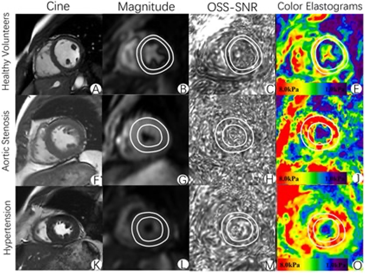
3.0-T MR single frequency elastography of the heart demonstrated excellent long-interval reproducibility for quantifying myocardial stiffness, distinguishing true longitudinal changes from measurement variability.
@RSNA@OxfordJournals@SusannaLeeRad
https://t.co/MnMF7EzDIm
A deep learning model using abdominal contrast-enhanced CT accurately predicted the severity of acute pancreatitis and outperformed traditional prognostic tools (e.g. BISAP and mCTSI).
@RSNA@OxfordJournals@SusannaLeeRad
https://t.co/N3RWRzbebr
A fully automated nnUnet-based model accurately segmented and quantified wrist synovial tissue volume on postcontrast MRI in patients with rheumatoid arthritis.
@RSNA@OxfordJournals@SusannaLeeRad
https://t.co/EWG2E56DfG
"The brain emerges as both battleground and masterpiece—a place where beauty and struggle become inseparable." created by Fatt Yang Chew, MD of Taichung, Taiwan. Discover this “Art of Imaging” article in the latest issue of #RadAdv.
@RSNA@OxfordJournals@SusannaLeeRad
https://t.co/exWh7B8AhP
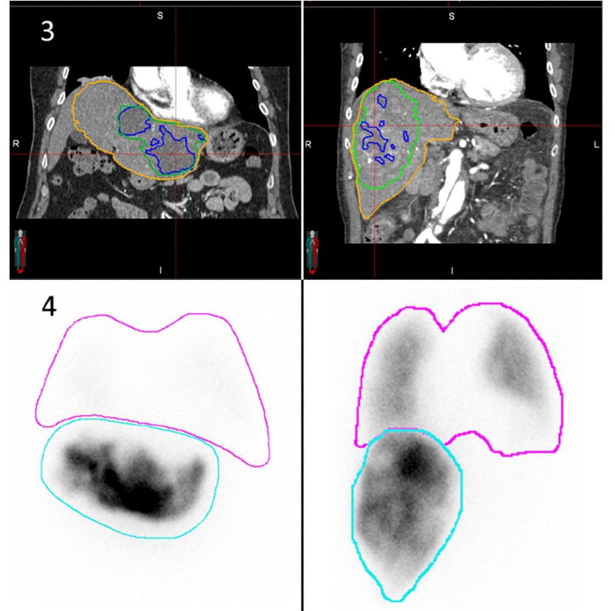
A novel lung shunt fraction (LSF) metric can reliably identify 90Y-SIRT cases where MAA-based LSF determination may be omitted while preserving treatment planning accuracy.
@RSNA@OxfordJournals@SusannaLeeRad
https://t.co/7etT4OSlSC
DeepPNP demonstrated superior accuracy for pulmonary nodule malignancy risk prediction compared with established malignancy risk models, with modest performance gains from enriched training data.
@RSNA@OxfordJournals@SusannaLeeRad
https://t.co/hyyAxyyT15
Fetal growth restriction/small for gestational age pregnancies. Machine learning integration of MRI, ultrasound, and clinical data improves outcome prediction over conventional ultrasound only approaches.
#RadAdv@RSNA@OxfordJournals@SusannaLeeRad
https://t.co/yJsmlD24TN
Clinical implementation of deep learning on MR neuroimaging cut scan times by over 50% while maintaining image quality
#RadAdv@RSNA@OxfordJournals@SusannaLeeRad
https://t.co/ri5EIPS2hN
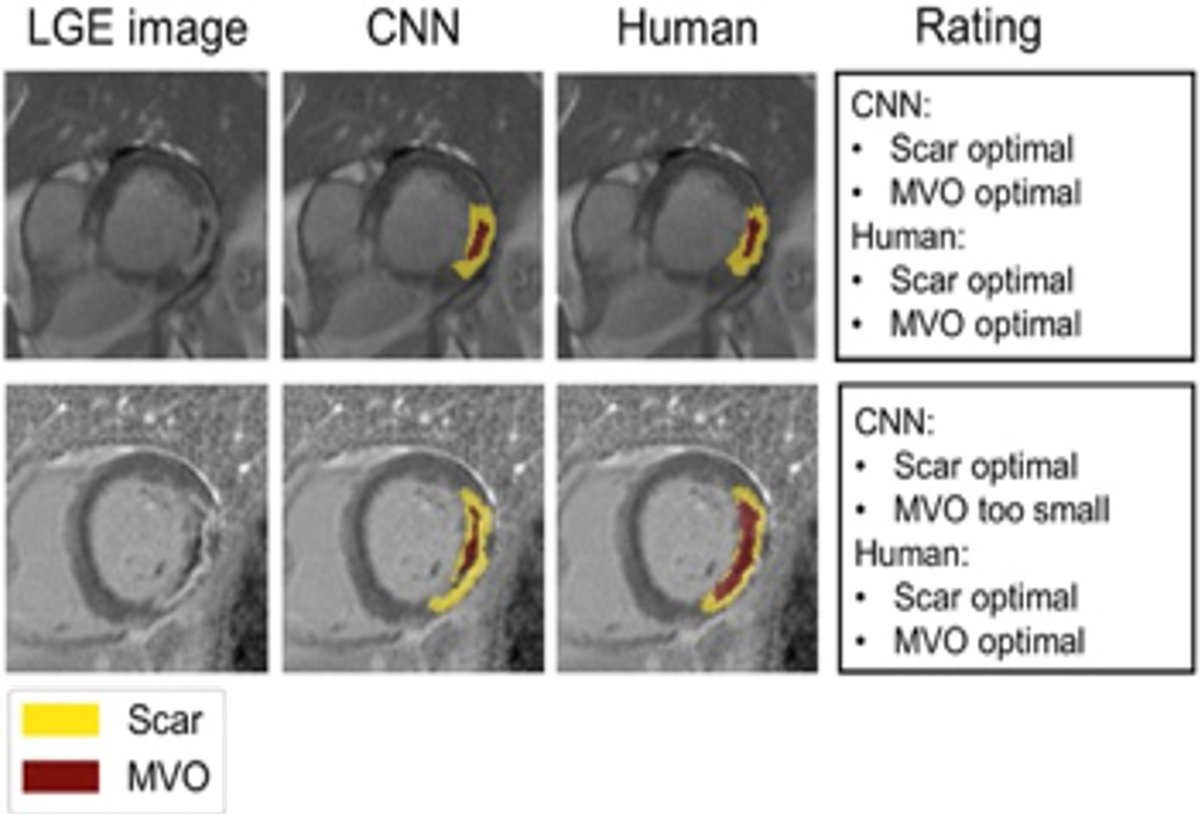
Deep learning tool for automated segmentation of myocardial infarcts on cardiac MRI outperforms trained readers and requires no pre-processing.
@RSNA@OxfordJournals#RadAdv
https://t.co/HNXEjUjgr1
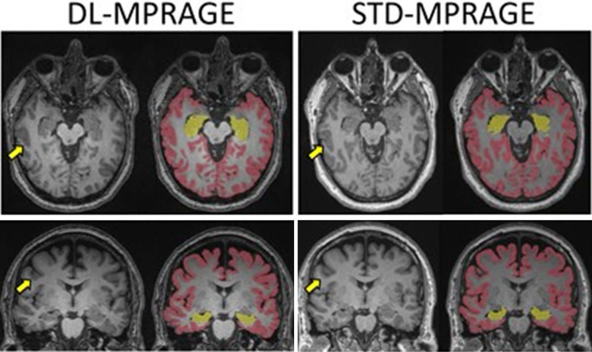
Deep learning-accelerated T1-MPRAGE cuts brain MRI scan time, improves spatial resolution, and delivers equivalent volumetric estimates with similar delineation of sulci and gyri .
@SusannaLeeRad@OxfordJournals#RadAdv
https://t.co/5t4gcI6soq
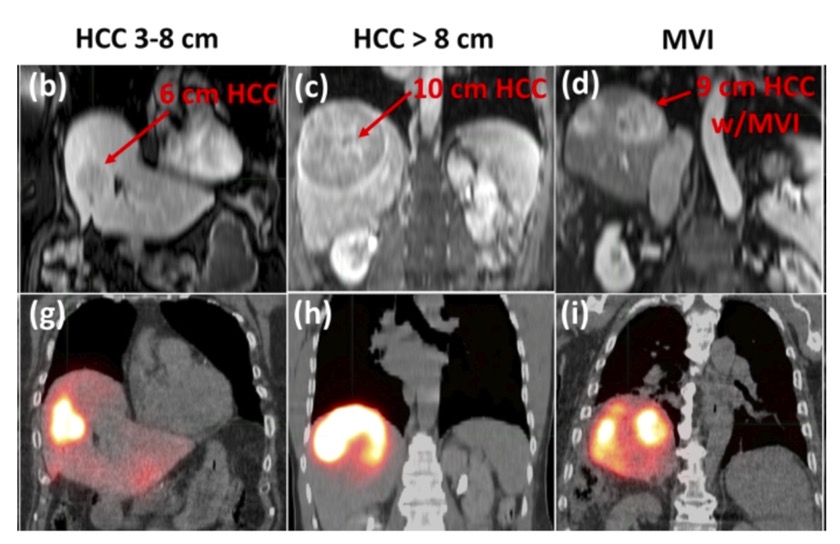
Contrast enhanced CT-derived hypervascular-tumor-to-perfused volume ratio may offer a non-invasive alternative to 99mTc-MAA for lung shunt fraction estimation in hepatic transarterial radioembolization @RSNA#RadAdv@OxfordJournals@SusannaLeeRad https://t.co/Gshm4yy97k
Adhesive capsulitis, commonly known as frozen shoulder. Discover this “Art of Imaging” and its creator in the latest issue of #RadAdv.
@RSNA@OxfordJournals@woojinrad@SusannaLeeRad
https://t.co/QkVaobxeu2