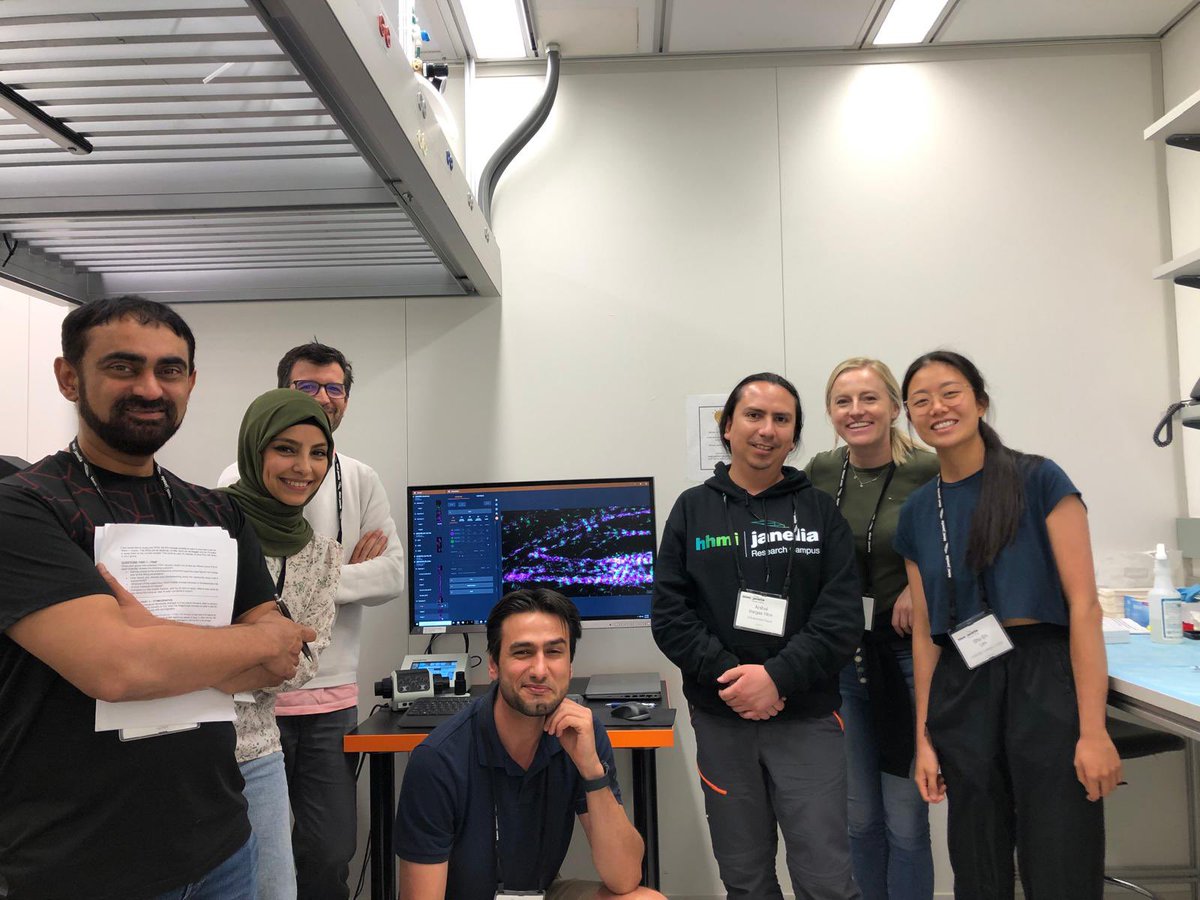
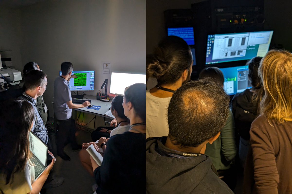
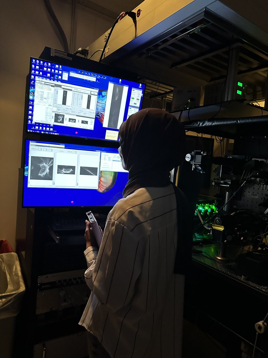
We are back at the light sheet microscopes this afternoon at #IALSJanelia! Our SiMView system provides a large-scale view of migration, while this data from our MOSAIC system gives a closer look at macrophages (magenta) dancing around a blood vessel (green).
This afternoon at #IALSJanelia we are exploring organism-scale imaging! This includes looking at macrophages responding to a zebrafish tail fin injury, as shown in this data from our SiMView light sheet microscope.
The latest AIC Call for Proposals is now open! Deadline is October 4, 2024. Come use @HHMIJanelia imaging technology and expertise for FREE. Visit https://t.co/zv2m8prcrN for details. Before applying, contact us to schedule a technical consultation!
Having some fun in AGAVE with the #IALSJanelia expansion microscopy data. This cross section of an expanded zebrafish embryo shows muscle fibers on either side of the notochord, with some patterned skin on top of the structure.
This afternoon at #IALSJanelia we are exploring expansion microscopy! This piece of an expanded zebrafish embryo was imaged on a lattice light sheet microscope.
Today we are celebrating our 10th anniversary! It is wonderful to have current @HHMIJanelia colleagues as well as some of our @AICjanelia alumni here for #AIC10
Our first Inductee, Harald Hess, has taken the stage! He is being honored tonight for his development of PALM (photoactivated localization microscopy) alongside his co-inventor @Eric_Betzig! #NIHF2024#Microscopy
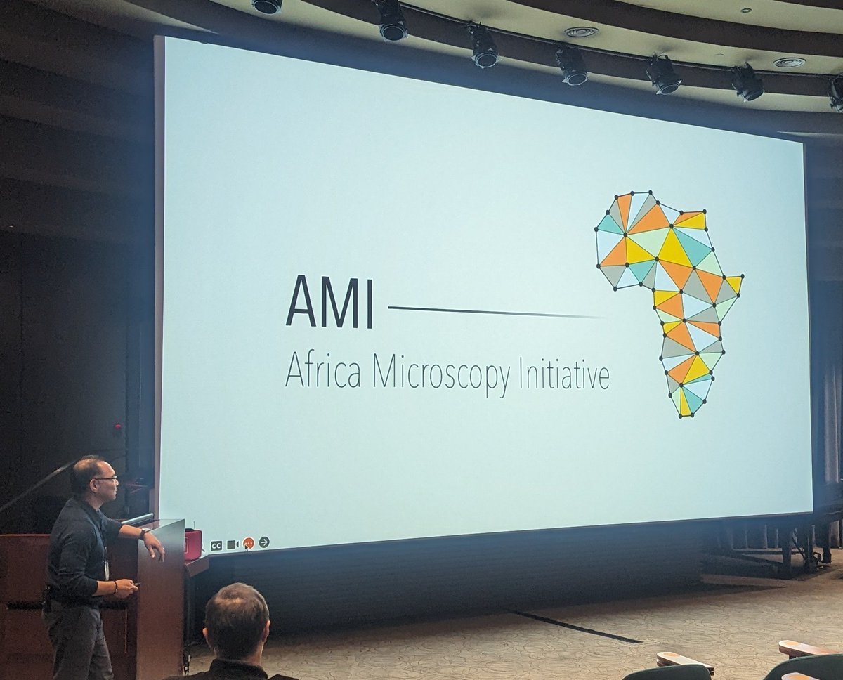
Great day the @janeliaconf on microscopy dissemination! I am learning so much and having really interesting conversations. Awed by projects promoting microscopy in the global south.
Also found kindred spirits lamenting how many contemporary/custom/open scopes lack eyepieces😄