I provides interactive ultrasound training that stimulates learners to achieve the next level of skill and confidence in answering the clinical question.
Excellent correlation of Lt CIV compression via duplex ultrasound and IVUS, matched by location, extend, severity and diameter.
#seeingisbelieving#UUM#AHVI
I am excited to announce this introductory podcast of Universal Ultrasound Management. My intention is to stimulating learners to achieve the next level of skill and confidence in answering the clinical question.
https://t.co/JzKrKbLpsV
Clear visualization of spontaneous reflux in color Doppler. Reversal of flow in the opposite direction is illustrated by red flow. Thus, venous insufficiency will be indicated by blinking between blue flow and red flow in the color box.
#UUM, #AHVI#SEEINGISBELIEVING
Assessing the ovarian vein for reflux, measure the diameter of the proximal, mid, and distal segments using B-mode, using color flow and pulsed wave Doppler.
#ultrasound#uum#veins#ahvi
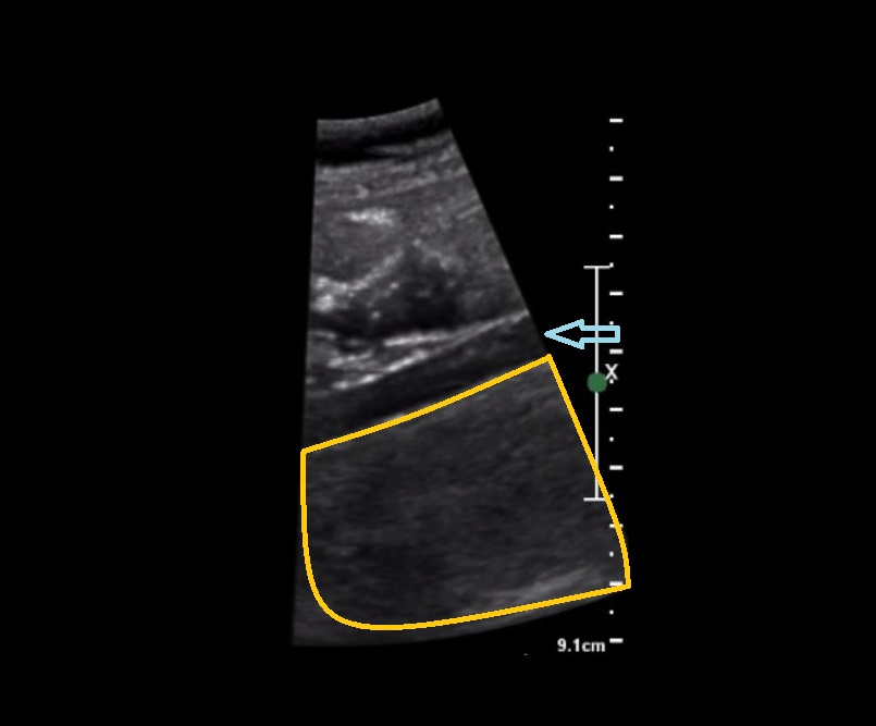
How to find the Ovarian Vein:
The first and second ultrasound image demonstrates the ovarian vein imaged via B-mode in transverse and longitudinal view. The blue arrow indicates the ovarian vein. The orange outline indicates the psoas muscle.
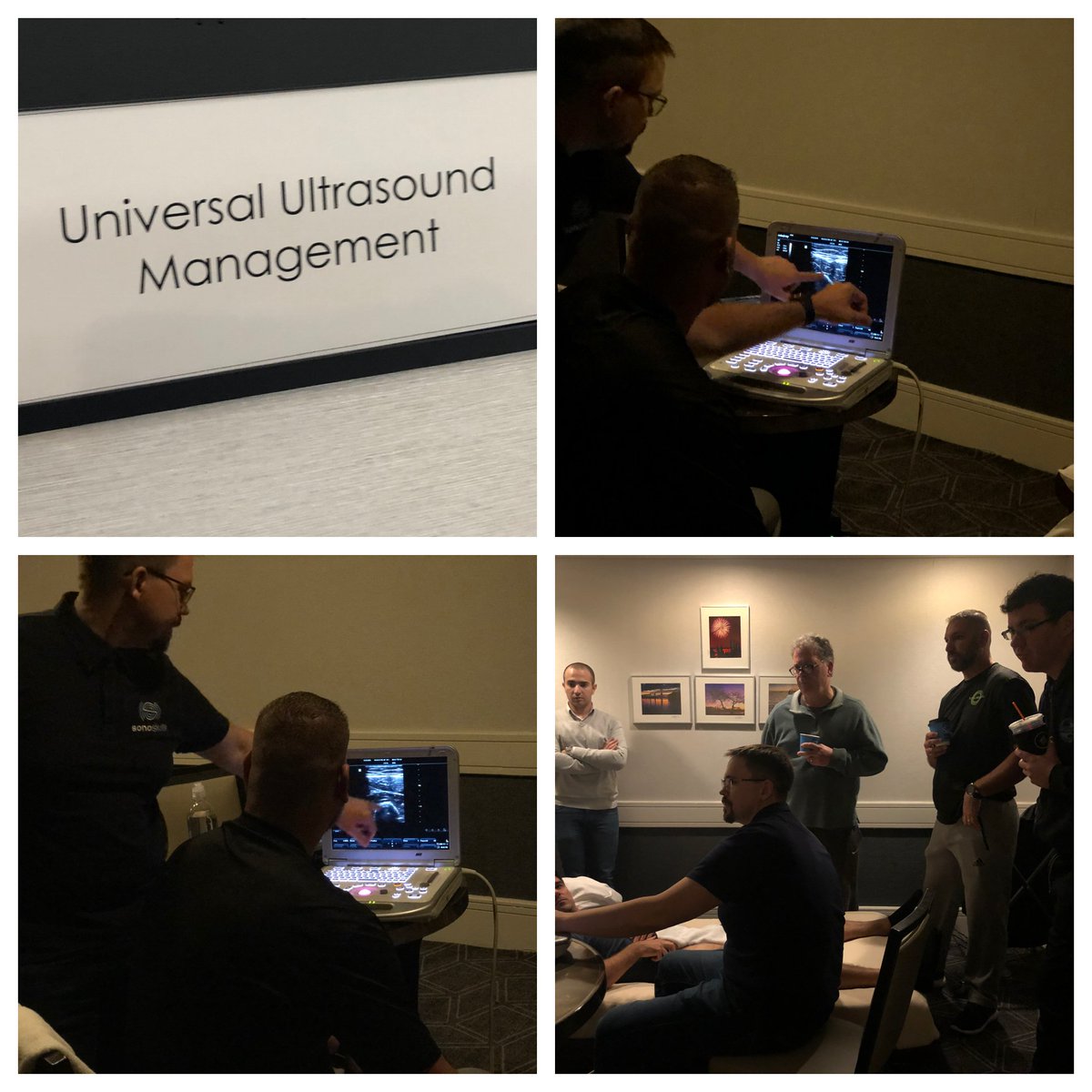
Pictured here is Saulius Rutkauskas, PhD, teaching didactic and interactive sessions during our Foundations of MSK Ultrasound course.
#UUM#sonoskills#MSKultrasound
To assess for patency during lower extremity stent surveillance, additional color Doppler and spectral Doppler images are also essential . The minimum segments that should be assessed via Doppler include: pre-stent, proximal stent, mid-stent, distal stent, and post-stent.
#UUM
To all my Twitter contacts attending American Vein & Lymphatic Society Annual Congress.
Do not forget to stop by booth number #605 and speak with me to learn about our IAC Accreditation consultative services, Ultrasound HOT Course, Venous Symposium and Expert Venous Management.
Some of the things we see in ultrasound can be quite frightening. Since its Halloween, I want to show you some pathologies that are quite spectacular. Below is an image of a prosthetic bypass graft, which has a chainsaw-like appearance when visualized via grayscale ultrasound.
Same pseudoaneurysm of the common femoral artery only imaged with a curved linear transducer in b-mode and color flow Doppler.
#UUM#AHVI#seeingisbelieving