Was the FINALS between Rcb and CSK ?
Did they defeat CSK in the Play offs ?
Or did csk gave them a Hard to digest Defeat in the last two or three years ?
Inspite of none of the above happening what was the real need of them DISRESPECTING CSK jersey like this ?
I mean like seriously ?
This is y the hate for these bitches increases day by day !
Csk has literally treated them like a bitch for 17 years and finally its their time and they cant even celebrate without bringing our jersey !
This has to end , if not RCB is gonna spread their virus to the next generation kids in the name of CELEBRATIon !
The hardest breakups aren’t the toxic ones.
They’re the ones where you really love the person… and they didn’t do anything wrong…
No cheating, huge fights or anything like that .
Just that gut feeling you can’t shake… even though everything looks good on paper, it’s not the right fit. You know it, but you tell yourself it’s not true.,
And that’s what messes you up. Some people never act on it, but the ones who do will be hurt for awhile.
You don’t just lose the person. You lose a friend. The routine. The texts. The inside jokes. The version of your day that included them.
It’s honestly WAY easier when someone cheats or turns it toxic, at least the choice gets made for you.
Walking away from something good because your intuition says “this isn’t it” is brutal…
But ignoring that feeling is how a lot of people wake up years later wondering what went wrong and miss out on life.
Case Discussion for Colleagues 👇
Here is an HRCT case of a 39-year-old female with known scleroderma.
The pattern is challenging, and I’m interested to hear your interpretation.
🔽 HRCT video is attached.
Using the 4-Step Approach, choose the conclusion you think best fits this case and explain your reasoning in the comments:
1️⃣ Probable usual interstitial pneumonia
2️⃣ Fibrotic nonspecific interstitial pneumonia
3️⃣ Probable usual interstitial pneumonia versus fibrotic nonspecific interstitial pneumonia
4️⃣ Indeterminate usual interstitial pneumonia versus fibrotic nonspecific interstitial pneumonia
Important: In your comment, please mention which step or steps helped you reach your conclusion.
For example:
•Step 1: Is honeycombing present?
•Step 2: Is the pattern UIP or non-UIP?
•Step 3: Are there non-IPF features?
•Step 4: Are there CTD-UIP clues?
This will help enrich the discussion and enhance the teaching value for everyone.
Looking forward to your insights!
VPN set to 🇮🇳 = cheaper flights
VPN set to 🇹🇭 = cheaper hotel bookings
VPN set to 🇧🇷 = cheaper subscriptions
VPN set to 🇹🇷 = cheapest Netflix, Spotify, YouTube Premium
VPN set to 🇦🇷 = cheapest digital tools & apps
VPN set to 🇵🇭 = cheaper airline baggage fees
VPN set to 🇲🇽 = cheaper software deals
VPN set to 🇸🇬 = best prices for gadgets
VPN set to 🇹🇭 = cheaper booking sites & tours
VPN set to 🇿🇦 = cheaper clothing stores & brands
VPN set to 🇵🇱 = cheaper online games
VPN set to 🇨🇴 = lowest price for Canva & Adobe plans
VPN set to 🇪🇬 = cheaper Facebook/Instagram ads
VPN set to 🇲🇾 = cheaper learning platforms (Udemy, Skillshare)
VPN set to 🇮🇩 = cheaper cloud services & domains
VPN set to 🇰🇪 = lower prices on some airline routes
VPN set to 🇹🇼 = cheaper Apple App Store purchases
VPN set to 🇹🇷 = cheaper streaming services
VPN set to 🇦🇷 = cheaper Netflix subscriptions
VPN set to 🇿🇦 = cheaper video games
VPN set to 🇵🇭 = cheaper online music
VPN set to 🇪🇨 = cheaper travel deals
VPN set to 🇲🇾 = cheaper app purchases
VPN set to 🇵🇰 = cheaper e-books
VPN set to 🇻🇳 = cheaper digital downloads
👁️🗨️ Sometimes MRI findings are subtle.
Radiologists don’t get the luxury of a bedside exam — instead, they develop hawk eyes (and, depending who you ask, very expensive high-resolution monitors 😅).
Yesterday, I showed you what’s normal:
Optic nerve = white matter tract
On T2: gray matter = light, white matter = dark
So the optic nerve should match cerebral white matter → dark signal.
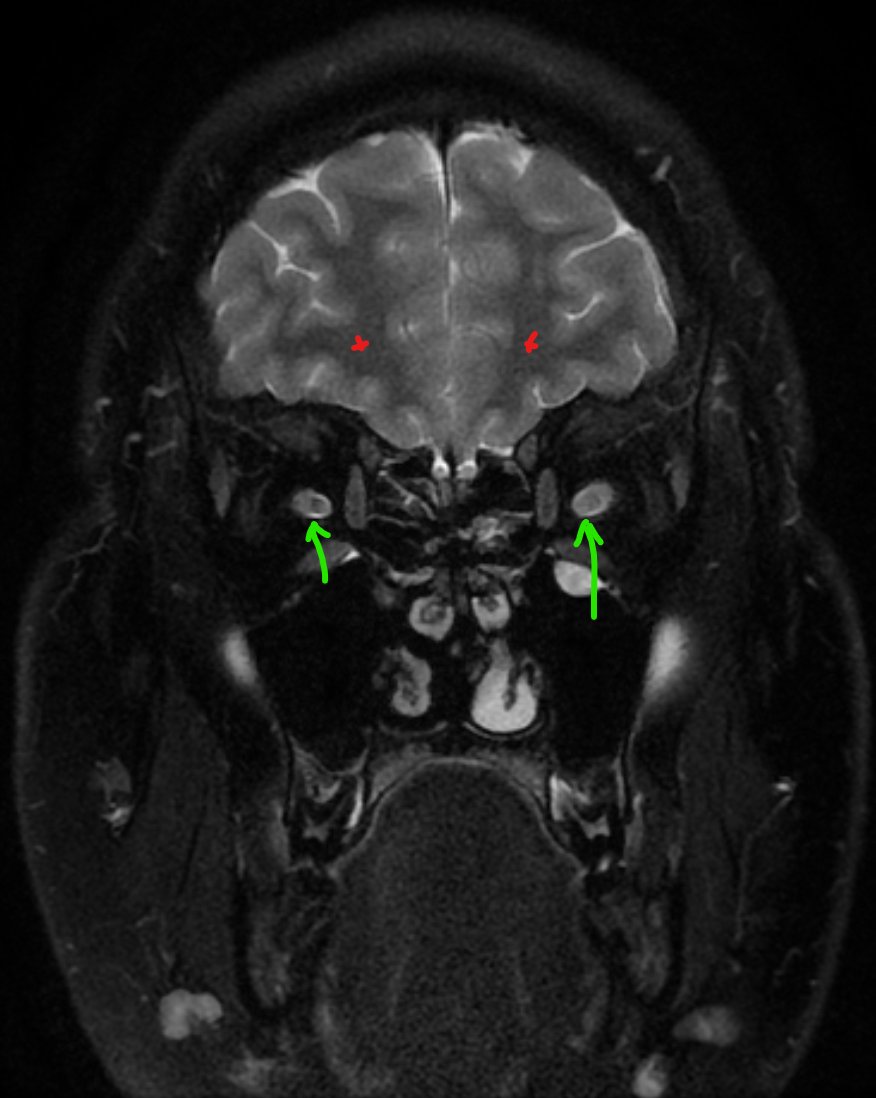
Now look at this coronal MRI T2 of the orbits with fat suppression (fat sat):
T2: gray matter lighter, white matter dark, CSF bright
Orbital fat = normally bright on T2 → here it’s suppressed (fat sat), so it appears dark and pathology stands out
Red asterisks = cerebral white matter (dark, normal)
Green arrows = optic nerves → only one matches white matter intensity (normal)
🧑⚕️ Clinical vignette:
20-year-old with family history of MS, 2-day history of monocular blurry vision.
❓Which eye is affected?
📌 Courtesy: Radiopaedia – Optic neuritis (MS)
#Neuroradiology #MultipleSclerosis #MRI #Neurology #Neuroophthalmology
See yesterday's explanation of MRI coronal optic nerve anatomy in the comment section of this tweet... below 👇👇