Instead of watching an hour of Netflix, watch this 2 hour hour Stanford lecture will teach you more about how LLMs like ChatGPT and Claude are built than most people working at top AI companies learn in their entire careers.
👩🏻💻 #DFLab | Startup Spora debuta con piezas decorativas de micelio de hongos en tienda de lujo de primo del rey Carlos III en Londres
https://t.co/1VmZFucZKT
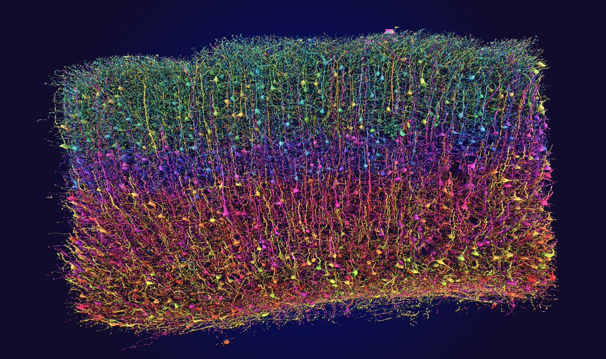
In this piece of brain tissue the size of sand grain are 200,000 neurons and nearly 2.5 miles of axons.
While it may seem small in size, it was previously thought impossible to map. The road to this beautiful image is both a scientific and human triumph: https://t.co/RtWiMgq75y
Hans Krebs identified the citric acid cycle - also known as the Krebs cycle. The cycle is a series of reactions that convert nutrients into other molecules with a large amount of chemical energy.
Do you know why it is so important?
Microbes found buried deep in Siberian permafrost may be able to survive over extremely long timescales using protein repair genes https://t.co/rbO7leonP2
This 1998 paper is, without question, one of the most beautiful in the history of biology.
It answers two questions:
First, how does a potassium channel let in K+ ions while excluding Na+ ions? And second, how does it funnel 100 million of those ions through each second?
These questions are interesting not only because potassium channels are so deeply involved in our brain's electrical signals, but also because K+ and Na+ both carry a positive charge and have similar sizes! A potassium ion has a Pauling radius (a measure of how far its electron cloud extends from the nucleus) of 1.33 Angstroms, compared to 0.95 for the sodium ion. So a potassium ion is slightly larger, but both ions carry exactly the same charge! And yet, despite these similarities, the potassium channel is “at least 10,000 times more permeant” to K+ than Na+.
How did evolution sculpt such an exquisitely-tuned machine?
To find out, scientists crystallized potassium channel proteins and solved its structure using X-ray crystallography. From this structure, a few things became immediately clear: First, the potassium channel’s interior measures 12 Angstroms long. And second, the channel's interior is lined with oxygens.
Normally, in a cell, ions are surrounded by water molecules. They must shed these waters to pass through the pore, but that's energetically expensive to do! The oxygens inside the channel are positioned at PERFECT locations to make this totally feasible; the K+ ions shed their waters and grab onto the oxygens instead.
The structure also revealed why Na+ cannot pass through. Because it is slightly smaller, Na+ ions cannot form contacts with all the oxygen atoms at once. The geometry is slightly off, giving potassium a decisive advantage.
Now onto the second question. Namely, how does this channel allow 100 million K+ ions to pass each second? That is very quick, considering these ions get "held" by their contacts with oxygen, presumably slowing them down a great deal.
Again, the scientists turned to structure. They again crystallized the potassium channel protein. But this time, they soaked those crystals in a liquid containing rubidium (Rb⁺) and cesium (Cs⁺). These ions behave like potassium but scatter X-rays more strongly, because they are heavier. Thus, they show up more brightly on the X-ray diffraction data.
When the scientists compared electron density maps with and without Rb⁺ or Cs⁺, they could literally see peaks where the ions bound inside the channel.
From this structure, they discovered that TWO K+ ions sit inside of the channel at once, separated by precisely 7.5 Angstroms. This distance is close enough that the ions "feel" each other’s electric repulsion (like charges repel!), but not so close that they destabilize the protein.
This repulsion is used by the channel as a feature, rather than bug! When a third K⁺ ion comes in from the top, the electrostatic “push” kicks the ions forward through the filter. In other words, instead of ions having to crawl through the pore one at a time, the channel uses their mutual repulsion to keep the flow moving; 100 million ions per second.
Beautiful paper. A classic in using 3D structures to reveal biophysical mechanisms.
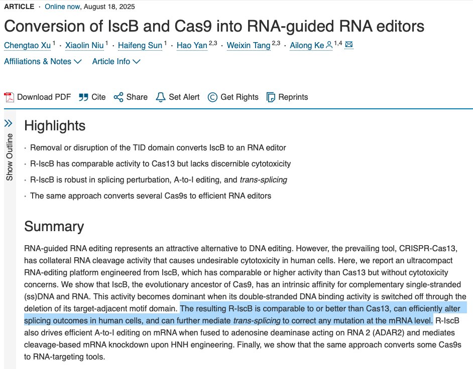
A new paper in Cell shows that it is possible (simple, really) to convert DNA-editing CRISPR proteins, such as Cas9, into RNA editors.
All you have to do is delete a chunk of them.
Deleting 55 amino acids from a DNA editor called IscB (the ancestor of Cas9) turned it into a “robust and versatile” RNA-guided RNA editor in human cells. This modified protein was used to cut mRNAs, knockdown gene expression levels, and even make an A-to-I base editor for RNA.
There are existing RNA-editing proteins, of course, with Cas13 being the most commonly used. Cas13 uses a short snippet of RNA to seek out, and then cut, target RNA molecules. But it also has a major problem: After it cuts its target RNA, Cas13 causes “collateral damage” by also randomly chopping up nearby, bystander mRNAs. This is toxic to bacterial and mammalian cells.
So in this paper, the authors deleted a part of the IscB protein (called the target-adjacent motif interaction domain, stretching from amino acids 433-487), which is normally responsible for its grabbing tightly onto DNA. And that deletion alone converted this protein into an RNA editor. The reason this works is because IscB naturally grabs onto both DNA and RNA, but is heavily biased toward DNA. So this deletion just removes its ability to grab onto DNA, thus biasing it to RNA.
The resulting RNA editor is not only “more active than Cas13,” according to the paper, but it also “has no discernible cytotoxicity in human cells.” The author also show that the same exact approach works for other Cas proteins, including Cas9. Lots of gene editors can be converted into RNA editors, in other words.
Good paper. Gene editors are even more versatile than we appreciated.
The only rule in biology is that there are exceptions to every rule. This is what makes biology infinitely exciting; even when you think you’ve got the complete view, the floor can drop out from underneath you at any given moment.
Case-in-point: The nucleus is the thing that makes eukaryotes...well, eukaryotes. It's the part of the cell that stores the genome, separating DNA from the cytoplasm and other organelles. (Bacteria do not have nuclei.) For decades, scientists thought that each nucleus contains one or more haploid sets of chromosomes.
But there are exceptions. Red blood cells, for example, don’t have nuclei at all. (They expel their nuclei during maturation to maximize hemoglobin concentrations.) Cells in the eye lens, too, lose their nuclei and organelles during differentiation, thus becoming transparent. And so on.
But now there is yet ANOTHER exception to this rule, and it’s one I hadn’t seen before. For a study in Science, researchers discovered that two types of pathogenic fungi that infect plants, called Sclerotinia sclerotiorum and Botrytis cinerea, have two different nuclei. And instead of storing a full set of chromosomes in each nuclei, they instead “distribute their chromosomes such that each of their nuclei contains only a subset of the haploid chromosomes.” The authors confirmed this by throwing a kitchen sink of methods at these cells; chromosome counting, DNA measurements using flow cytometry, single-nucleus PCR, and more.
Nobody knows why the fungi do this, but the scientists claim (in their discussion) that it could enable them "to respond and adapt more effectively to local environmental stresses within their extensive mycelial networks. Nuclear shuffling may facilitate the rapid generation of new genotypes, enhancing adaptability to changing environments.” There is also evidence that the chromosomes within each nucleus may briefly collide during cell division, before going back into their separate nuclei.
This is a great paper. It is simple, to the point, and challenges the status quo. It has serious potential to become a “classic” of the genre.
Link: https://t.co/AtsN5Kufpl
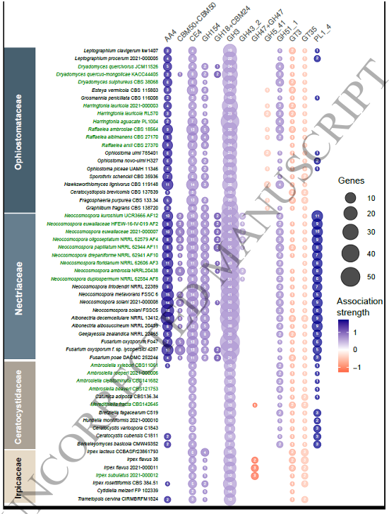
"changes are lineage-specific, not convergent"
CAZyme Expansions: "AA4 in Nectriaceae, CE4 in Ophiostomataceae, and GH3 in Ophiostomataceae and Ceratocystidaceae"
Genome diversification of symbiotic fungi in beetle-fungus mutualistic symbioses https://t.co/wpEQFr4Wgm
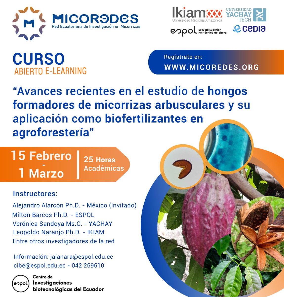
𝘾𝙪𝙧𝙨𝙤 𝙀-𝙡𝙚𝙖𝙧𝙣𝙞𝙣𝙜:
Avances Recientes en el Estudio de Hongos Formadores de Micorrizas y su Aplicación como Biofertilizantes en la Agroforestería
Inscripciones: https://t.co/QXlClsQyqh
En pocos días comienza "Avances Recientes en el Estudio de Hongos Formadores de Micorrizas y su Aplicación como Biofertilizantes en la Agroforestería"
Expertos de la @espol, @UniYachayTech y nuestro docente investigador @Leorange69 transferirán sus conocimientos en el curso.
👇
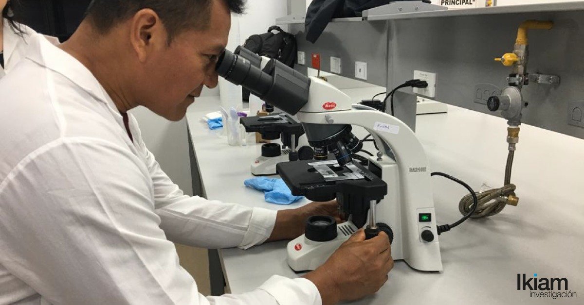
@UniYachayTech@espol@CEDIAec@AgriculturaEc@GoberNapo@EduSuperiorEc En la segunda jornada del proyecto. Se trabaja en la determinación de la frecuencia e intensidad de colonización micorrízica arbuscular en raíces de plantas de interés agroforestal. A cargo de los investigadores Verónica Sandoya, Jaime Naranjo y Leopoldo Naranjo-Briceño