Step 1 – Incubation with 2 nM Cellaris™ dyes for 2–4h
Step 2 – Seeding in the Culture-Insert 2 Well
Step 3 – Time-lapse imaging every 20 minutes for 72 hours
✨ Cellaris™ delivers biocompatible and photostable fluorescence – no transfection needed.
We’re thrilled to share the results of our collaboration with ibidi GmbH! Together, we developed a simple and robust live-cell tracking method for a co-culture invasion assay.
👉 Read the Application Note: https://t.co/ntBad4Be90
👉 Explore Cellaris™: https://t.co/S5ULh3dq75
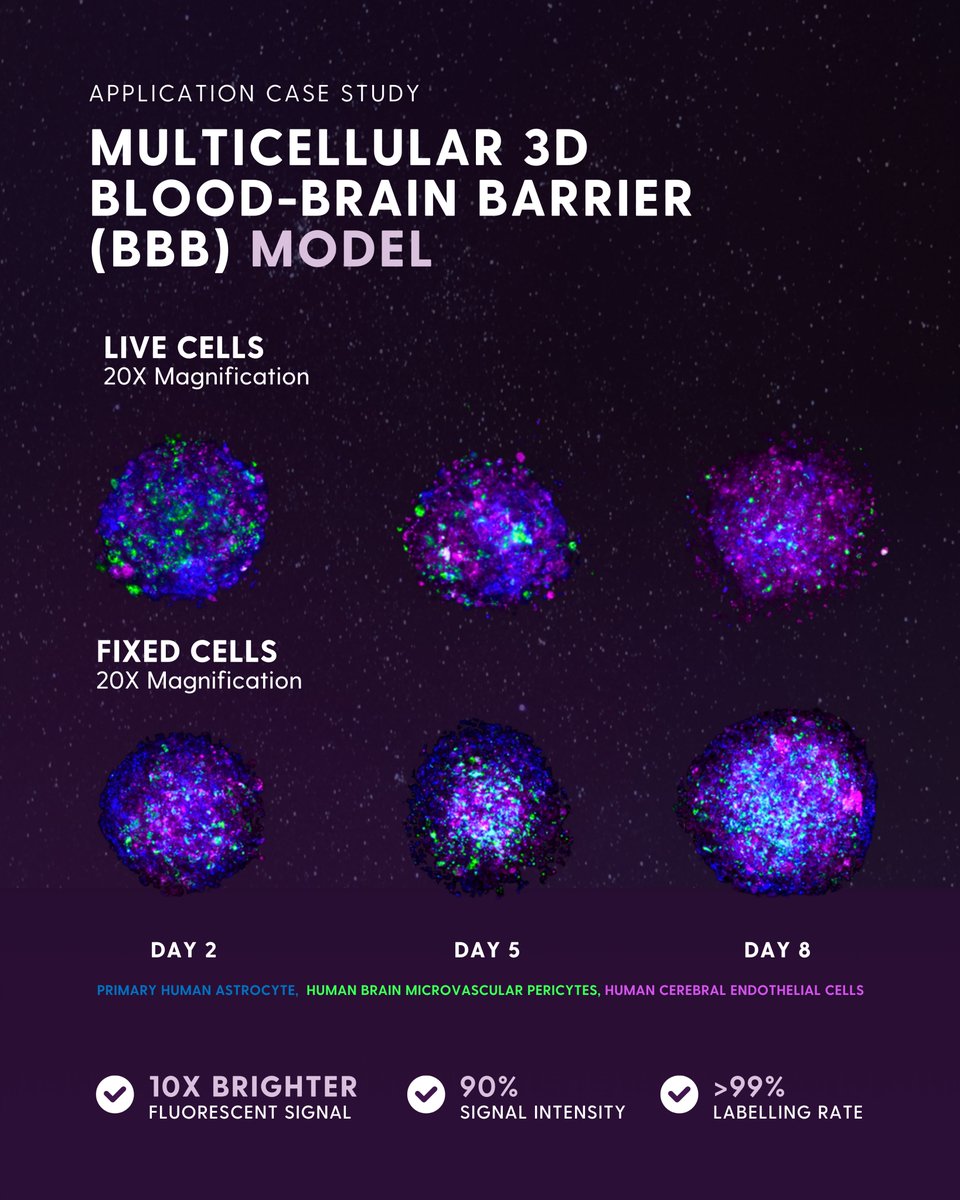
𝐑𝐞𝐬𝐮𝐥𝐭𝐬?
✨ Detailed visualisation of BBB cellular organisation and dynamics
🌟 Stable labelling with no fluorescence loss over 8 days of steroid culture
⚡️ High labelling efficiency across all BBB cell types
#BloodBrainBarrier#Organoids#Fluorescence#CellTracking
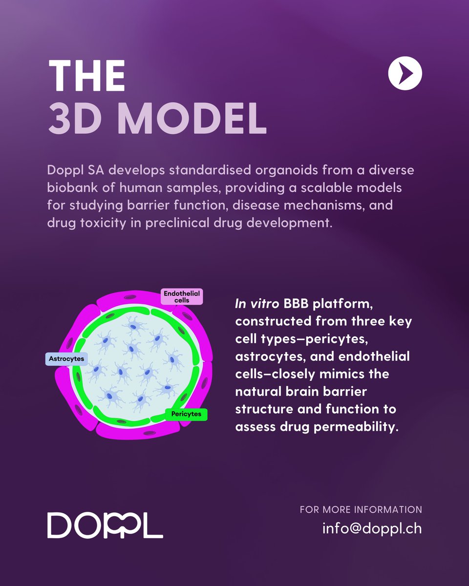
We’re excited to share our latest study using Cellaris™ dyes to label a 3D Blood-Brain Barrier (BBB) spheroid model of astrocytes, pericytes, and endothelial cells – ideal for assessing the permeability of brain-penetrating drugs!
👉 https://t.co/S5ULh3dq75
Final Call: 25% OFF!
Why Choose Cellaris™?
✨ Up to 30 minutes of continuous laser irradiation with no fluorescence loss
🔬 Monitor up to 10 cell generations in vitro
🐁 Over 21 days of in vivo tracking (with reports up to 8 weeks!)
The NIR-II imaging method, together with our product Cellaris™ 1010 enables real-time, non-invasive imaging, reducing the need for repeated procedures while improving both data quality and ethical research practices.
#NIRII#TumourImaging#CancerResearch#InVivoImaging
NIR-II long-term in vivo tumour imaging is unlocking new possibilities in tracking tumour distribution and dynamics!
👉 Find out more about Cellaris™ 1010: https://t.co/dEDncitMnE
Happy #FluorescentFriday! ✨
New Year, New Gear! 25% OFF for 2025!
Enhance your research with 25% off our next generation fluorescent probes.
🧫 Biocompatibility
✨ Ultra-Bright Fluorescence
💪 Unparalleled Photostability
👉 Claim your discount now: https://t.co/0rFS8S3Rqr
We gratefully acknowledge the contributions of our collaborators: Dr. Stefanie Kiderlen and Dr. Lukas Krainer (@ProspectiveInst), as well as Yasemin Geiger and Dr. Stefanie Sudhop (CANTER, University of Applied Sciences Munich) for collaborating on this study!
Things Just Got Brighter with @Luminicell's Arrival as a Trusted Supplier Partner on @Labscoop Marketplace !
Through this new partnership, Labscoop customers will have access to Luminicell’s state-of-the-art flagship products.
Read more on https://t.co/W7Ng7xZOzl
#Biotech
🚀 Labscoop is thrilled to announce our new partnership with @Luminicell , a cutting-edge subsidiary of @nanolumi ! 🌟
Together, by combining Labscoop's innovative lab supply marketplace with Luminicell's advanced bio-luminescent technologies, we will empower labs to achieve stellar imaging results in long-term cellular and vascular visualization.
🤝 Through this new partnership, @Labscoop #Marketplace users will have access to Luminicell’s innovative product line, which includes its state-of-the-art flagship products: Cellaris™ , next-generation cell culture system labeling kits, and Vascular Tracker™, next-generation vessel labeling kits. These advanced fluorescent nanoparticles are 10X brighter, exhibit unparalleled photostability, are highly biocompatible, and require no genetic modification or lengthy transfection process.
Read the full press release here: https://t.co/qUYPi2NAJ4
Learn more about Luminicell technology at https://t.co/7Jv7UV5FXh
#LifeSciences #CellBiology #vascular #cellular #labsupplies #research #biotech
#Cellaris, our flagship cell labelling kits, are well-characterised for their unparalleled photo-physical properties (signal longevity and photo-stability), designed to be the best-in-class reagent for long-term live cell experiments.
Our flagship product #Cellaris 670 and 506 were used to label (@Luminicell) Hep3b cancer cells respectively during seeding stage (Day 0), cultured in #OrganiX (@AIMBiotech).
Spheroids (~500 µm) were imaged at Day 5 and 11, with homogenous signals and brightness.
NIR-II fluorescent probes penetrate deep into tissue with significantly reduced background noise, delivering an exceptional signal-to-noise ratio for precise imaging.
Happy #FluorescentFriday!
We are excited to showcase the results of NIR-II vessel imaging performed on a whole mice using our Vascular Tracker 1010!
Learn more about Vascular Tracker:
https://t.co/J5YKf1e8V6
Contact our experts to discuss your application:
[email protected]
A special thanks to Dr. Xavier Le Guevel and Dr. Veronique Josserand from IAB Grenoble (Université Grenoble Alpes) for their outstanding work, where Montecarlo Restoration improved image contrast and spatial resolution.
Upon IV injection, Vascular Tracker remains in the bloodstream for several hours, offering high photo-stability.
This enables researchers to conduct high-quality, long-term imaging with remarkable clarity for their studies.