Is trying to remember toxic encephalopathy patterns making you toxic?
Does memorizing metabolic encephalopathy patterns put you into a metabolic coma?
Don't become encephalopathic!
Here is the cheat sheet of the encephalopathy patterns you NEED to know & how to remember them!
➡️Corpus Callosum:
🔸Lesions here are called cytotoxic lesions of the corpus callosum or CLOCC.
🔸Usually transient from cytotoxicity, especially viral infections.
🔸Remember they are commonly viral & transient bc CLOCC lesions are on the CLOCK
➡️Central Pons:
🔸Usually osmotic demyelination
🔸Remember this bc the T2 hyperintensity is in the shape of O for Osmotic
➡️Dentate:
🔸Usually flagyl toxicity
🔸Remember this bc the wavy appearance of the dentate looks like a FLAG for FLAGyl
➡️Corticospinal tract:
🔸Usually from cobalamin deficienty
🔸Remember cortiCOspinal for CObalamin
➡️Symmetric Gray Matter:
🔸Differential is EXTENSIVE
🔸Remember the differential list for deep gray is deep!
➡️Cortical Gray Matter:
🔸Typically hypoglycemia & hyperammonemia
🔸Remember it looks like the brain has been coated in shimmering sugar = related to glucose
🔸Hyperammonemia has about 1000 Ms in it & the undulated cortex looks like an M
➡️Symmetric White Matter:
🔸Commonly from chasing the dragon (heroin) & methotrexate
🔸Remember that the puffy white matter signal looks like the smoke from a dragon
🔸Remember methotrexate as methoTRACTSate = affects many white matter TRACTS
➡️Posterior subcortical edema:
🔸This is PRES or posterior reversible encephalopathy
🔸So if it’s all POSTERIOR, think POSTERIOR reversible encephalopathy
➡️Asymmetric White Matter:
🔸This is usually demyelinating from chemotherapy
🔸Demyelinating lesions have a C shaped pattern of enhancement
🔸So if you see a C enhancing think of C-hemotherapy
Now you know all the toxic/metabolic encephalopathy patterns.
So hopefully now remembering the patterns of toxic/metabolic won’t seem so diabolic!
An easy way to differentiate these midbrain syndromes is to remember that, moving from anterior to posterior, all three involve oculomotor nerve damage, with additional distinct features:
Weber’s syndrome – affects the anterior midbrain, involving the corticospinal tracts, leading to contralateral weakness.
Benedikt’s syndrome – involves the mid-midbrain, including the red nucleus, causing abnormal involuntary movements such as tremor and chorea.
Claude’s syndrome – localized to the posterior midbrain, where superior cerebellar peduncle fibers (after decussation) are affected, resulting in cerebellar signs (think Claude = Cerebellar).
#Neurotip
Chater's point
6cm above the EAM on a line
perpendicular to the orbitomeatal line;= 1.5cm above the squamosal suture along the same perpendicular line.
A 4cm craniotomy centered at Chater's point provides access to recipient vessels in the angular gyrus for EC/IC bypass surgery
The snow isn't stopping our residents!
Dr. Ziqi Wang, PGY-1, and Dr. Elizabeth Bridwell, PGY-2, performed bilateral burr holes for a chronic subdural hematoma during the snowstorm.
#Neurosurgery#UofLNeurosurgery@uofl@uoflmedschool@UofLHealth
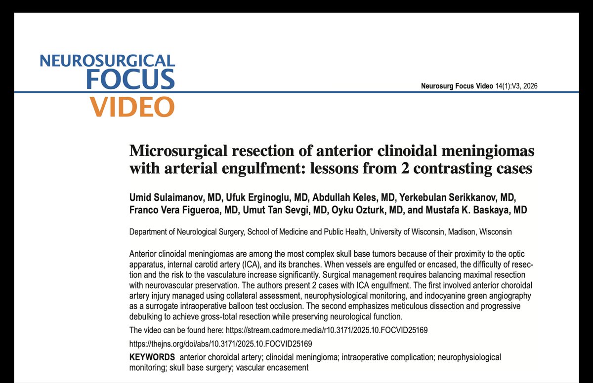
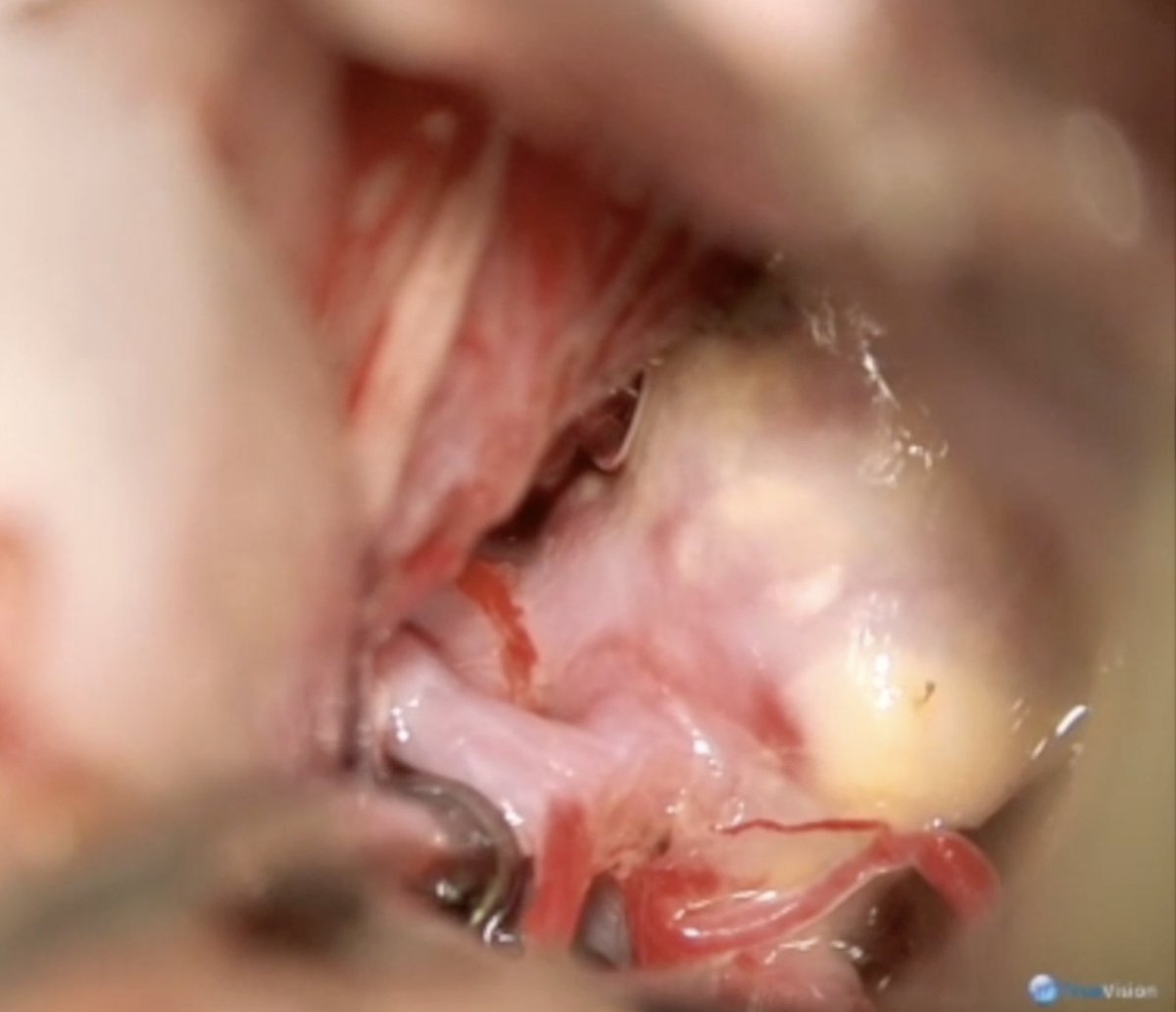
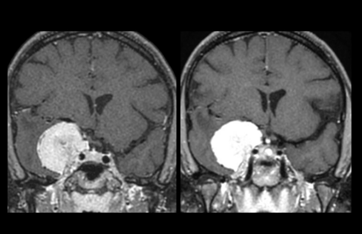
🧠🎥 New operative video in JNS Reports Microsurgical resection of diaphragma sellae meningioma with extradural clinoidectomy and preservation of superior hypophyseal arteries.
📄 DOI: 10.1055/a-2765-5582 🔗 https://t.co/jf00RfK4gK
#Neurosurgery#SkullBase#UWHealth#UWMadison