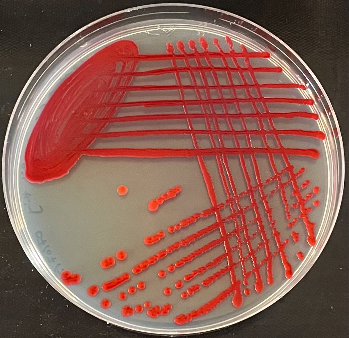
We saw a lot of diversity on these plates!
This photo is a great snapshot.
In just this small area, we can see at least 10 different colony types!
(Each structure is a colony)
It’s hard to identify microbes from their colonies alone, but here's a thread of our thoughts:
@Micro_Mad@IMIBirmingham These are awesome! Whilst I'm not expecting one for every plate, could we have a description of some of the things we can see?
@ClaireW36827493 That's what we like to hear!
We've posted a thread today with a bit more detail. Plate 140 has a couple of those hairy colonies that are probably Bacillus!
@TheRealStu1@IMIBirmingham We're glad to hear the boys enjoyed it!!
Check out today's thread, it covers the hairy stuff!
As for plates 59 and 60, it's interesting that they look more like one another than other plates in the collection! People who spend a lot of time together can share their microbes.
We're about to start sharing photos of everyone's plates from #CoCoMAD ! 🧫🧫
Our plate photo studio is set up here at @IMIBirmingham , and we'll be posting throughout today - keep an eye out for your number!
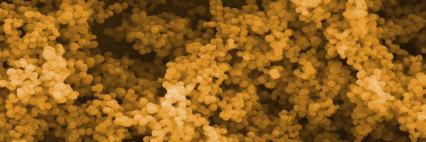
We looked at four colonies from this snapshot under the microscope!
In colony 2 there are two types of cells - large purple and small pink. In colony 3, there are three types! The biggest purple cell in 3 is probably a yeast.