@SkinMD13 🙂Answers C,C,A. Reticulohistiocytoma has "ground glass"/"muddy rose" cytoplasm. Can be solitary but multicentric reticulohistiocytosis (MRH) presents with lesions, often on hands, with a "coral bead" appearance. Non-Langherhans' origin = CD1a neg. Arthritis common with MRH.
40 y/o F. Finger papules.
BEST answers.
Dx:
A) giant cell tumor
B) granuloma annulare
C) reticulohistocytoma
D) xanthogranuloma
IHC + in cells of interest:
A) CD1a
B) CD3
C) CD68
D) Sox10
Important to ? pt about:
A) arthritis
B) DM
C) HTN
D) NHL
#Dermquiz#Dermpathquiz#Derm
My answers:
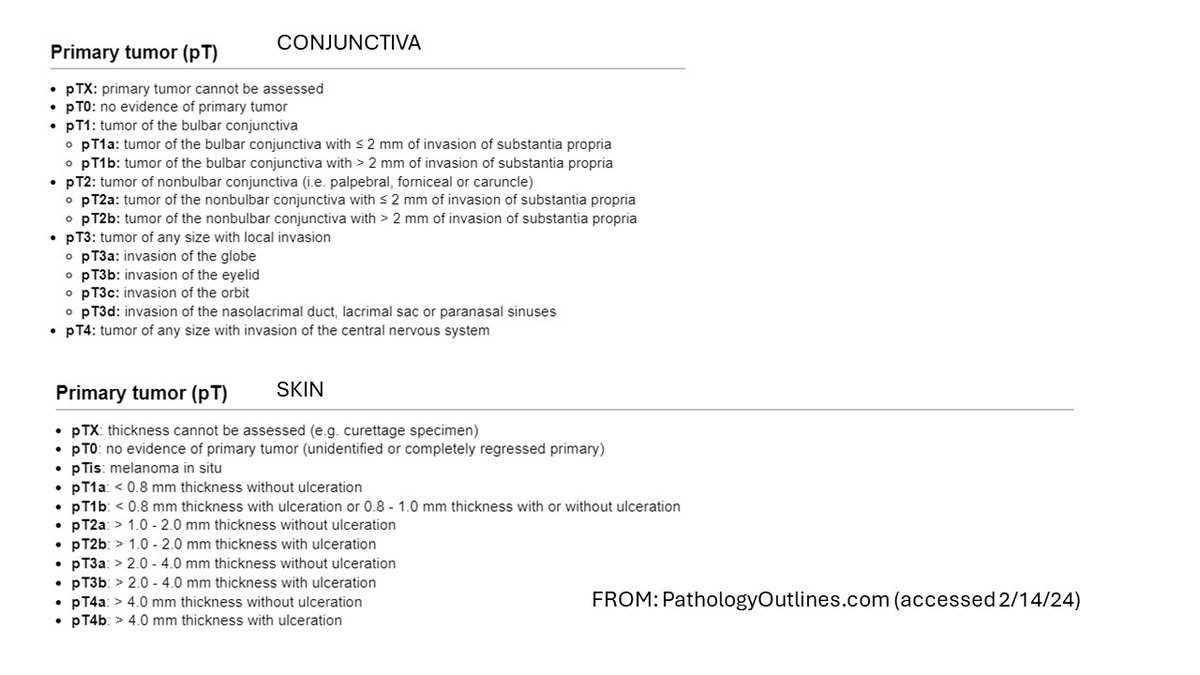
TRUE -pT criteria differ for conjunct MM, some in dermpaths own group unaware;
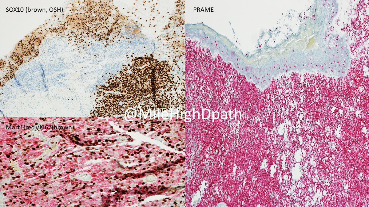
TRUE -subjectivity here, no fighting, but IMHO the utility is about same;
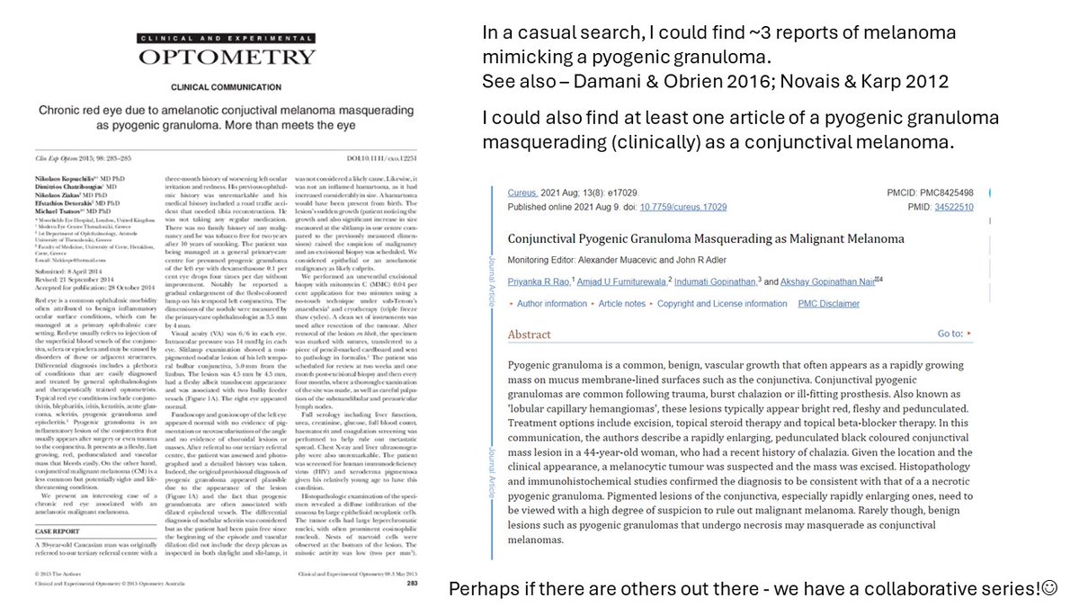
TRUE -reports of conjunctival MM confused with PG and vice/versa
Refs attached.
Hope that expands ur knowledge!🙂
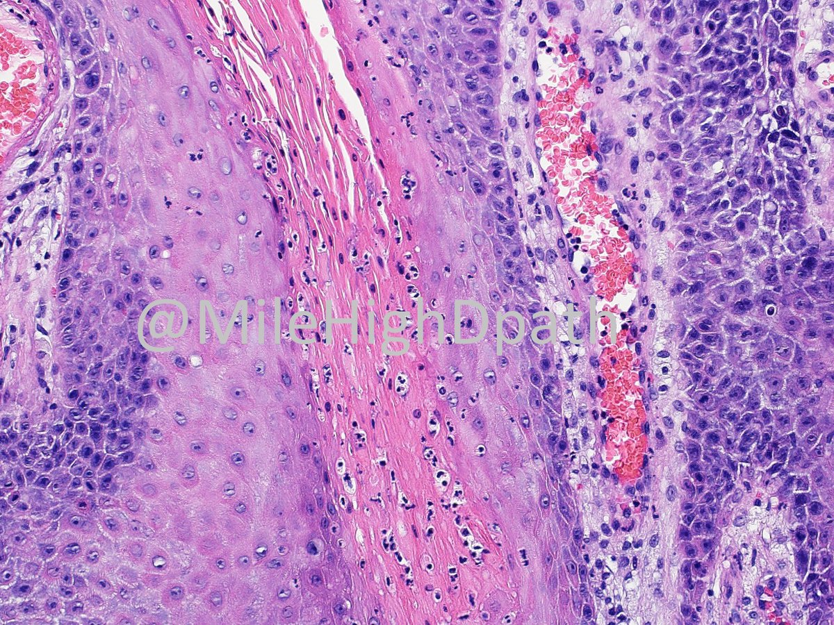
Young man (<30 y/o). Noted lesion for several years. Dx as pyogenic granuloma at some point, but continued to grow. Bx performed.
The best dx is:
A) cavernous hemangioma
B) Kaposi sarcoma
C) melanoma
D) pyogenic granuloma
#Dermquiz, #Dermpathquiz, #ConjunctivalLesion
Things learned from case:
T/F - pT criteria for conjuctival MM differ from those of skin
T/F - evidence re: utility of PRAME for conjuctival melanoma is similar to skin MM
T/F - there are several case reports in idex med lit. of conjuctival MM being confused (clinically) with PG
@Dermpathl All evolved this week.
The morning specimen reached my desk, I heard (vaguely) NPR summary of the CDC announcment re: syphilis rates reaching all time highs during my commute!
Wish every provider, especially, non-dermatology related providers, could see case. Seems ominous!
Part 1. 39 y/o. Rash since 11/23. Also c/o joint/muscle pain. Chart states "saw derm last week, dx???" Derm did bx. FP APP did many blood tests.
You now want:
A. Want more blood tests
B. Want IHC
C. Want special stain
D. Want to see more skin
E. >/=3 of above.
#Dermpath#Derm
@Dermpathl Yes. I also wanted a RPR/VDRL and FTA-ABS, and asked for more detailed pictures (none exist) I had to call both the dermatology office, and the family practice office, before the blood tests were performed (pt difficult to reach) but they were both POSITIVE. Thanks reply.
Last week CDC stated syphilis infections at highest rate since 50's!
Here is what was done:
1) asked for pics of palms/soles - none
2) obtained IHC for spiros (Steiner is other option)
3) I recommended RPR/VDRL and FTA-ABS (+/+)
#syphilis,#dermpathquiz, #dermquiz
Hints:
(1) Fam Med focused on CTD, but in limited pics, there is not photodisribution and a "boiled ham" color to pap squamous rash.
(2) There are granulomatous elements (uncommon in CTD), but also tracking of inflammation down N/V bundles and curious number of plasma cells.
IMHO best theories on foam cell origin go something like this:
- unknown event damages keratinocytes, perturbations in repair mechanism(s) yield cytokines that attract histiocytes, histiocytes ingest lipids from degen keratinocytes, form foam.
(Hu Ja et al. 2005, among others)
34 y/o. Papule on scrotum. "R/O condyloma." No other history known. What is the best diagnosis?
A. Condyloma acuminatum
B. Condyloma lata
C. Epidermolytic hyperkeratosis of the genitalia
D. Verruciform xanthoma
#Dermpathquiz,#Dermquiz
Yes, verruciform xanthoma. Common on oral mucosa and genitalia. Histo features:
1) papillomatous architecture
2) "crypt-like" parakeratosis
3) neutrophils in parakeratosis
4) lipid laden foamy histiocytes in shallow dermis
Associated in literature with CHILD syndrome.
For MCQ examination purposes:
Which of the following syndromes is most strongly associated with the presence of verruciform xanthomas?
A. CHILD syndrome
B. LAMB syndrome
C. PHACE syndrome
D. SAPHO syndrome
IHC below represents "Adipu" stain (or combination of Pu.1 (brwn) - a histiocytic marker, and adipophilin (red) - a lipid marker). It highlights the lipid laden histiocytic cells in the shallow dermis.
There are 3 proposed mechanisms for lipid accumulation.