Before making a diagnosis of invasive breast cancer in a core needle biopsy, be sure it is truly a primary breast tumor, particularly in the era of neoadjuvant chemotherapy
Metastatic serous carcinoma and metastatic melanoma to breast (pic 1)
Metastatic lung adenocarcinoma (pic 2)
Metastatic medullary thyroid carcinoma (pic3)
Of 2423 TNBC submitted to Caris for molecular profiling over 19 months there was a misdiagnosis of TNBC in 73 cases (3%). Half of those cases were non-small lung cancer (data submitted for publication)
Dr. Schnitt #USCAP2026 #pathology #PathX #PathTwitter
Low-grade B-cell lymphomas may present with prominent reactive inflammatory infiltrates or coexist with autoimmune disorders
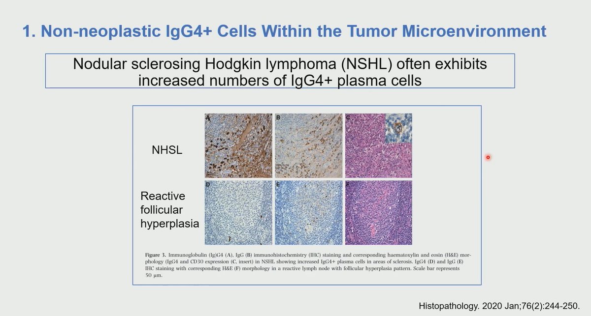
⚠️ Increased IgG4:IgG ratio? Stop and think before diagnosing IgG4-related disease. Always exclude an associated lymphoproliferative disorder
What is the cause of abundant IgG4 in lymphomas?Possible explanations...
1. Non-neoplastic IgG4 cells within the tumor microenviroment
2. Monoclonal IgG4 production by lymphoma
3. Lymphoma can coexist with IgG4-related disease and other autoimmune disease
Dr. Herrera Hernandez #USCAP2026 #pathology #PathX #PathTwitter
Cutaneous tumors - Head and neck tumors
***Same histology - Different terminology***
Cylindroma: Adnexal tumor in scalp/face/ rarely lip. Malignant transformation rare
Basal cell adenoma: Salivary gland tumor, >80% on parotid/can involve lip. High rate of local recurrence. Malignant transformation 3-4%
Dr. Ivan #USCAP2026 #pathology #PathX #PathTwitter
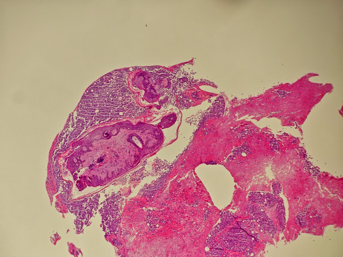
Incidental polyp in the terminal ileum of a colon cancer right hemicolectomy. IHC negative apart from patchy CD34. This is a benign calcifying fibrous tumour. Can be found anywhere in the GI tract and do not have any syndromic associations (IgG4 IHC was less than 40%) #GIpath
WHO 2021 Thymoma Classification in a Glance! 🔬
Master Type A, B (B1-B3), AB, and Metaplastic variants with our simple memory tricks. Essential for surgical pathology!
Follow us for more daily high-yield tips! 👇
#Thymoma#WHO2021#Pathology#Oncology#MedEd#SurgicalPathology
Nodal nevus in the capsule vs Melanoma Metastasis (subcapsular). Same patient. Nice contrast in cellular morphology and IHC #dermpath#pathology#IHCpath
📣Curso online gratuito Workshop de Patología Intervencionista, mañana 1 de octubre.
✍🏻15:00 hora española.
Ponentes españoles con ponencias en español.
Organizado por el Ipatimup - Oporto.
Link Inscripción: https://t.co/65AYXsIU8L
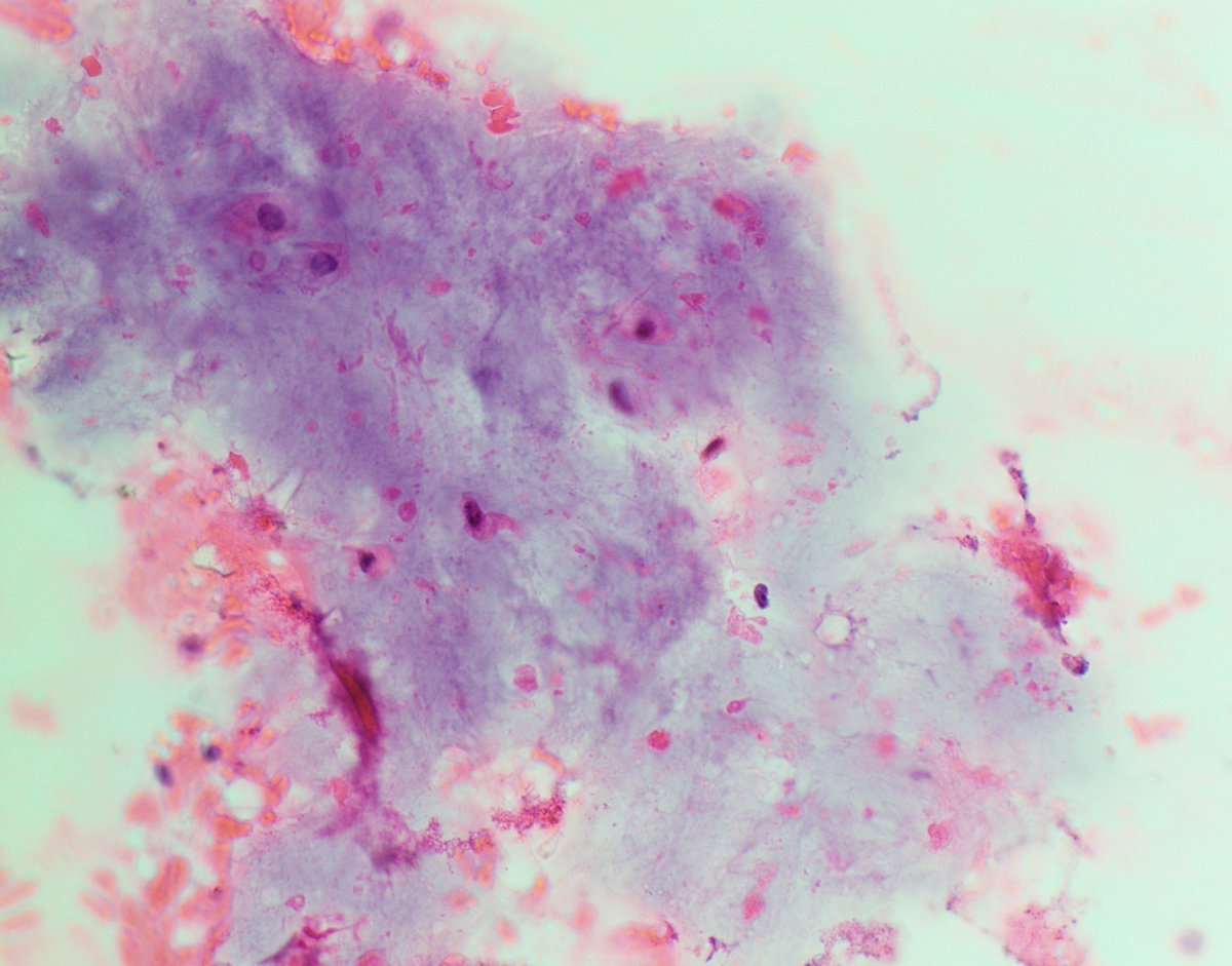
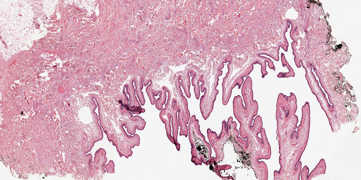
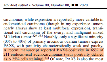
My colleague Matt just walked into my office.
Matt: How often is PAX8 positive in endocervical adenocarcinoma?
Me: It's about 50/50...could be 40%...could be 60%. I can't remember. Let's look it up in "my review."
Matt: I didn't think you would have covered that.
Me: 😂😂😂
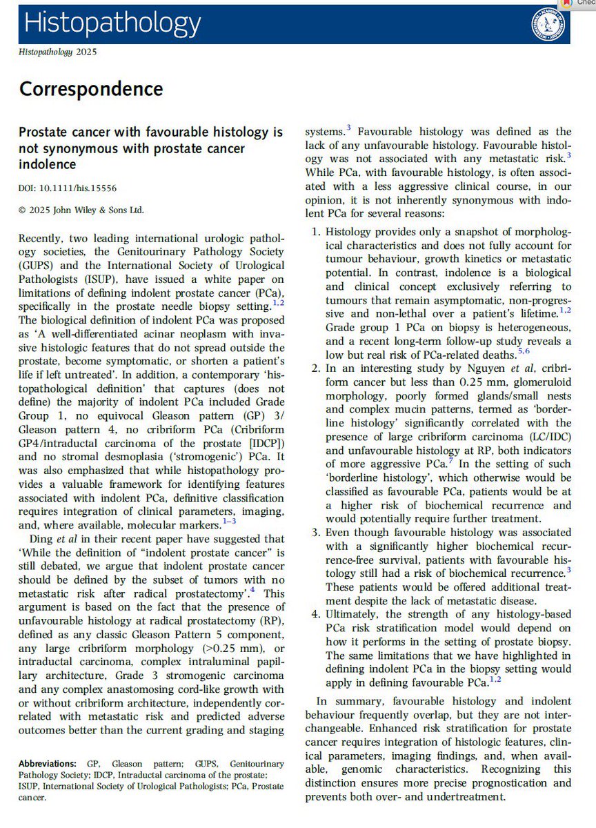
Hot from press and right in time for Prostate Cancer Awareness Month: we explain why prostate cancer with favorable histology is not same as prostate cancer indolence! Must to understand the concept! 5 minutes read! #ProstateCancer#gupath
Morphologic correlation with molecular findings
72 yo F with calf mass and history of high grade sarcoma 6 years ago s/p radiation
Now presenting with myxofibrosarcoma
Is this a recurrence or a radiation-associated sarcoma?
.
Both specimens were sent for targeted sequencing
and they have identical missense variants of uncertain significance (VUS)
-Usually we don't pay attention to VUS but in this case the VUS are less than 50% (they are somatic) and it doesn't make sense for two different tumors clonally unrelated to have 3 identical VUS with a somatic allele frequency.
Final diagnosis: This is a recurrent sarcoma and not a de-novo radiation associated sarcoma
Dr. Dermawan -Comprehensive and Immersive Soft Tissue Pathology Course-May 2025 #pathology #BSTPath #PathX
Hodgkin Lymphoma is a great example of how important it is to accurately report immunohistochemical stains. Simply ‘Positive’ and ‘Negative’ isn’t enough! #PathX#HemePath
For PGY1s: compared to ordinary macrophages, Langerhans cells have far more irregular nuclear contours. See this side by side comparison
@grok@AskPerplexity tell us something about Langerhans cells and Paul Langerhans