#POCUS#MedTwitter#Nephpearls
Many #VExUS enthusiasts asked for a #tweetorial on image acquisition pearls. Did one b4 but time for an updated one 🧵
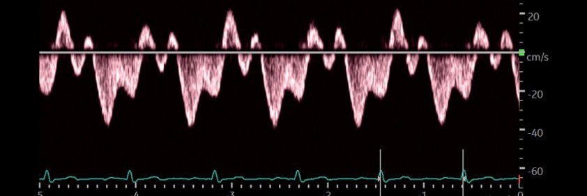
#1 Let's start with basics
Color Doppler identifies the flow + tells the direction (blue is away & red towards the probe [BART])
@NephroP@norman_sukmadi Also premature closure is severe AR or Av block. I'm an mode lover Always use it. Always gives useful information.
I'm in my 70s, 40 years using echo. My younger colleagues does not use it, I always teach many times with clinical cases
How to do SVC #POCUS 📹 + an illustration of abnormal SVC Doppler patterns.
#VExUS#eVExUS#Nephpearls
From 🔗J Am Soc Echocardiogr. 2023;36(5):447-463. doi: 10.1016/j.echo.2023.01.017
Pleased to have had the opportunity to write this CJASN editorial @asnpublications on the USE-the-FORCE-for-Acute Kidney Injury trial by our Canadian colleagues.
🔗https://t.co/3ouwPLv2wZ
I'm especially glad to see mainstream #nephrology journals taking interest in multi-organ #POCUS. Even better, I managed to sneak my proprietary hemodynamic circuit illustration into the article!!
High-output heart failure associated with arteriovenous fistula remains an underrecognized, albeit well-described, clinical entity. This is a nice case with illustrative images - doi: 10.1016/j.jaccas.2026.107193. PMID: 41770183
#POCUS#Nephpearls
Yes.
#POCUS is so heterogeneous right now that someone who only looks at the IVC is considered to be doing POCUS, while someone performing a critical care echocardiogram with spectral Doppler is also doing POCUS.
That’s why teaching learners to pay attention to subtle grayscale and M-mode clues still has value - helps as more people gradually adopt advanced POCUS techniques. Moreover, what’s considered ‘advanced’ keeps evolving.
It depends on what you’re using it for. For example, it’s very helpful for assessing RVOT collapse in tamponade when there is no simultaneous ECG tracing available. It can also aid in the detection of systolic anterior motion and dynamic LVOT obstruction.
In the image above, there’s a B-bump, which should prompt a closer evaluation of diastolic function. Likewise, a flattened EF slope should raise suspicion for mitral stenosis.
These subtle clues are particularly important in the #POCUS world because focused examinations do not include all views and measurements obtained during a comprehensive echocardiogram. Paying attention to these small findings can help identify important pathology.
@NephroP@TransplantJrnl It has been a true pleasure working with you on this paper. You have a unique talent for adding valuable #POCUS expertise to any clinical discussion and translating physiology into practical bedside care. Excited to see where this work leads next. @UFNephrology@UFMedicine
It's always tricky to comment on left ventricular diastolic function from an EPSS tracing because even a slight off-axis view can easily lead to misinterpretation. For the same reason, it's hard to say much about the posterior mitral leaflet above. I was mainly referring to the morphology of the anterior mitral leaflet tracing.