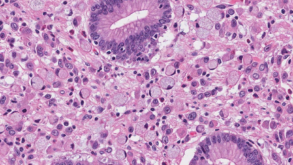
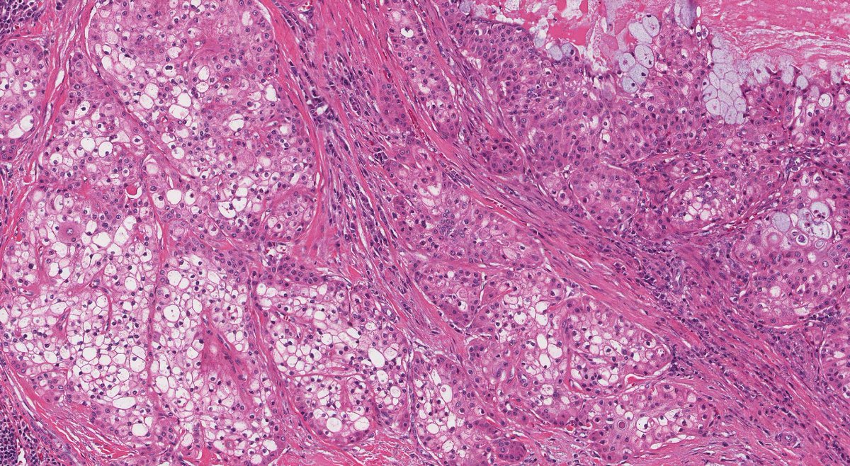
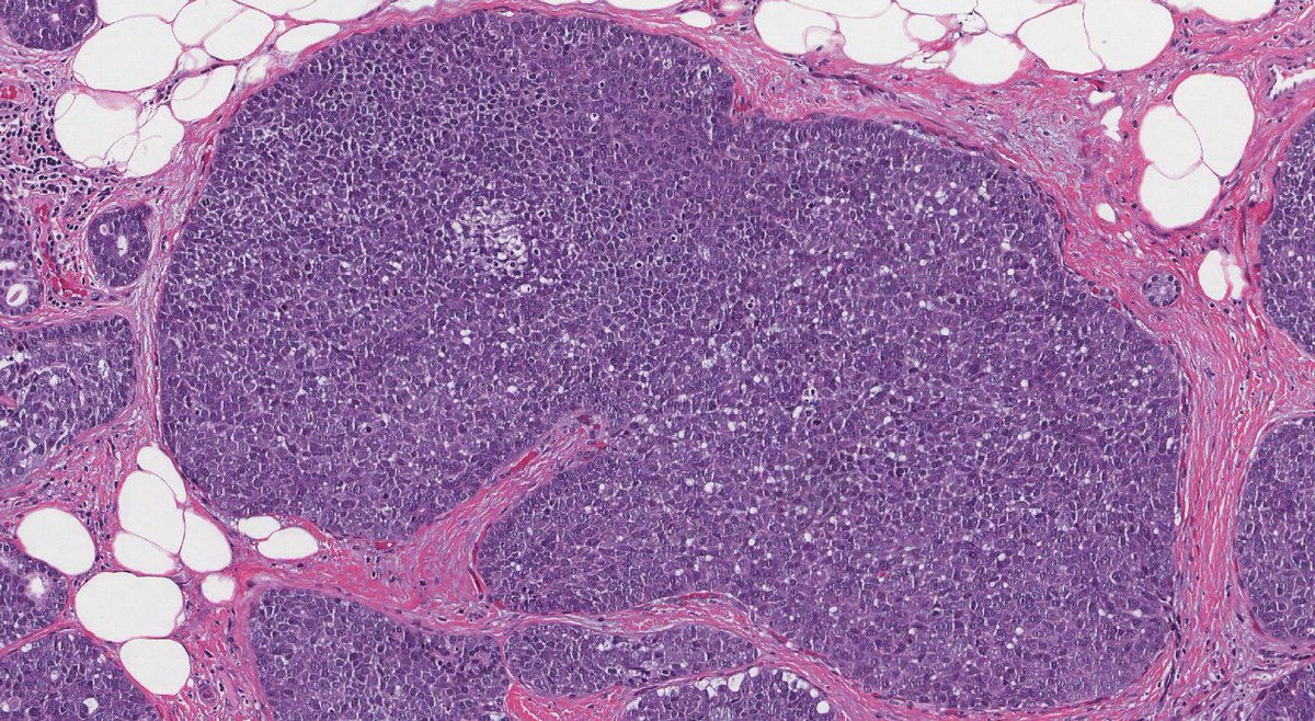
Chronic active ileitis is a pattern of inflammation and injury often associated with Crohn's disease, but other causes must also be considered. #IBD#patients
Learn more about your #pathology report in this new article on https://t.co/KuYsGsMmiz
https://t.co/fRFQBQSRyh
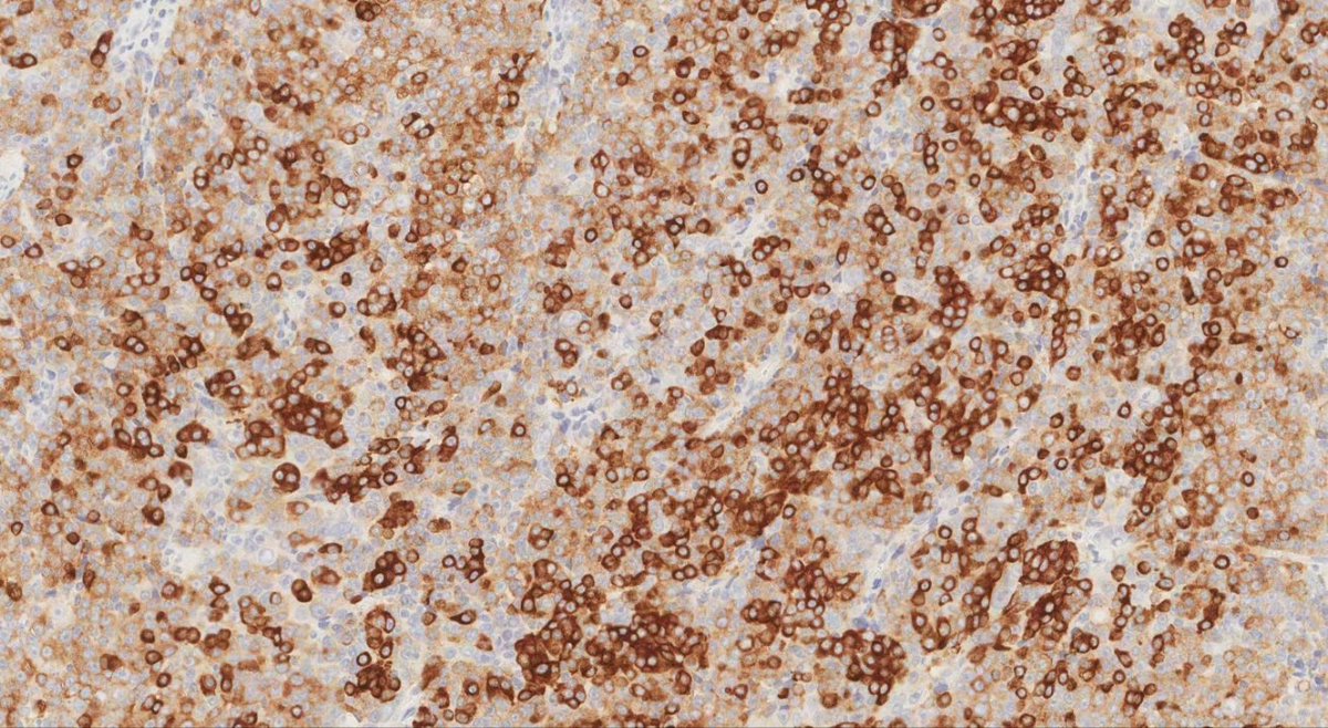
A rare case of intrathyroidal thymic carcinoma identified on ultrasound examination as a thyroid nodule. The tumour cells show strong expression for CD5, synaptophysin, and CK5.
#cancer#pathology#raretumour#pathologists#thyroid
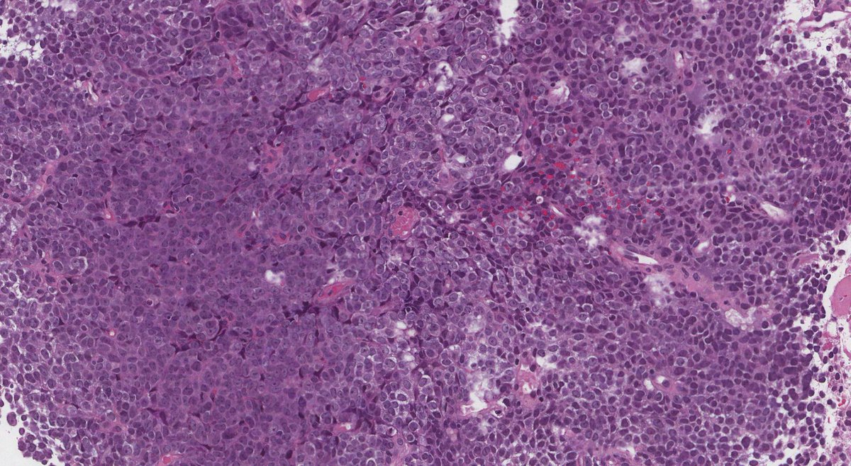
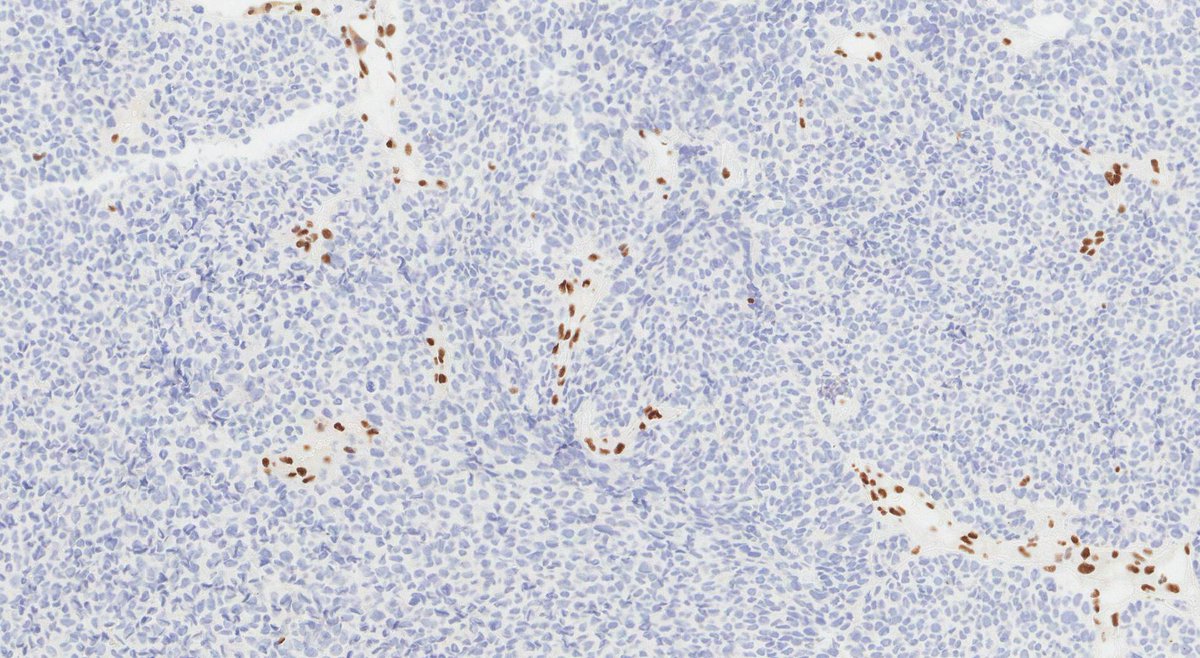
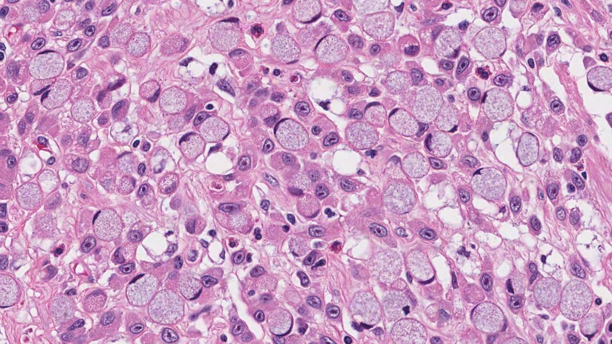
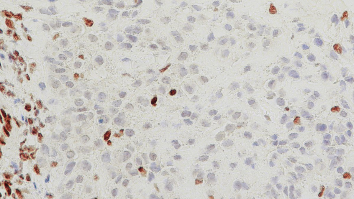
SMARCB1-deficient sinonasal carcinoma is an aggressive malignancy characterized by the loss of SMARCB1 (INI1). Finding plasmacytoid/rhabdoid cells in an otherwise malignant basaloid tumour aids in the diagnosis. #cancer#pathology#patientcare#MedEd#PathTwitter#medicine
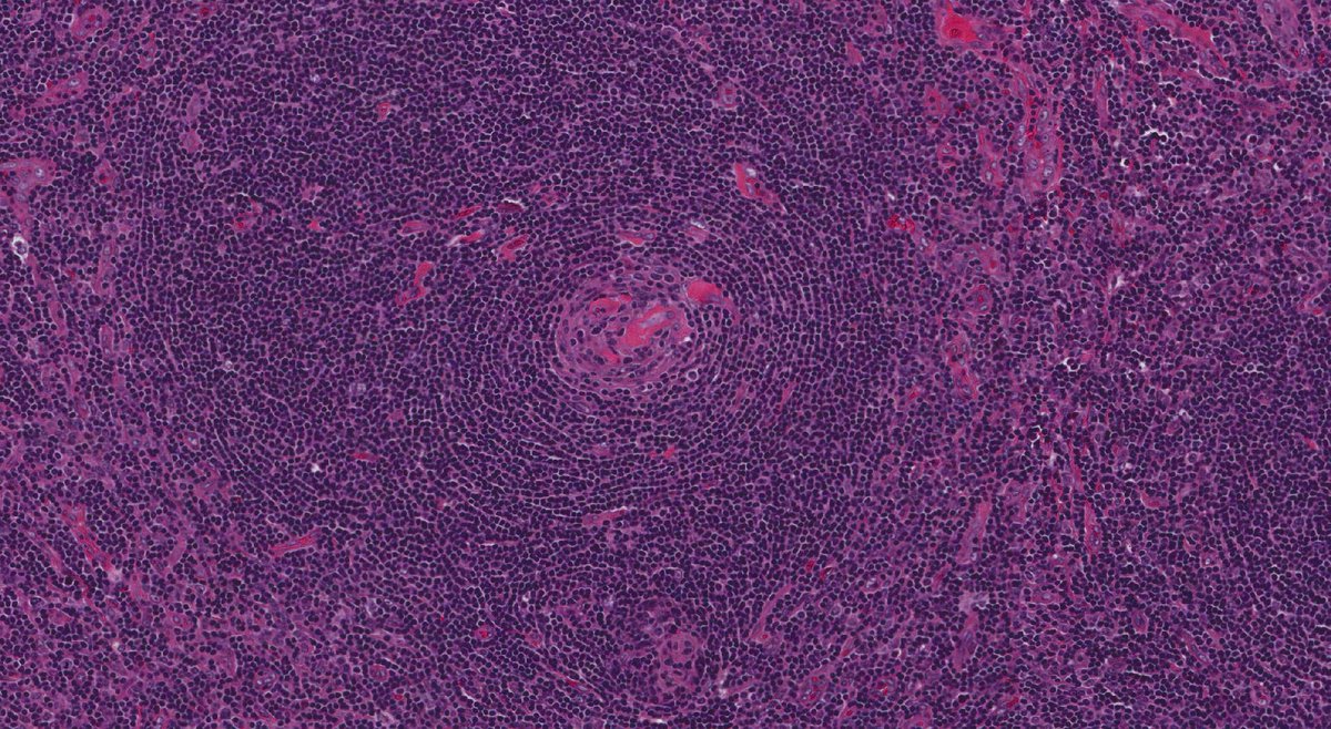
Unicentric Castleman's disease, hyaline vascular subtype. The H&E stain shows "onion skinning" around a germinal center while CD21 highlights the "twinning" of two germinal centers surrounded by a single mantle. #pathology#pathologist#medicine
https://t.co/S17yd6UYW9
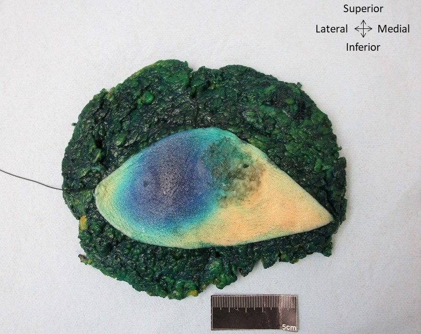
The Ottawa Atlas of Pathology is back with a new feature: Gross Image of the Week.
Can you guess what this picture shows? Hint: The blue is ink injected to identify sentinel lymph nodes. #pathology#PathTwitter#medicine
https://t.co/diQctIFErF
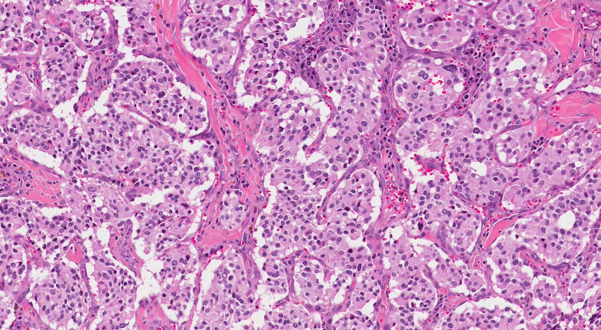
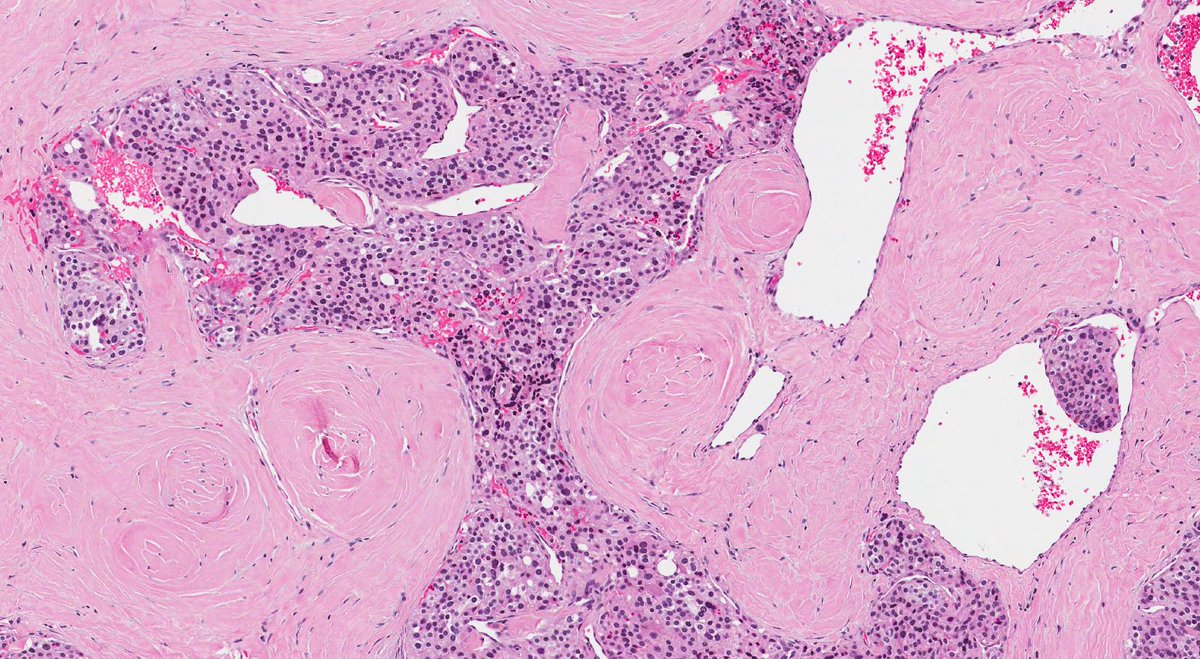
Carotid paraganglioma. It is composed of chief and sustentacular cells arranged in a nested or "zellballen" architecture with a prominent vascular network and areas of hyalinized stroma. IHC would stain chief cells chromogranin+/synaptophysin+ and sustentacular cells S100+
Gastric adenocarcinoma with signet ring morphology. Sections show benign glands with a diffuse stromal infiltrate of single cells. The neoplastic cells have abundant intracytoplasmic mucin and an eccentrically placed nucleus.
Barretts esophagus. This biopsy of the distal esophagus shows intestinal metaplasia of the normal stratified squamous mucosa into columnar epithelium with goblet cells. The most common risk factor is GERD. Endoscopically it appears a salmon colored plaque extending beyond the GEJ
Epithelioid sarcoma. This tumour is a malignant mesenchymal neoplasm with epithelial differentiation. It's composed of small-medium epithelioid cells with eosinophilic cytoplasm and mildly atypical nuclei. IHC is CD34+ CK8/18+ and loss of INI1 (associated with a SMARCB1 deletion)
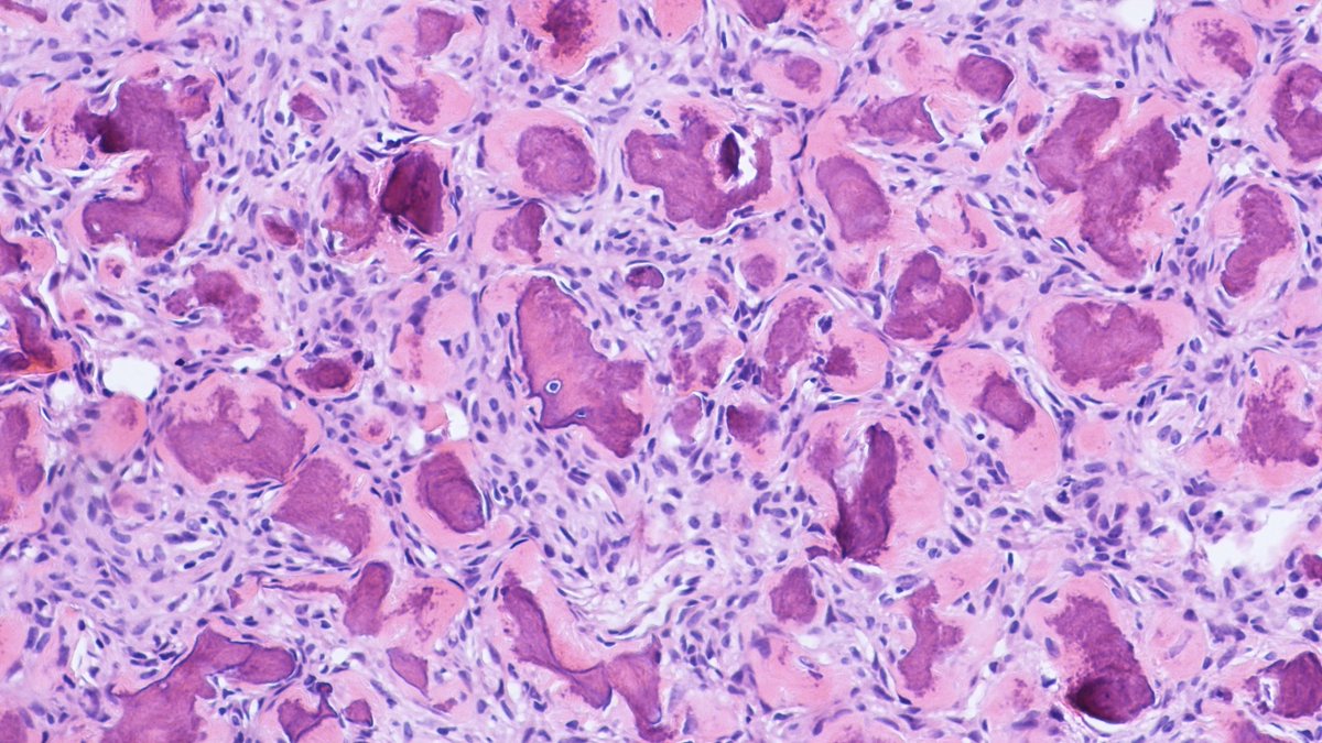
Psammomatoid juvenile ossifying fibroma. This tumour is a rapidly growing benign fibro-osseous lesion that affects the extragnathic craniofacial bones of young patients. It consists of psammomatoid bodies embedded in a cellular fibrous stroma and within bony trabeculae.
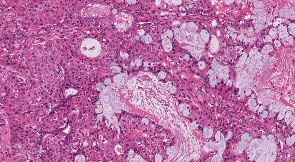
Mucoepidermoid carcinoma of the parotid. This is the most common malignant salivary gland neoplasm. It has a solid-cystic architecture with epidermoid, intermediate, clear cells, and mucin secreting cells. t(11;19) is seen in 55-65% of cases resulting in a CRTC1-MAML2 fusion.
Adenoid cystic carcinoma of the breast. This tumour is composed of luminal and myoepithelial cells with cribriform, solid, or tubular architecture, similar to its counterpart in salivary glands. A common genetic finding is a translocation resulting in an MYB-NFIB fusion.