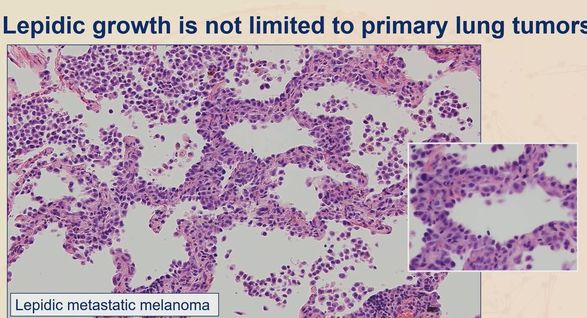
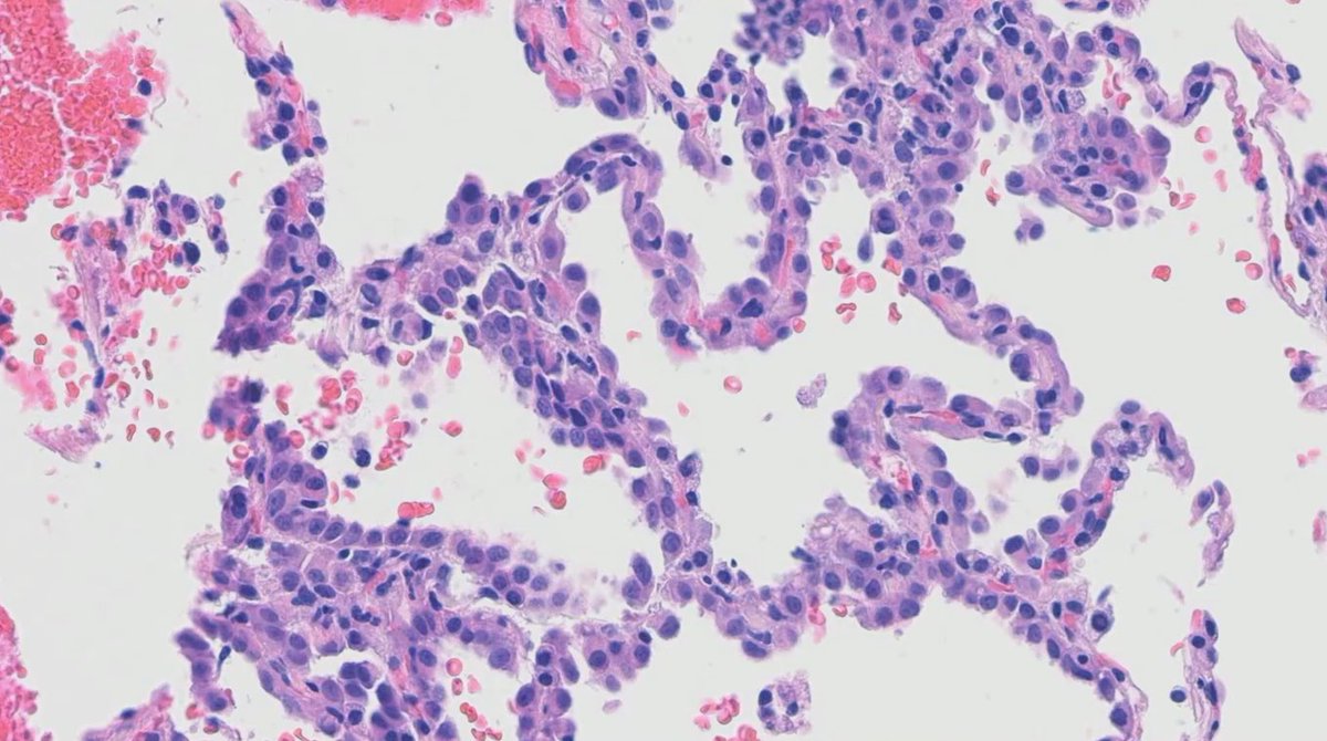
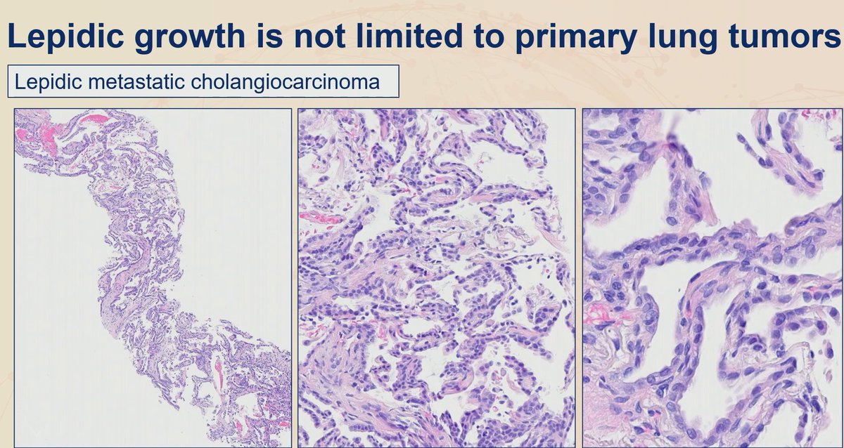
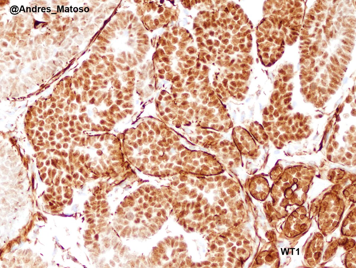
***Lepidic pattern is not limited to primary lung tumors***
- Female patient with history of breast cancer now with lung lesion (pic 1 and 2): Mesothelioma, epithelioid type
- Melanoma (pic 3)
- Cholangiocarcinoma (pic 3)
Practical lession: Lung tumors that looks like carcinoma but negative for TTF-1 and p40, add Claudin 4 and WT-1 to exclude the possibility of mesothelioma
Dr. Sauter #USCAP2026 #pathology #PathX #PathTwitter
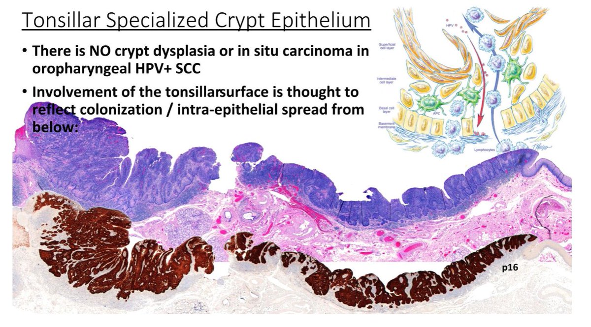
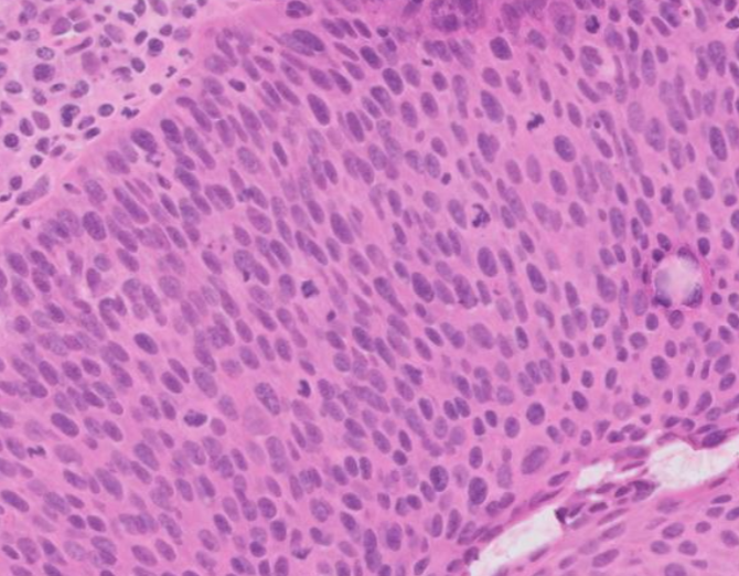
Oropharynx exophytic lesion: SCC?
Tonsillar crypts are lined by a unique reticulated epithelium. The basement membrane is disrupted and non-contiguous, allowing the epithelium easy access to lymphatics.
- There is no crypt dysplasia or in situ carcinoma in oropharyngeal HPV+ SCC.
- All HPV-related neoplasia of the tonsils should be regarded as malignant even in the absence of "classic" histologic features of invasion.
Dr. Cipriani #USCAP2026 #pathology #PathX #PathTwitter
Rosai-Dorfman disease in skin -somebody please call the police - the emperipolice! An S100 protein stain highlights the nuclei (arrow) and cytoplasm of abnormal histiocytes but not the engulfed inflammatory cells in the cytoplasm (emperipolesis - engulfment without destruction).
Ugh. Melanoma fooled me. Two initial immunostains (desmin and actin) were negative - I regrouped and sorted it out. Not the first time - won't be the last. Melanoma's always there to humble us. #UMiamiPath
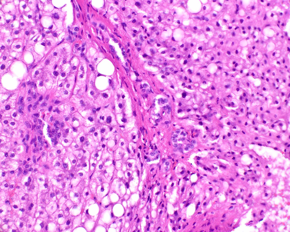
Such a perfect example of hepatic focal nodular hyperplasia with the classic "map-like" glutamine synthetase immunohistochemical staining pattern. Note that the adjoining normal liver tissue lacks that pattern. #UMiamiPath
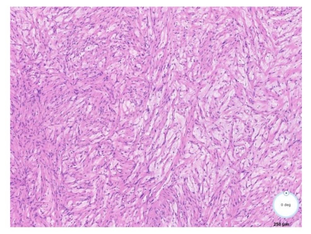
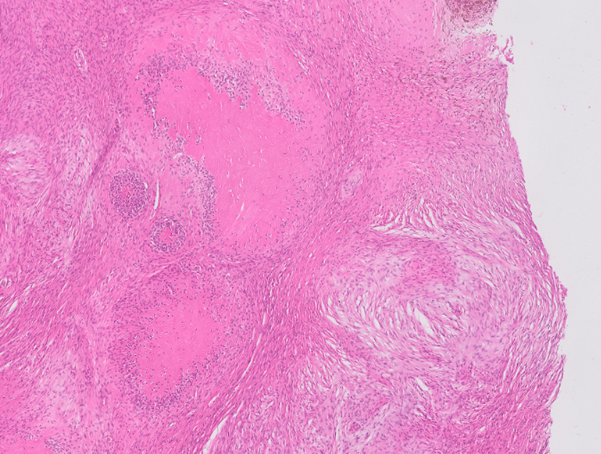
M 29 Y, Orchiectomy, 13mm nodule:
Large cell calcifying sertoli cell tumor (LCCSCT)
• Trabeculae of polygonal cells with eosinophilic cytoplasm in myxoid stroma.
• Calcifications of variable sizes and shapes.
• Neutrophilic infiltrate, lymphocytic rim.
#GUpath#Pathology
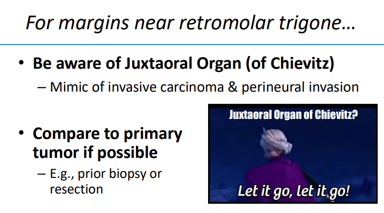
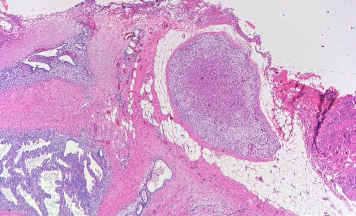
Patients with squamous cell carcinoma and margin evaluation in retromolar trigone -frozen sections pitfall ⚠️
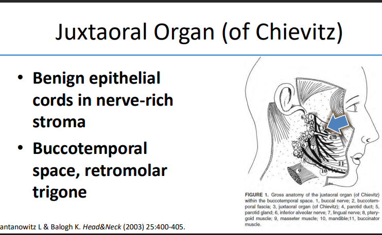
Juxtaoral organ of Chievitz:
- Benign epithelial cords in nerve rich stroma
- Lobulated
- Well circumbscribed nest, cytologically bland
Dr. Nishino- 2025 Diagnostic Pathologic Update #USCAP #pathology #PathX
Sinonasal biopsy showing necrosis and inflammation:
- GMS and PAS-F to rule out infection (invasive fungal sinusitis)
- IHC and EBER-ISH to rule out NK/T-cell lymphoma
- Recommend ANCA serology to rule out granulomatosis with polyangiitis
Dr. Nishino - 2025 Diagnostic Pathology Update #USCAP #pathology #PathX
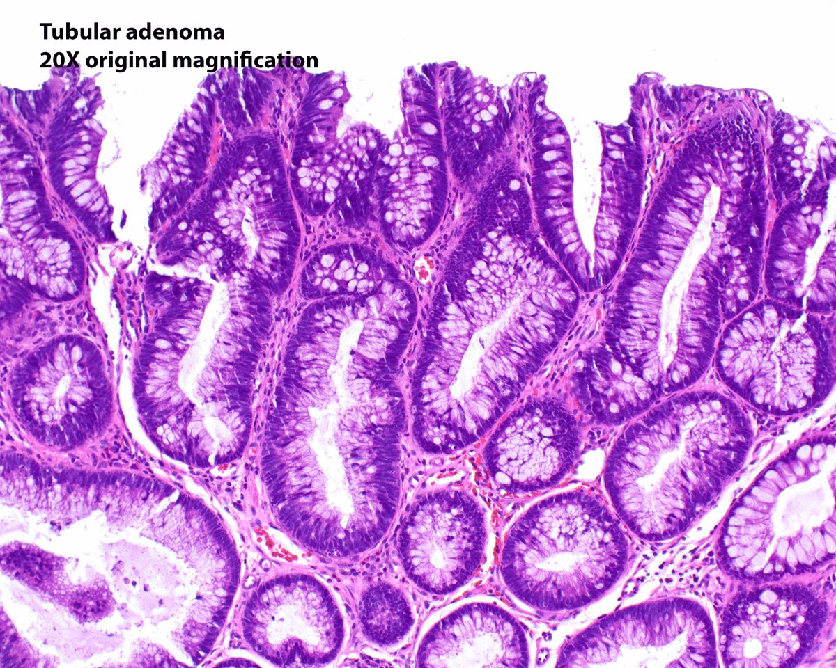
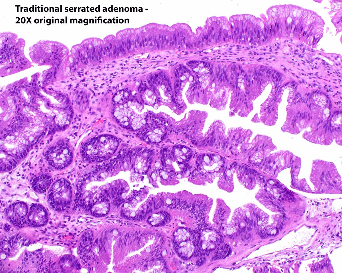
Traditional serrated adenoma (TSA) and tubular adenoma (TA) in the same biopsy cup. The nuclei in the TSA are much smaller and paler than those in the TA, even in the proliferation zones (the ectopic crypt formations).