Stay in the loop with https://t.co/ZyQQ0FN3V3!
»https://t.co/BeyD1XcQeE
»https://t.co/PEFiwrGEi6
»https://t.co/0Q7Pf6KSti
»https://t.co/J8TxLK54hN
»https://t.co/wJOxOK111m
»https://t.co/Ek3yYO5wR3
»https://t.co/JCqkifkAnw
»https://t.co/EUiFKJueYz
#PathologyOutlines#PathTwitter
@NicoloVianini@NicoloVianini Users can share *links* to our textbook images and topics in their social media posts/comments and to “repost”/“share” posts from our official social media profiles. Our Image Use Policy prohibits users from posting our actual images on social media.
@NicoloVianini@NicoloVianini Please promptly remove all https://t.co/ZyQQ0FN3V3 images from posts on your LinkedIn profile and X profile. Continued use of our images in your social media posts may prompt us to pursue further action to protect our copyrighted content.
Stay in the loop with https://t.co/ZyQQ0FN3V3!
»https://t.co/BeyD1XcQeE
»https://t.co/PEFiwrGEi6
»https://t.co/0Q7Pf6KSti
»https://t.co/J8TxLK54hN
»https://t.co/wJOxOK111m
»https://t.co/Ek3yYO5wR3
»https://t.co/JCqkifkAnw
»https://t.co/EUiFKJueYz
#PathologyOutlines#PathTwitter
@Raul_path@Vmm6633 Our site was knocked offline earlier today due to a large spike in harmful traffic from Brazil hitting our directory. To alleviate strain on the server and protect our data, traffic from Brazil is temporarily blocked. Our site is now more stable, and we will unblock Brazil ASAP.
Stay in the loop with https://t.co/ZyQQ0FN3V3!
»https://t.co/BeyD1XcQeE
»https://t.co/PEFiwrGEi6
»https://t.co/0Q7Pf6KSti
»https://t.co/J8TxLK54hN
»https://t.co/wJOxOK111m
»https://t.co/Ek3yYO5wR3
»https://t.co/JCqkifkAnw
»https://t.co/EUiFKJueYz
#PathologyOutlines#PathTwitter
Visit https://t.co/ZyQQ0FMw5v or the links below to stay in the loop!
LinkedIn: https://t.co/j70cr2DSau
Instagram: https://t.co/ZzbNt6FOeI
Facebook: https://t.co/jHrKHRq7M3
Threads: https://t.co/WFw6wQRksa
YouTube: https://t.co/6mKRe0fAVo
WordPress: https://t.co/rQfiI7gdKW
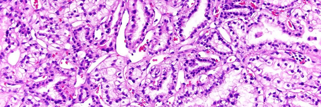
Which of the following is the most likely diagnosis?
A. ALK positive histiocytosis
B. Erdheim-Chester disease
C. Extranodal Rosai-Dorfman disease
D. Inflammatory myofibroblastic tumor
E. Juvenile xanthogranuloma
Answer: https://t.co/NZbqL0hmz8
#PathTwitter#PathologyOutlines
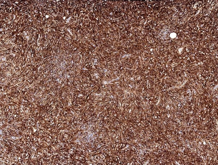
Image Quiz!
A 41 year old woman underwent a core needle biopsy for a 3 cm breast mass discovered on her first screening mammogram. The lesional spindle cells show the CD68 immunostaining pattern seen below; they are also positive for ALK and negative for SMA and desmin. (Cont.)
Mark your calendar for the Kameda Pathology Seminar, featuring Dr. Nat Pernick as a guest speaker, on December 25 at 18:00 JST! Participate online from anywhere or attend in person in Tokyo.
Register for online or onsite attendance: https://t.co/9uWhB8vO3K
#Pathology#Tokyo
View our new testimonials via the link in the comments!
We love hearing how our resources help people at all stages of their pathology careers. We welcome you to comment here or use the tutorial on the testimonials page to record your testimonial video.
#PathTwitter#CAP24
Case of the Month: A 32 year old woman with a clinical history of complex partial type seizures underwent a brain MRI, which showed a 2 cm solid cystic lesion without contrast enhancement in the left temporal lobe.
Although the lesion remained stable over a 1 year follow up, the patient underwent surgery due to an increased frequency of seizures. The lesion was completely removed; the patient is in good health with no evidence of recurrence 1 year after surgery. What's your diagnosis?