🚨 Anthropic just showed a 27-minute workshop on how to actually do prompts for Claude.
Taught by the people who built it.
Free. No registration. No paywall.
I've seen $300 courses that don't cover what they teach in the first 8 minutes.
Watch the video and bookmark it
One mistake in RLS which I think we should stop making.
A patient has been taking pramipexole for years.
Initially it worked very well.
Now he says:
Doctor, earlier the problem used to start only at night.
Now it starts in the evening.
After a few months, it starts in the afternoon.
Earlier it was only in the legs.
Now sometimes the arms are involved.
So we increase pramipexole.
It helps for some time.
Then the symptoms return even earlier.
This may not be worsening RLS. This may be augmentation.
And increasing the dopamine agonist further can make the problem worse.
This is something worth asking every RLS patient on long-term pramipexole or ropinirole:
What time did your symptoms start before treatment, and what time do they start now?
Also ask:
Have the symptoms become stronger?
Have they spread beyond the legs?
Are you needing the medicine earlier or in higher doses?
Earlier onset + increasing severity + spread to arms/trunk should immediately make us think of augmentation.
This is one major reason why dopamine agonists are no longer preferred as routine first-line long-term treatment for RLS.
If augmentation has already occurred, do not suddenly stop the dopamine agonist.
Check iron properly, address other aggravating factors and sleep disorders, stabilise the RLS with alternative treatment, and then taper the dopamine agonist gradually.
The 2026 Continuum review suggests tapering no faster than:
Pramipexole 0.125 mg every 1–2 weeks
Ropinirole 0.5 mg every 1–2 weeks
For me, the most useful clinical lesson is very simple:
When an RLS patient on a dopamine agonist starts getting worse, don't immediately increase the dose.
First ask whether the treatment itself has started worsening the RLS.
#NeuroX #MedX #RLS
Today resident said :
Sir, this patient has tongue fasciculations.
I ask only one question.
Where did you see them?
While the patient was protruding the tongue.
That is the first mistake.
You cannot confidently diagnose spontaneous activity in a muscle that you have already asked the patient to activate.
The tongue must be examined twice.
First as a resting muscle.
Then as a working organ.
Ask the patient to open the mouth gently and keep the tongue relaxed inside.
Do not begin with protrusion.
A protruded tongue is voluntarily active. It may show tremulousness, corrective movements and difficulty maintaining posture.
Every movement is not a fasciculation.
Now look beyond movement.
Is the tongue full or wasted?
Are both halves symmetrical?
Is there focal furrowing?
Are the movements occurring inside an atrophic segment?
A fasciculation tells you the motor unit is irritable.
Atrophy tells you motor units are being lost.
Then look at laterality.
Unilateral tongue wasting + focal resting fasciculations + deviation toward the weak side
Think hypoglossal localisation first.
Trace the XII nerve:
Medulla
↓
Hypoglossal canal
↓
Skull base
↓
Carotid space
↓
Upper neck
↓
Tongue
Do not write ALS before completing the anatomy.
Now ask the tongue to work.
• Protrude
• Move side to side
• Push against each cheek
• Repeat ta-ta-ta, da-da-da, la-la-la
• Assess speech, swallowing and cough
A tongue may move and still be weak.
The patient may not say weakness.
They may say:
Food collects inside the cheek.
Speech becomes unclear.
Water occasionally goes the wrong way.
Cough has become weak.
These complaints matter more than the visible twitch.
Then examine the whole motor system.
Jaw jerk.
Palate.
Facial strength.
Neck flexion.
Limb wasting.
Reflexes.
Plantar responses.
Progression.
The patterns are different.
Normal bulk + normal power + movements mainly during protrusion
Not enough to call tongue fasciculations.
Unilateral atrophy + resting fasciculations + deviation
Think focal hypoglossal lesion.
Bilateral wasting + resting fasciculations + progressive dysarthria, dysphagia, weak cough and limb signs
Now bulbar motor neuron disease becomes a serious possibility.
The most important word is not fasciculation.
It is progressive.
Even tongue EMG must be interpreted carefully.
Relaxation is difficult.
Insertional activity may confuse interpretation.
Voluntary motor-unit activity may contaminate the tracing.
So fasciculation potentials in genioglossus is not a diagnosis.
Ask:
• Is there chronic neurogenic change?
• Is there active denervation?
• Is recruitment reduced?
• Is the abnormality unilateral and localisable?
• Or is it multiregional?
• Does it match a progressive clinical syndrome?
Never diagnose ALS from a moving tongue.
First make the tongue rest.
Then examine its shape.
Then test its force.
Then place it inside the entire motor system.
That is the difference between seeing tongue movements and understanding tongue fasciculations.
#ALS #MotorNeuronDisease #MedX #BulbarPalsy #ClinicalNeurology #NeuroX
🩺🇮🇳 CME INDIA CASE FILES
When an Insulin Pump Should NOT Stay On During Surgery
A 63-year-old man with type 1 diabetes was admitted for elective thoracolumbar spine fixation under general anesthesia. He had excellent glycaemic control using a hybrid closed-loop insulin pump with continuous glucose monitoring (CGM).
The planned surgery was expected to last approximately 4 hours in the prone position. On preoperative assessment:
Capillary glucose: 212 mg/dL
Mild peripheral edema was present.
The anesthesiology team anticipated the need for low-dose norepinephrine to maintain blood pressure during surgery.
The insulin pump infusion set was located over the upper gluteal region, close to the operative field.
The surgical and endocrine teams discussed whether the insulin pump could safely remain in place throughout the procedure.
CME INDIA Clinical Discussion
This patient has multiple contraindications to continuing continuous subcutaneous insulin infusion (CSII) during surgery.
These include:
Major surgery lasting >2–3 hours
Prone positioning, making the pump inaccessible
Infusion site located near the surgical field
Expected vasopressor therapy, which may reduce subcutaneous tissue perfusion and impair insulin absorption
General anesthesia, preventing the patient from recognizing pump malfunction or hypoglycaemia.
Under these circumstances, subcutaneous insulin delivery becomes unreliable, increasing the risk of both severe hyperglycaemia and diabetic ketoacidosis in a person with type 1 diabetes.
Recommended Management
The safest approach is to:
Remove the insulin pump and CGM before surgery
Initiate an intravenous regular insulin infusion with dextrose and electrolyte monitoring according to institutional protocol
Perform hourly blood glucose monitoring intraoperatively.
Resume pump therapy only when the patient is awake, hemodynamically stable, and able to manage the device.
Why not continue the pump?
Simply moving the pump away from the operative field does not address:
Reduced insulin absorption during vasopressor use
Inaccessibility of the device in the prone position
Inability to troubleshoot infusion failure
Increased risk of rapid ketosis in type 1 diabetes if insulin delivery is interrupted.
CME INDIA Clinical Pearls
🔹 Type 1 diabetes requires uninterrupted insulin delivery—even brief interruption can precipitate diabetic ketoacidosis.
🔹 Intravenous insulin is preferred for major surgery, prolonged procedures, hemodynamic instability, or vasopressor use.
🔹 Prone positioning and inaccessible pump sites are important reasons to discontinue CSII during surgery.
🔹 CGM values should not be relied upon as the sole basis for intraoperative insulin adjustment; point-of-care blood glucose testing remains the standard.
🔹 Transition back to insulin pump therapy only after the patient is fully alert, clinically stable, and capable of safely operating the device.
Take-home message:
Major surgery + prone position + vasopressor requirement = discontinue insulin pump and CGM, and manage with intravenous insulin infusion.
Pondering how to remember pontine syndromes?
Don’t become despondent about syndromes of the pons!
Here’s an easy way remember the basic parts of pontine syndromes:
Pontine lesions are usually vascular and are either from an infarct in the AICA territory (lateral) or paramedian branches of the basilar artery (medial)
Lateral & medial pontine syndromes can be remembered by the easy mnemonic THE PONS, w/ “THE” representing lateral syndromes & “PONS” representing medial syndromes
LATERAL SYNDROMES:
T = Temperature (spinothalamic = loss of pain & temp contralateral body, trigeminal nucleus = ipsilat face)
H = Horner’s (sympathetic pathway)
E = Equilibrium problems (spinocerebellar = ataxia of ipsilateral arm & leg)
MEDIAL SYNDROMES:
P = Proprioception (medial lemniscus = loss of contralateral vibration & proprioception)
O = Ophthalmoplegia (MLF = internuclear ophthalmoplegia)
N = Nonmobile (corticospinal tract = weakness of contralateral body)
S = Sixth nerve (Sixth nerve nucleus = loss of ipsilateral CN 6)
There are many different syndromes that vary by what exact structures are involved, but if you always remember THE PONS, you can easily pontificate about pontine lesions!
Need a better strategy to find strategic infarcts?
Strategic infarcts are infarcts located in structures directly related to cognition.
Damage to these structures results in dementia without any other significant volume loss.
These infarcts are like a ninja. Just one shot right to the heart to take it down.
Just one infarct in one of these important structures can cause dementia like a shot to the heart.
So which structures are these?
Well, there are many structures that have been implicated in strategic infarct dementia.
But the main ones are hippocampus, internal capsule (ant & genu), thalamus (paramedian) & caudate.
I remember this w/the mnemonic:
One HIT CAUses dementia
H = Hippocampus
I = Internal Capsule
T = Thalamus
CAUD = Caudate
Now you can be a ninja looking for these ninja infarcts!
1/Time is brain! But what time is it?
If you don’t know the time of stroke onset, are you able to deduce it from imaging?
Here’s a thread to help you date a stroke on MRI!
How will you approach a hyperkinetic movt disorder in an OPD?
A jerk is not a diagnosis.
It is simply the beginning of the differential.
When you see a hyperkinetic movement, ask:
Does it have a script?
⚡ Shock-like, lightning fast
→ Myoclonus
🎲 Random, flowing, unpredictable
→ Chorea
🔁 Repeated in the same way every time
→ Tic
That single question solves a surprising amount of OPD confusion.
Now the interesting part.
A patient repeatedly grimaces.
Repeatedly pulls the neck.
Repeatedly performs the same movement.
The movement is recognisable.
Predictable.
Stereotyped.
It has a script.
So it belongs to the tic family.
But there is another clue.
The movement also creates an abnormal posture.
A pull.
A twist.
A dystonic flavour.
Now the diagnosis becomes:
Dystonic tic
Not myoclonus.
Not chorea.
Not pure dystonia.
A dystonic tic.
This is how movement disorder specialists think.
They don’t start with the disease.
They start by identifying the movement family.
Tremor is rhythmic.
Myoclonus is shock-like.
Chorea is unpredictable.
Tics have a script.
Dystonia creates a posture.
First identify the family.
Then identify the disease.
#Neurotwitter #Movementdisorder #MedX
🧠 FOR RESIDENTS | HOUSE OFFICERS | CONSULTANTS
(STROKE LOCALIZATION MADE SIMPLE)
When a stroke patient arrives, don’t start with scans first.
👉 First question at bedside: Which vascular territory is involved?
This single step predicts the full neurological deficit.
1️⃣ Middle Cerebral Artery (MCA) Stroke
📍 Most common stroke territory
Contralateral face & arm weakness > leg
Contralateral sensory loss
Dominant hemisphere → Aphasia
Broca: non-fluent speech
Wernicke: fluent but meaningless speech
Non-dominant hemisphere → Hemispatial neglect
💡 Key clue: Face + arm > leg = MCA
2️⃣ Anterior Cerebral Artery (ACA) Stroke
Contralateral leg weakness > face/arm
Sensory loss (leg predominant)
Frontal lobe features:
Personality change
Urinary incontinence
💡 Key clue: Leg > face/arm = ACA
3️⃣ Posterior Cerebral Artery (PCA) Stroke
Contralateral homonymous hemianopia
No motor weakness
Memory impairment (hippocampus)
💡 Key clue: Isolated visual field defect = PCA
4️⃣ Basilar Artery Stroke
⚠️ Neurological emergency
Locked-in syndrome
Conscious but quadriplegic
Vertical eye movements preserved only
Bilateral motor deficits ± cranial nerve palsies
💡 Key clue: Locked-in = basilar until proven otherwise
5️⃣ Lacunar Strokes (Small vessel disease)
Seen in HTN & diabetes
Deep brain involvement:
Pure motor hemiparesis (internal capsule)
Pure sensory stroke (thalamus)
Ataxic hemiparesis (pons)
Dysarthria–clumsy hand syndrome
❗ No cortical signs:
No aphasia
No neglect
No visual field defects
💡 Key clue: Pure motor OR pure sensory = lacunar
🔑 ONE-LINE PATTERN RECOGNITION:
Face/arm > leg → MCA
Leg > face/arm → ACA
Visual field cut only → PCA
Locked-in → Basilar
Pure motor/sensory → Lacunar
🧠 If you can localize, you can diagnose before imaging.
A tremor patient enters OPD.
The biggest mistake?
Diagnosing the disease before observing the movement.
The easiest way to assess tremor in OPD is this:
First ask:
WHEN does the tremor appear?
That single question simplifies most tremor clinics.
I use a simple 5-gate approach:
REST
Hands on lap.
Rest tremor + bradykinesia/rigidity
→ Think Parkinsonism
But remember:
Tremor alone ≠ Parkinson disease.
Always look for:
• decrement
• rigidity
• reduced arm swing
• masked facies
And never forget dopamine-blocking drugs.
POSTURE
Arms outstretched.
Fine symmetric tremor?
Think:
• caffeine
• anxiety
• thyroid
• β-agonists
• lithium
• valproate
• SSRIs
• tacrolimus
Many neurology tremors are actually metabolic or drug-induced.
ACTION
Writing
Spiral
Pouring water
Bilateral action tremor for years without other neurological signs
→ Essential tremor likely.
But this is the trap:
Action tremor is NOT always essential tremor.
Look carefully for:
• dystonia
• cerebellar signs
• asymmetry
• gait imbalance
TARGET
Finger-nose test.
Tremor worsening near target + ataxia/dysarthria/nystagmus
→ Cerebellar/structural until proven otherwise.
These patients deserve attention, not reassurance.
PATTERN
This is where many diagnoses hide.
• Position-specific
• Null point
• Sensory trick
→ Dystonic tremor
• Variable
• Distractible
• Entrainable
→ Functional tremor
Young patient + tremor + psychiatric/liver/dystonic clues?
Do not miss Wilson disease.
The real OPD pearl:
Do not start with:
Is this ET or Parkinson’s?
Start with:
Rest?
Posture?
Action?
Target?
Pattern?
Classification first.
Disease label later.
That single shift improves tremor diagnosis dramatically.
MDS Tremor Classification + modern movement disorder practice have made one thing very clear:
Phenomenology is still the most powerful investigation in neurology.
What not to miss:
• acute onset
• rapid progression
• focal deficits
• severe ataxia
• encephalopathy
• age <40 unexplained tremor
These patients may need urgent imaging/work-up.
Final rule:
Treat the tremor you have PROVEN.
Not the tremor you guessed.
#Neurotwitter #Medtwitter #Tremor
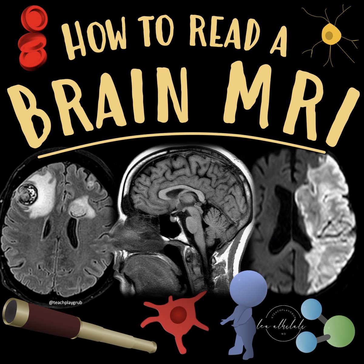
1/Do radiologists sound like they are speaking a different language when they talk about MRI?
T1 shortening what? T2 prolongation who?
Here’s a translation w/an introductory thread to MRI.
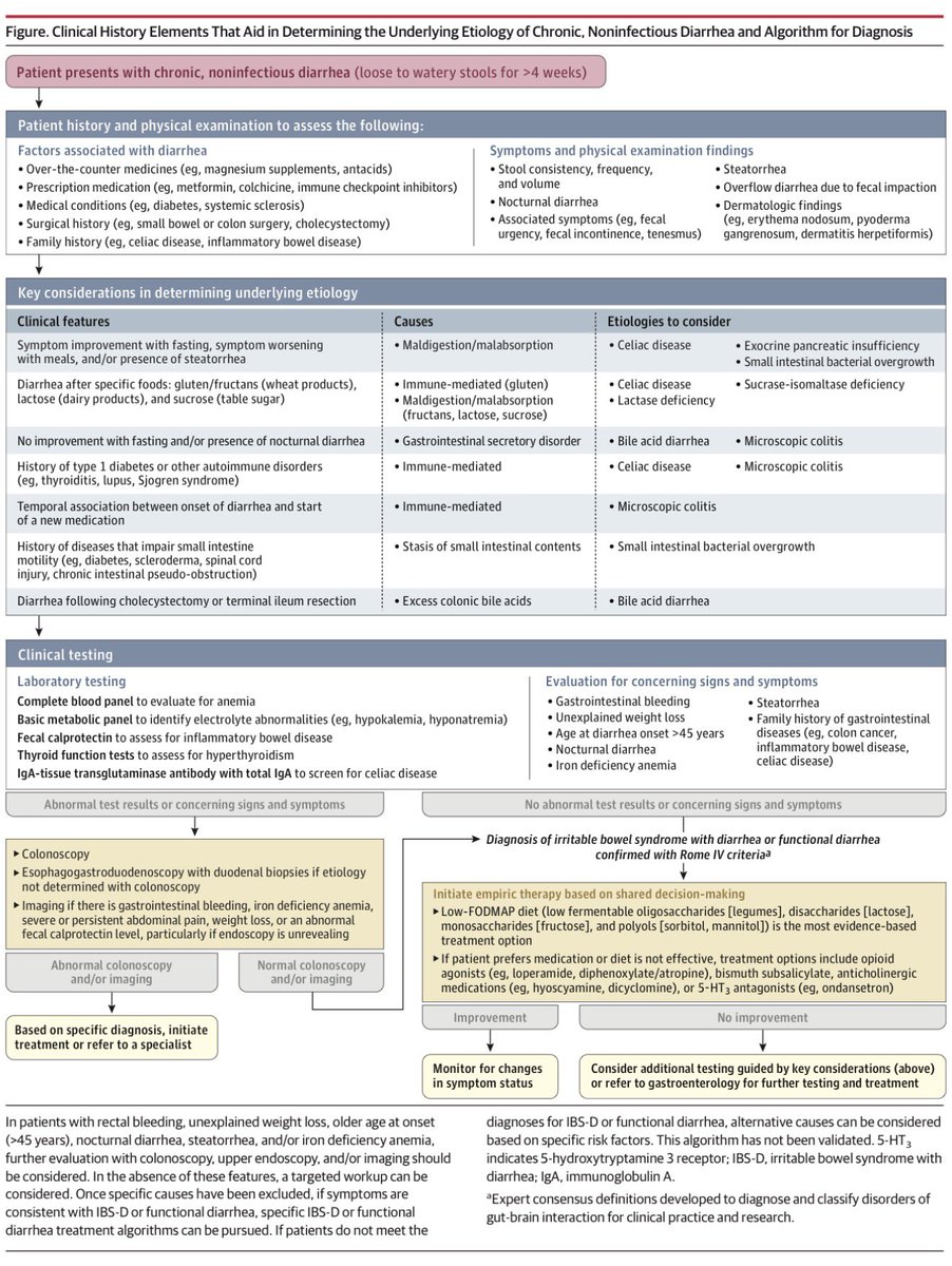
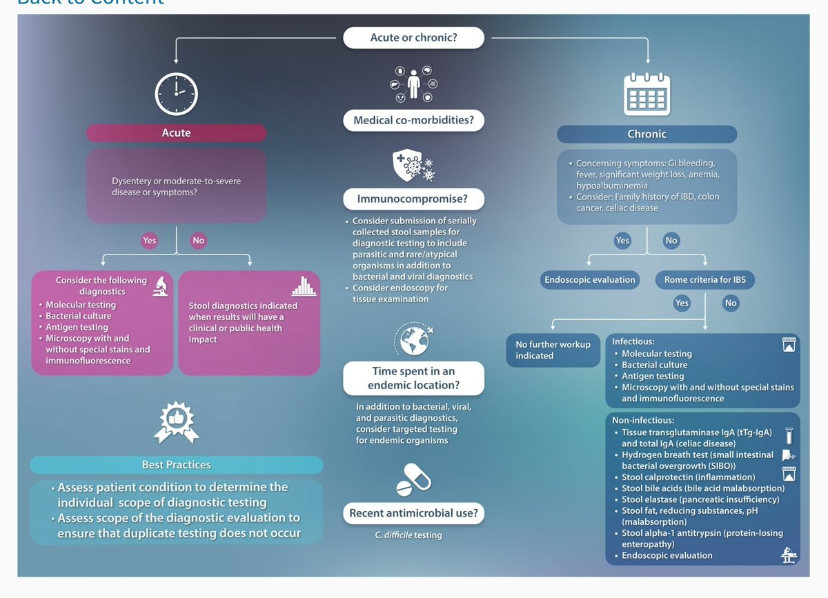
One of the must follow accounts if you are medical is @JAMA_current. They publish state of the art reviews, but unlike other journals, provide links that bypass paywalls. See their review on management of chronic diarrhoea below 👇
📸: https://t.co/WhDCsASuH2
🧠 Neurology - Concepts you should never forget : 2️⃣
🟢Cortical vs Subcortical stroke
Let’s break it down step-by-step. 👇
🟦 1️⃣ What is a Cortical Stroke?
A cortical stroke involves the cerebral cortex the outer layer of the brain responsible for higher neurological functions.
Key cortical areas include:
🔻 Frontal lobe → motor control, executive function, speech production (Broca)
🔻 Parietal lobe → sensory perception & spatial awareness
🔻 Temporal lobe → auditory processing, language comprehension (Wernicke), memory
🔻 Occipital lobe → vision
When these higher cortical functions are affected → it strongly suggests cortical involvement.
Most commonly affected vessels:
🔻 Middle Cerebral Artery (MCA) mc
🔻 Anterior Cerebral Artery (ACA)
🔻 Posterior Cerebral Artery (PCA)
These are typically large vessel strokes.
🟦 2️⃣ Clinical clues of a Cortical Stroke
Because the cortex handles complex processing, strokes here produce cortical signs.
Look for:
🔻 Aphasia (dominant hemisphere language dysfunction)
🔻 Neglect (classically right parietal cortex)
🔻 Visual field defects (homonymous hemianopia)
🔻 Gaze deviation
🔻 Apraxia or agnosia
🔻 Cortical sensory loss (astereognosis, agraphesthesia)
🔻 Early seizures
Weakness may occur, but cortical signs usually accompany it.
🟦 3️⃣ What is a Subcortical Stroke?
A subcortical stroke affects deep brain structures beneath the cortex.
Common locations include:
🔻 Internal capsule
🔻 Basal ganglia
🔻 Thalamus
🔻 Pons
These areas mainly carry motor and sensory pathways but do not control higher cortical functions like language or spatial awareness.
🟦 4️⃣ Blood vessels involved in Subcortical stroke
These strokes involve small penetrating arteries, such as:
🔻 Lenticulostriate arteries (branch of MCA)
🔻 Thalamoperforators (branch of PCA)
🔻 Anterior choroidal artery (branch of ICA)
🔻 Paramedian pontine perforators
This is why they are called lacunar strokes.
Most commonly caused by:
➡️ Chronic hypertension or diabetes
➡️ Small vessel disease due to lipohyalinosis or microatheroma
🟦 5️⃣ Lacunar strokes: The classic “3 NO’s”
A quick way to localize small vessel (subcortical) stroke is remembering the 3 NO’s:
❌ No cortical symptoms
Meaning no:
• aphasia
• neglect
• visual field defects
• gaze deviation
❌ No higher cortical dysfunction
Meaning:
• language intact
• cognition preserved
• no behavioral changes
Because the cortex is spared, deficits are usually pure motor or sensory.
🟦 6️⃣ Clinical patterns of Subcortical (lacunar) stroke
Classic lacunar syndromes include:
🔻 Pure motor hemiparesis (internal capsule)
🔻 Pure sensory stroke (thalamus)
🔻 Sensorimotor stroke
🔻 Ataxic hemiparesis
🔻 Dysarthria–clumsy hand syndrome
🟦 7️⃣ The 10-second bedside trick
When a patient presents with weakness, ask yourself 4 quick questions:
1️⃣ Is language affected?
2️⃣ Is there neglect?
3️⃣ Is there visual field loss?
4️⃣ Is there gaze deviation?
If any of these are present → think cortical stroke.
If none are present → think lacunar / small vessel stroke.
🟦 8️⃣ Language test
Ask the patient to repeat a sentence or name objects.
Example:
🗣 “No ifs, ands, or buts.”
If speech is impaired → aphasia → cortical stroke
Language centers lie in the dominant cerebral cortex.
🟦 9️⃣ Neglect test
Ask the patient to raise both limbs or perform simultaneous sensory stimulation.
If they ignore one side → hemispatial neglect
This is classic for right parietal cortical stroke.
Subcortical strokes rarely produce neglect.
#MedTwitter #MedX #NeuroTwitter
🚨 Cardio pearls you should never miss 6️⃣
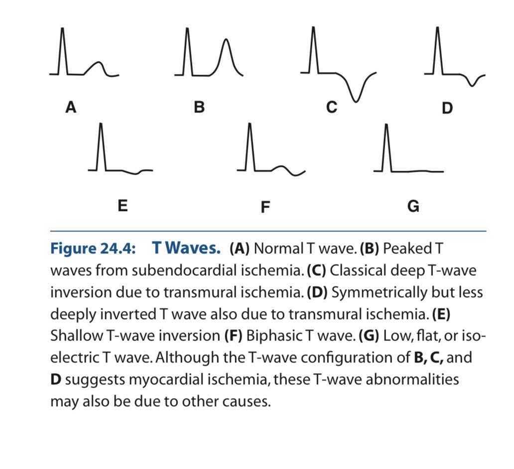
The T wave in the ECG is a story about repolarisation not a random squiggle.
When it inverts, something changed in the direction or timing of ventricular recovery.
Let’s clear the confusion once for all & understand in depth.
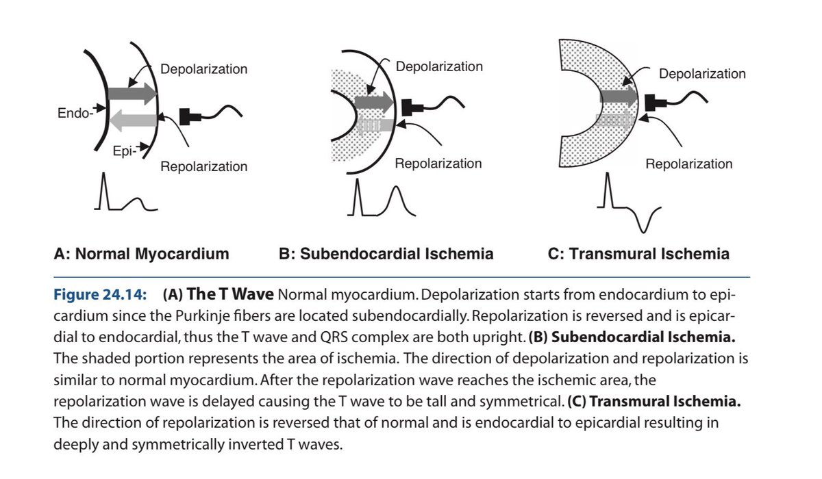
1️⃣ Why is the normal T wave upright?
✔ Purkinje fibers are subendocardial in location.
So:
Depolarization → Endocardium → Epicardium
Repolarization → Epicardium → Endocardium
Epicardial cells:
🔻Shorter action potential duration
🔻Larger transient outward K⁺ current (Ito)
🔻Faster phase 3 repolarization
Repolarization is a negative wave moving opposite depolarization → vector aligns with QRS.
👉 Upright T wave.
2️⃣ Subendocardial ischemia (early)
Inner myocardium + Purkinje network suffer first.
Ischemia causes:
🔻ATP depletion
🔻KATP channel opening
🔻Extracellular K⁺ accumulation
🔻Shortened action potential in ischemic zone
🔻Slowed Purkinje conduction
ECG:
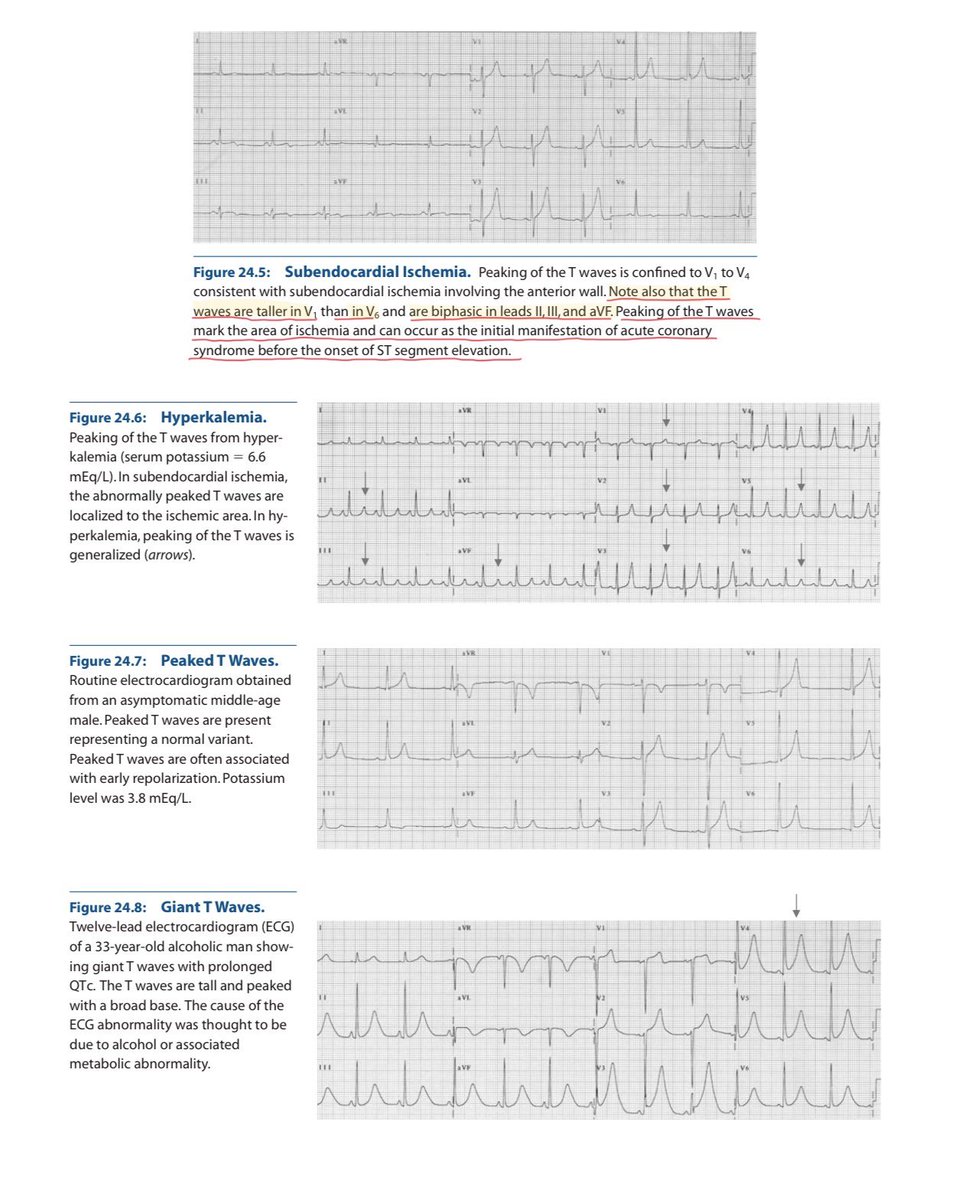
→ Tall, symmetric, broad-based T waves
(Hyperacute phase)
3️⃣ Transmural ischemia
Repolarization sequence reverses abnormally.
Vector flips.
→ Deep, symmetric T-wave inversion
Seen in:
🔻Evolving MI
🔻Wellens pattern
🔻Reperfusion
Deep + symmetric + territorial + dynamic = ischemia until proven otherwise.
4️⃣ Hyperkalemia
Global extracellular K⁺ elevation.
ECG:
🔻Narrow, tented T waves
🔻Short QT
Sharp, narrow spikes ≠ broad ischemic T waves.
5️⃣ Hypokalemia
Delayed repolarization.
ECG:
🔻Flat T waves
🔻ST depression
🔻Prominent U waves
U waves help differentiate from ischemia.
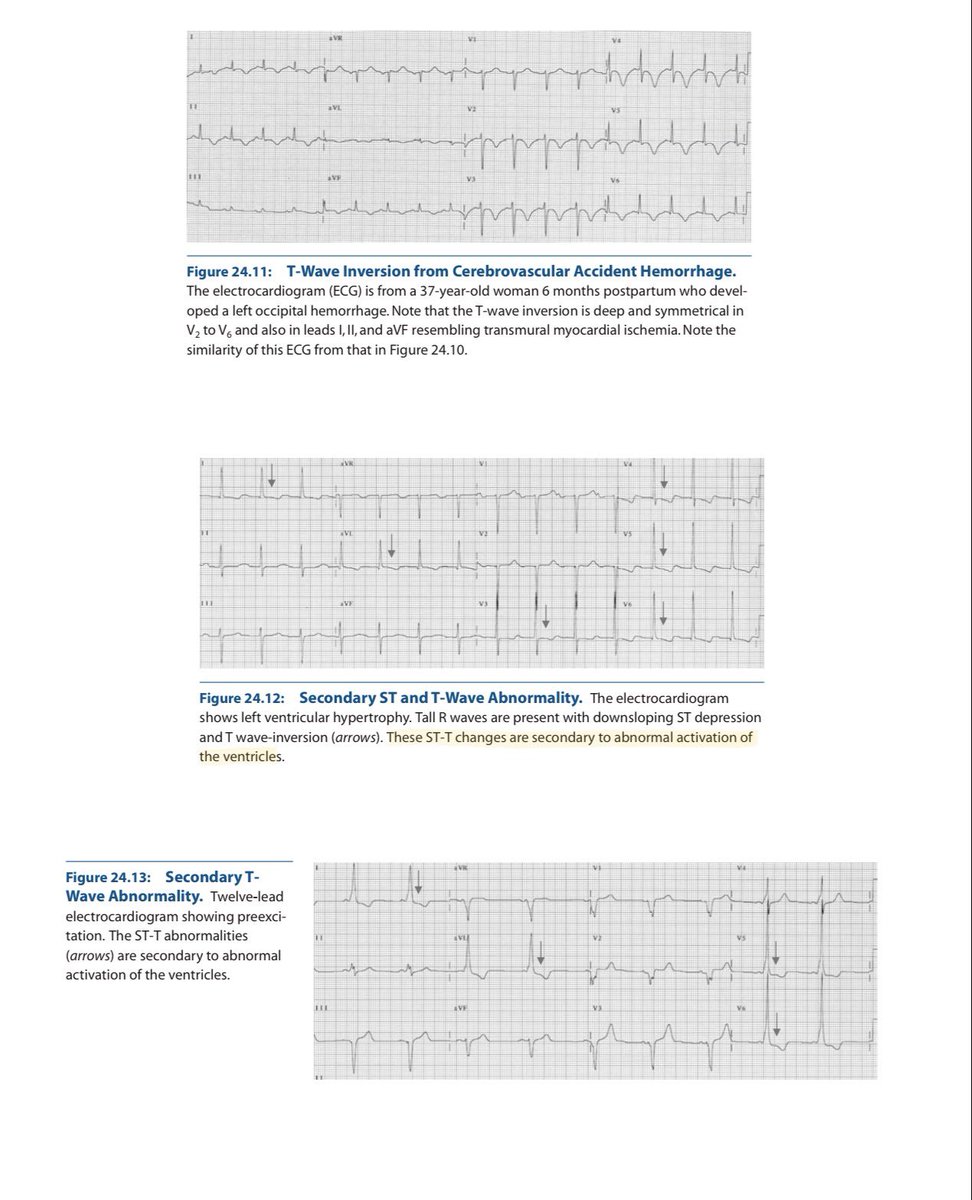
6️⃣ CNS / Neurogenic T waves
Catecholamine surge → myocardial stunning.
ECG:
🔻Deep, diffuse T inversions
🔻QT prolongation
🔻Not vascular territory specific
Distribution matters.
7️⃣ LVH & Strain Pattern
This is not primary ischemia.
It’s repolarization abnormality secondary to abnormal depolarization.
In LVH:
🔻Increased myocardial mass
🔻Prolonged depolarization
🔻Delayed repolarization in hypertrophied wall
ECG:
✔ High-voltage QRS
✔ Downsloping ST depression
✔ Asymmetric T-wave inversion
(Usually lateral leads: I, aVL, V5–V6)
Key point:
Strain T waves are asymmetric, gradual downstroke + rapid return.
Ischemic T waves are symmetric.
8️⃣ Cardiomyopathies
Hypertrophic cardiomyopathy (HCM)
🔻Deep T inversions (often in precordial leads)
🔻May mimic ischemia
🔻Often associated with large voltages
Apical HCM
🔻Giant negative T waves in precordial leads
Dilated cardiomyopathy
🔻Nonspecific ST-T changes
🔻Often diffuse
Mechanism:
Structural remodeling alters repolarization gradients.
Not vascular. Not dynamic like ACS.
9️⃣ Final Differentiation Framework
✔ Symmetric + territorial + dynamic → Ischemia
✔ Narrow tented + short QT → Hyperkalemia
✔ Flat T + U waves → Hypokalemia
✔ Diffuse deep T + long QT + neuro event → CNS
✔ High voltage + asymmetric lateral T inversion → LVH strain
✔ Giant precordial inversions + echo changes → HCM
✔ Stable V1–V3 inversion in young → Benign variant
T waves reflect:
• Repolarization timing
• Potassium currents
• Myocardial thickness
• Conduction sequence
• Autonomic tone
If you understand vectors + ionic physiology, the ECG becomes logical.
Electrophysiology > memorized patterns.
(Ref: Marriott’s Practical Electrocardiography; Surawicz & Knilans)
#MedTwitter #MedX #ECG #Cardiology
I scraped every single NotebookLM prompt that blew up on X, Reddit, and academic corners of the internet.
Turns out most people are using NotebookLM like a fancy note-taker.
That's insane.
It's a full-blown research assistant that can compress 10 hours of analysis into 20 seconds if you feed it the right instructions.
Here's what actually works:
The Clinical Approach to a Positive Antinuclear Antibody (ANA):
A positive ANA is one of the most common consults in Internal Medicine, yet it is widely misunderstood.
Positive ANA ≠ Lupus.
It causes significant patient anxiety and unnecessary referrals.
Here is the evidence-based approach to interpreting a positive ANA for the busy clinician. 🧵
#MedEd #Rheumatology #MedTwitter @DrAkhilX @IhabFathiSulima #InternalMedicine #Lupus #MedicalEducation
Infectious Diarrhea: Practical Bedside Approach
🔹 Definition & Burden
Diarrhea is defined as ≥3 loose or watery stools per day or a clear change from baseline bowel habit.
Infectious diarrhea continues to be a major global cause of morbidity across all age groups, with significant impact in low- and middle-income countries.
🔹 Clinical Evaluation: Think Before You Test
Careful history is the most important diagnostic tool: duration, stool character, fever, blood, travel, food exposure, antibiotics, and comorbidities.
Most acute diarrheal illnesses are self-limiting and do not require extensive investigations.
Red flags needing evaluation: severe dehydration, high fever, bloody diarrhea, immunocompromised state, extremes of age.
🔹 Stool Testing: Use Judiciously
Stool tests should be ordered only if results will change management.
Best yield comes from stool collected during active symptoms.
Multiplex molecular tests are rapid and sensitive but may detect colonization rather than true infection—clinical correlation is essential.
Traditional culture and microscopy remain relevant for epidemiology, antimicrobial sensitivity, and public health reporting.
🔹 Core Management Principle
Hydration and electrolyte replacement are the cornerstone of therapy—oral rehydration for most patients, IV fluids for severe dehydration.
Early correction of dehydration significantly reduces complications and mortality.
🔹 Antibiotics: Not for Everyone
Empiric antibiotics are not routinely indicated in uncomplicated acute diarrhea.
Antibiotics should be reserved for:
Severe disease with systemic toxicity
High-risk patients (elderly, immunocompromised)
Specific pathogens where benefit is proven
Unnecessary antibiotic use increases antimicrobial resistance and risk of complications.
🔹 Special Populations
Immunocompromised patients may have atypical or opportunistic infections and require lower thresholds for testing and treatment.
Chronic or persistent diarrhea warrants evaluation for non-infectious causes and post-infectious sequelae.
🔹 Emerging Concepts
Rising antimicrobial resistance mandates strong antibiotic stewardship in diarrheal illness.
Microbiota-based therapies and probiotics are being explored as adjuncts, especially in recurrent or refractory cases, but should be evidence-guided.
🔹 Prevention: Often Overlooked
Hand hygiene, safe food and water practices, and sanitation remain the most effective preventive strategies.
Vaccination (e.g., rotavirus in children) has significantly reduced severe diarrheal disease.
🔹 Bottom Line
Most infectious diarrhea is self-limited—treat the patient, not the stool report.
Hydration first, tests selectively, antibiotics sparingly.
Clinical judgment remains the most powerful diagnostic and therapeutic tool.
📌 CME India Take-Home Message:
Rational evaluation, early rehydration, and antimicrobial stewardship are the pillars of safe and effective management of infectious diarrhea.
https://t.co/MXlrGsDxiB