Forget me nots and chicory compress whole sky blue into millimetre scales. Microscopy emphasizes powdery epidermal wax and diffuse anthocyanin gradients baby blue as gentle vacuolar dilution, not weak saturation.
#BabyBlue#ForgetMeNot#Chicory#Pollen#TheFloralLens
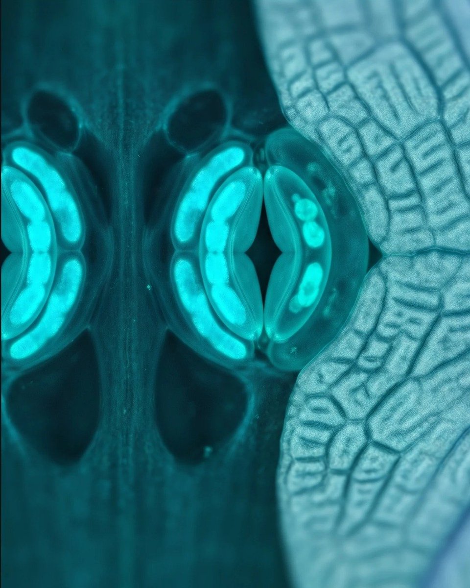
Cyan in flowers often reads where chlorophyll rich tissue meets pale petal bases. Iris abaxial stomata show vivid cyanoplast glow against teal substomatal cavities; plumbago’s papery petals supply cool aqua ridges a combo that reads microscope first, garden second.
#Cyan #BlueIris #Plumbago #Stomata #TheFloralLens
Delphinium and cornflower share some of the richest delphinidin based blues in the plant kingdom. Under petal transverse section, conical cells act as light-trapping prisms while vacuoles hold saturated navy pigment; cornflower pollen adds indigo, spiky exine sculpture at the margin of the field.
This palette anchors the floral corpus: botanical microscopy as couture color theory deep blue as structural petal architecture, not flat dye.
#DeepBlue #Delphinium #Cornflower #BotanicalMicroscopy #TheFloralLens
Glioblastoma’s signature is necrosis with nuclear palisading. Central tau grey coagulative necrosis is encircled by hyperchromatic steel blue tumor cells and focal coral pink endothelial proliferation echoing the variegated grey and pink macroscopic cut surface of high grade glioma.
#Glioblastoma #GBM #BrainTumor #Neuropathology #TheCellularLens
Diabetic nephropathy thickens glomerular filtration barriers. PAS highlights magenta mesangial nodules and lilac basement membranes the same sugary pink cast of glycoprotein rich matrix seen in renal cortex scarring.
#DiabeticNephropathy#KidneyPathology#Nephrology #PathologyArt #TheCellularLens
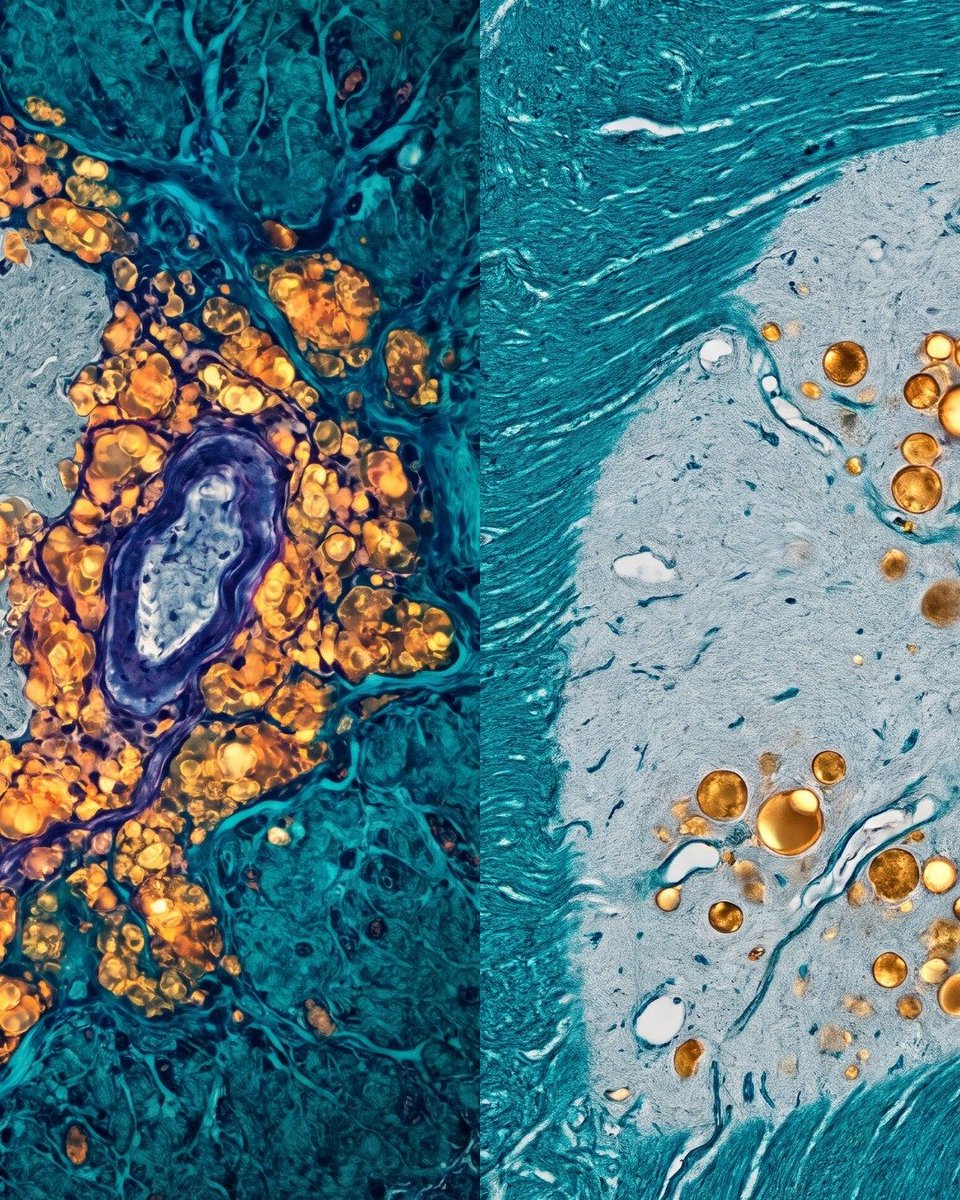
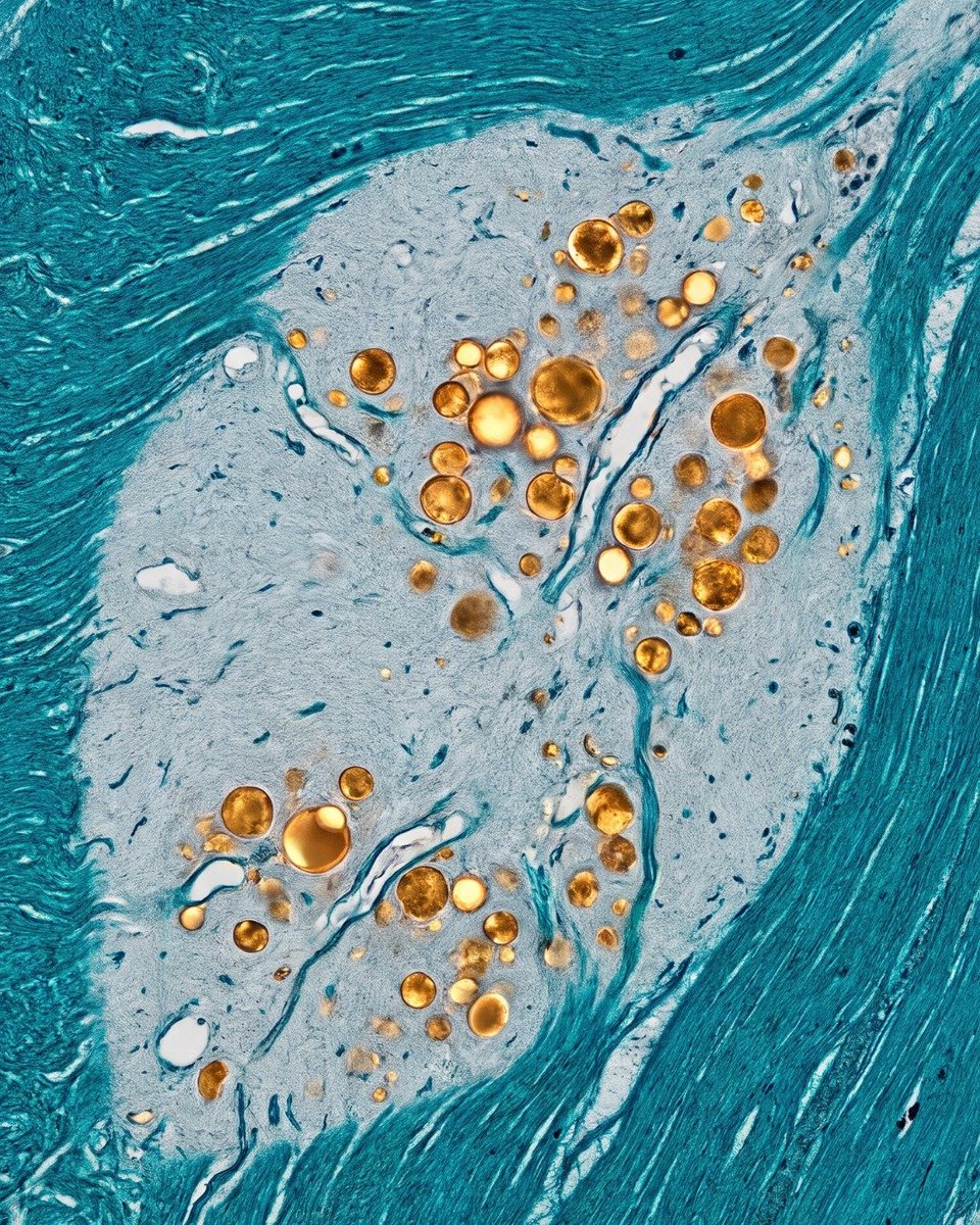
MS creates sharply bordered plaques of myelin destruction in white matter. Luxol fast blue leaves demyelinated zones washed grey blue while intact tracts stay teal analogous to the dull grey white appearance of plaques on fresh brain section.
#MultipleSclerosis#MS #Demyelination #Neuropathology #TheCellularLens
Polymerized hemoglobin S deforms erythrocytes into rigid sickle forms. On Wright-Giemsa, sickled cells take a deeper burgundy than biconcave discs, echoing the dark red brown hue of deoxygenated venous blood on serosal surfaces.
#SickleCellDisease#Hematology#Hemoglobinopathy #PathologyArt #TheCellularLens
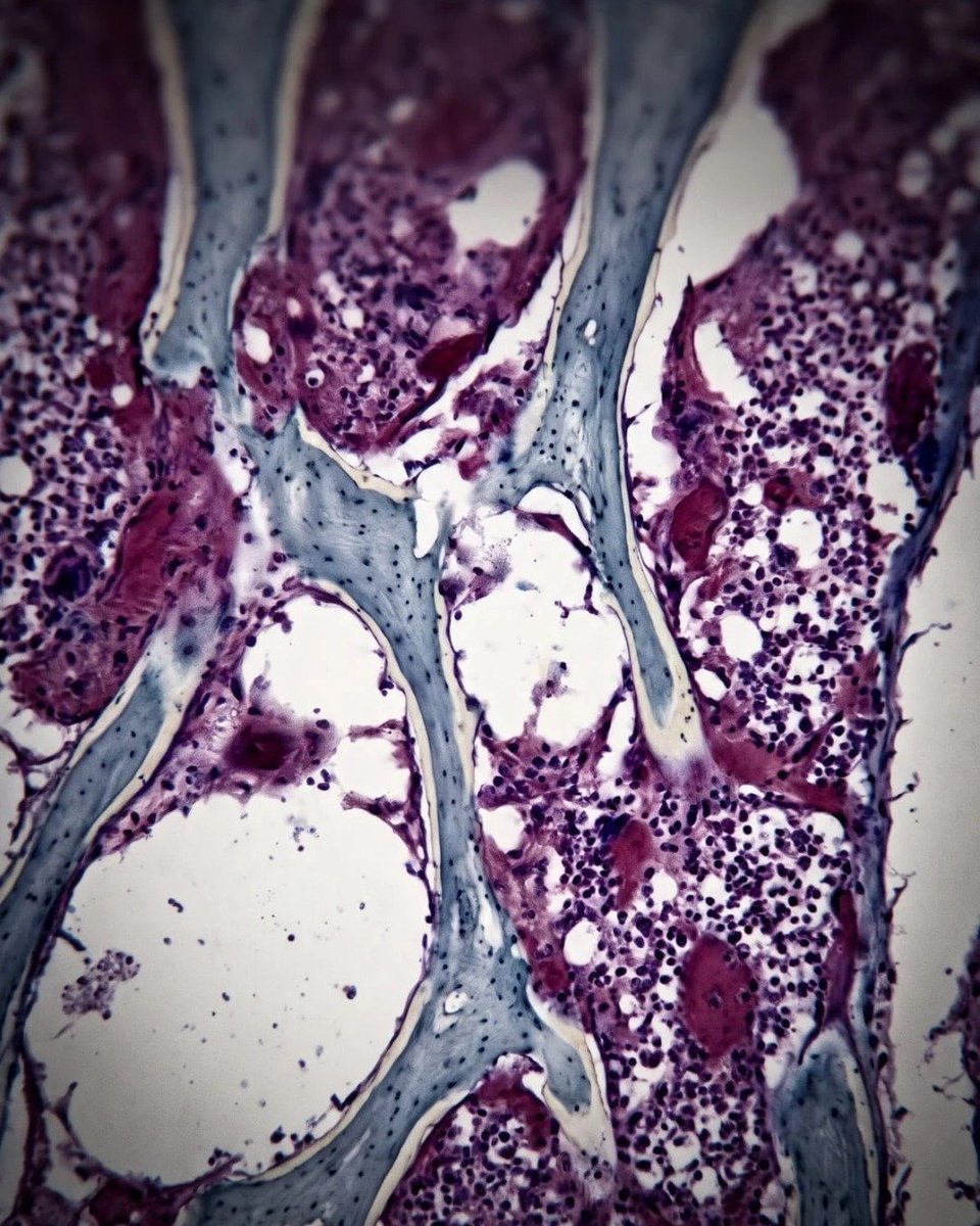
Osteoporosis thins the load bearing trabecular lattice. Toluidine blue metachromasia stains cartilage and osteoid in cool blue while mineralized bone reads ivory to blue grey the same pallor visible on dry trabecular cut surfaces in gross anatomy specimens.
#Osteoporosis #BonePathology #Musculoskeletal #PathologyArt #TheCellularLens
Alzheimer’s disease reorganizes cortical cytoarchitecture through tau phosphorylation and extracellular amyloid deposition. Silver based impregnation highlights dark violet flame tangles inside surviving pyramidal neurons against a desaturated blue-grey background matrix, evoking the cooled hue of oxygenated cortical ribbon on gross inspection.
#AlzheimersDisease #Neurodegeneration #Neuropathology #PathologyArt #TheCellularLens
Psoriasis remodels the epidermis into a high turnover architectural stack. Under routine histology, the plaque shows compact parakeratotic scale (the clinical “silver” surface) overlying a broadened spinous layer and tortuous capillary loops in the papillary dermis. The palette mirrors real skin optics: pale refractile keratin, warm pink living epidermis, and punctate vascular red at the surface.
#Psoriasis #Dermatopathology #PathologyArt #Skin #TheCellularLens
End stage cirrhosis partitions liver parenchyma into nodules trapped by dense collagen. Masson trichrome renders fibrous bands in blue green matching the gross fibrous caps of a cirrhotic liver while residual hepatocytes retain a muted rose cytoplasmic tone.
#Cirrhosis #LiverPathology #Fibrosis #PathologyArt #TheCellularLens
Osteoporosis thins the load bearing trabecular lattice. Toluidine blue metachromasia stains cartilage and osteoid in cool blue while mineralized bone reads ivory to blue grey the same pallor visible on dry trabecular cut surfaces in gross anatomy specimens.
#Osteoporosis #BonePathology #Musculoskeletal #PathologyArt #TheCellularLens
Alzheimer’s disease reorganizes cortical cytoarchitecture through tau phosphorylation and extracellular amyloid deposition. Silver based impregnation highlights dark violet flame tangles inside surviving pyramidal neurons against a desaturated blue-grey background matrix, evoking the cooled hue of oxygenated cortical ribbon on gross inspection.
#AlzheimersDisease #Neurodegeneration #Neuropathology #PathologyArt #TheCellularLens
Psoriasis remodels the epidermis into a high turnover architectural stack. Under routine histology, the plaque shows compact parakeratotic scale (the clinical “silver” surface) overlying a broadened spinous layer and tortuous capillary loops in the papillary dermis. The palette mirrors real skin optics: pale refractile keratin, warm pink living epidermis, and punctate vascular red at the surface.
#Psoriasis #Dermatopathology #PathologyArt #Skin #TheCellularLens
The Surfactant Enigma: Pulmonary Alveolar Proteinosis Unveiled
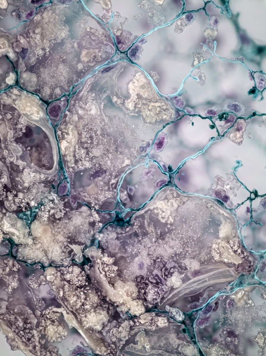
▫️ Within the delicate architecture of the lung, gas exchange relies on pristine, clear alveolar spaces. When pulmonary alveolar proteinosis disrupts this vital process, surfactant derived lipoproteinaceous material accumulates organically within the tissue. Captured through high resolution confocal fluorescence microscopy, this striking photomicrograph reveals a sweeping landscape of translucent alveolar spaces partially choked by heterogeneous foamy aggregates and fine granular clouds. Rendered in a refined palette of glacial cyan, pearlescent ivory, and muted amethyst, the delicate branching alveolar septa stand in stark, beautiful contrast to the sedimentary deposits. This visual synthesis transforms a complex respiratory disorder into an extraordinary piece of microscopic art, blending medical science with high end editorial aesthetics.
#PulmonaryAlveolarProteinosis #PathologyArt #MicroscopicPhotography #TheCellularLens #Histopathology
The Vibrant Threat: Pseudomonas aeruginosa and the Pyoverdine Glow
🔹 Within the complex landscape of a bacterial infection lies a stunning, yet sinister, display of microbial warfare. This high magnification photomicrograph, rendered as striking fine art, captures the vibrant reality of a Pseudomonas aeruginosa colonization. The image reveals dense clusters of rod shaped bacteria (seen here glowing in electric lime and yellow green) actively producing pyoverdine, a water soluble fluorescent pigment. This pigment casts an ethereal, neon like hue through the infected tissue, creating a mesmerising contrast against the deep, muted indigo and amethyst of the host cellular architecture. This visual interplay between the glowing pathogen and the dark stroma transforms a critical clinical threat into a captivating work of cellular art, highlighting the complex beauty hidden within infectious disease.
#PseudomonasAeruginosa #PathologyArt #MicroscopicPhotography #TheCellularLens #Microbiology
The Inflamed Canvas: Glomerulonephritis and the Loss of Filtration
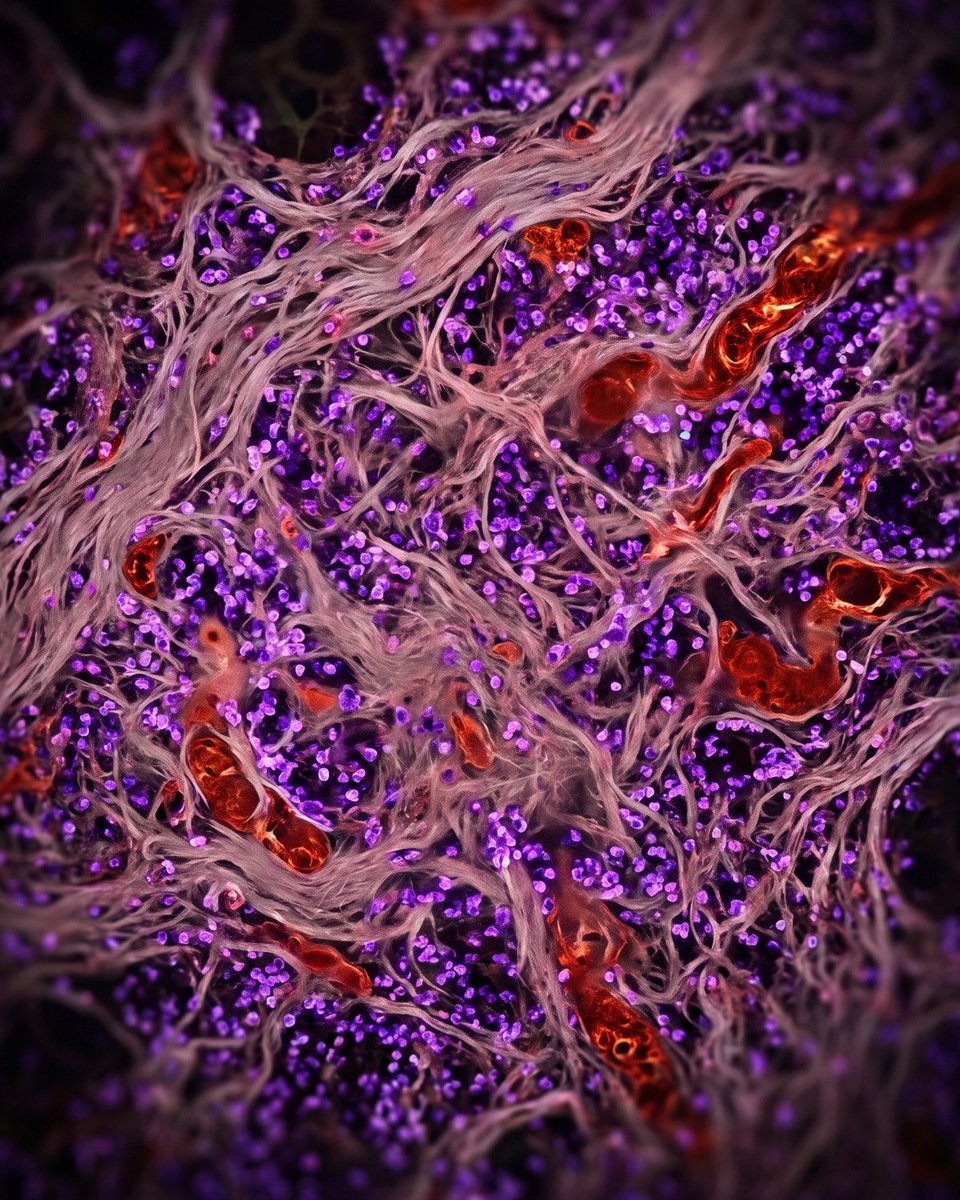
▫️ Within the intricate architecture of the kidney lies a delicate filtration unit, the glomerulus. When disease strikes, this masterpiece of biological engineering can become an inflamed, hypercellular tapestry, as seen in this high magnification photomicrograph. Rendered as striking fine art, this image captures the devastating pathology of glomerulonephritis. Once delicate capillary loops are now obliterated by a proliferation of inflammatory cells (dense purple nuclei) and an expanded mesangial matrix, which glows with an intense, vibrant magenta pink under Periodic acid Schiff (PAS) staining. This visual interplay between the glowing matrix and the dark nuclei transforms a critical clinical condition into a mesmerising and urgent work of cellular art, highlighting the complex and often beautiful architecture of disease.
#Glomerulonephritis #PathologyArt #MicroscopicPhotography #TheCellularLens #KidneyBiopsy
The Architecture of Resilience: Pulmonary Sarcoidosis Unveiled
🔹 Within the delicate labyrinth of lung tissue lies a silent, complex narrative of immune response: pulmonary sarcoidosis. In this striking high resolution confocal fluorescence photomicrograph, non caseating granulomas are revealed as radiant, emerald green architectural forms embedded within heterogeneous lung parenchyma. Luminous turquoise and icy cyan illuminate the surrounding branching alveolar and connective tissue networks, while subtle violet magenta signals and golden amber highlights trace the intricate cellular and inflammatory details. This masterful visual fusion transforms complex immunopathology into a mesmerizing and scientifically credible work of microscopic art.
#PulmonarySarcoidosis #Sarcoidosis #PathologyArt #MicroscopicPhotography #TheCellularLens