3D Maps Reveal Molecular Complexities of the Brain
Researchers made significant strides in understanding the brain's complexity through spatial omics and deep learning.
By integrating biochemical imaging with cutting-edge computational techniques, their team has developed 3D molecular maps that illuminate the brain's cellular activities in unprecedented detail.
This innovative approach, supported by a substantial NIH grant, enables precise analysis of how brain cells interact chemically, shedding light on the mechanisms underlying health and disease.
The research not only demonstrates the power of single-cell metabolomics but also highlights the essential role of collaborative, interdisciplinary efforts in pushing the boundaries of neuroscience.
1/3
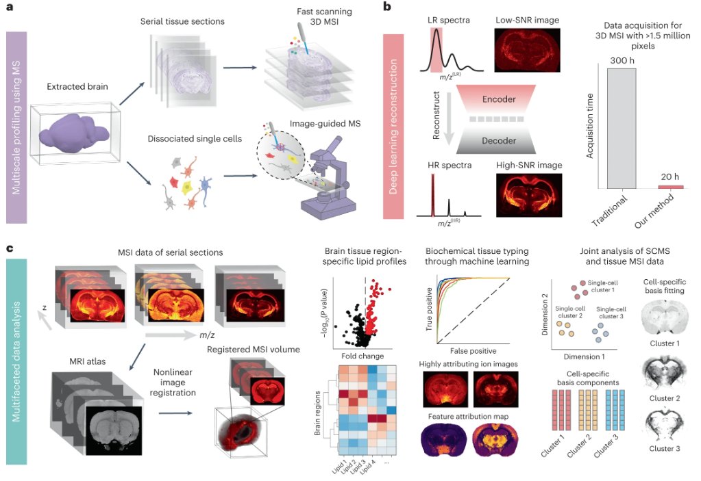
The major work during my PhD study is finally out today! We leverage deep learning based mass sepc imaging to enable integrative tissue and single-cell profiling. We bring some interesting data analysis components as well to jointly analyze MS imaging and SC data.
Out today from the Sweedler and Lam labs is MEISTER, an experimental and computational framework for mass spec that integrates 3D, organ-wide biomolecular mapping with single-cell analysis for multiscale profiling of spatial–biochemical organization. https://t.co/23sYiYnQDT
Really excited to present our new work for material discovery: we leveraged language representations to design a framework that can recommend, rank, and explore functional materials: https://t.co/OMImyjQrjx
Joint work with @RichardYuxuan#materialsscience#MachineLearning
1/3
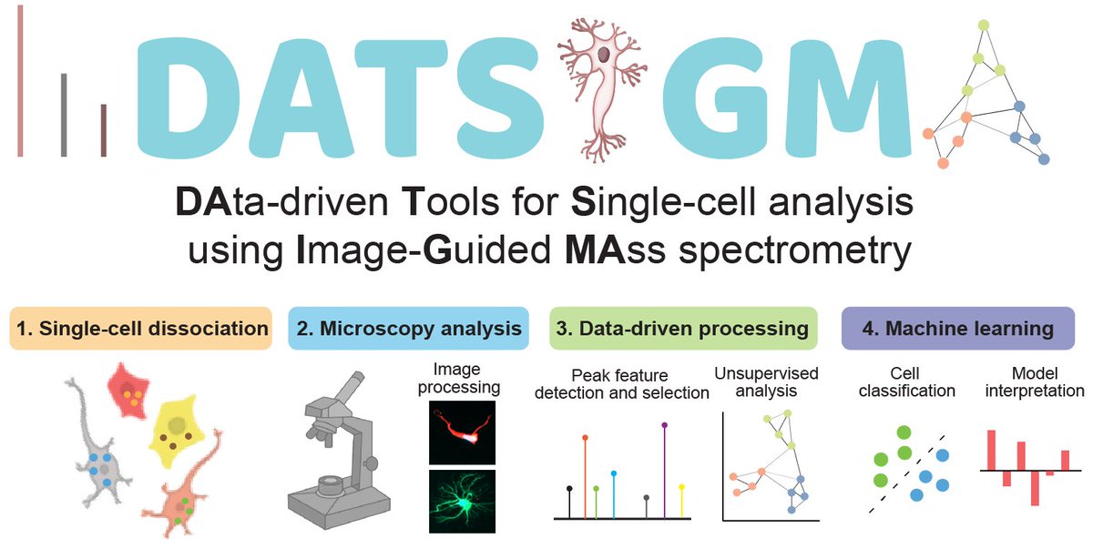
📖Our data-driven+#MachineLearning framework for open-source processing single-cell MS data are online @JProteomeRes. We benchmarked on six image-guided single-cell MS datasets across various instruments & species.
Article: https://t.co/QHyBaLNUAM
Code: https://t.co/xGh0aribWM
BREAKING: 48K University of Calfornia TAs, postdocs & other student workers just walked off the job.
It's the largest strike in the US since 2019.
Many UC workers earn < $24k/yr. A PhD student I spoke to said they donate blood plasma to make ends meet.
https://t.co/RVI7e1xchT
https://t.co/cardTgLqxE My personal view on Intelligence -- the most difficult jigsaw puzzle I have ever tried to understand my whole life. The paper is not an invitation for debate. Please no rushed comment or argument on this serious topic till you understand everything in it.
Thanks for sharing our work!😀
For anyone who wants to try out the tool, the computational implementation is available with a demo: https://t.co/SmgdI7Bo8b
I'll be excited to see (or help) to have this on others' FTMSI platforms.
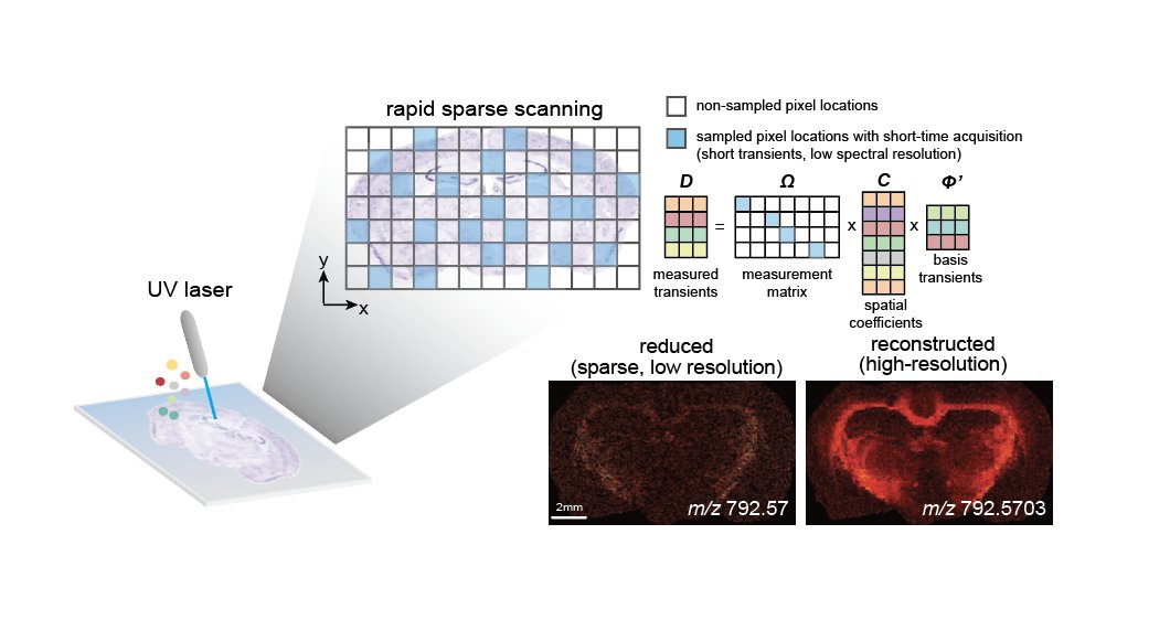
Fourier transform-based mass spectrometry imaging of tissues is accelerated through subspace modeling in conjunction with compressed sensing demonstrated in this work from @RichardYuxuan & the Sweedler lab. #UIUC https://t.co/TaTIoELyxB
Our work using computational imaging (compressed sensing+subspace imaging) for FT-mass spec imaging is out: https://t.co/vMF2KBnrMh
Reduced data are reconstructed for high-mass/spatial resolution w/ total of 10x higher throughput.
Implementation code: https://t.co/SmgdI7jNgD
I’m super excited to share our work, out today @NatureBiotech: a new deep learning algorithm to accurately identify cells in imaging data! Our method works across image platforms, tissue types, and species with no fine-tuning required 🧵 https://t.co/VqgyccAy4q
#DeepInterpolation from the @LecoqJerome lab is a denoising approach for calcium imaging, electrophysiology and fMRI data. The deep learning-based approach increases SNR and allows extracting more information from processed data. @natorlova@the_moliver
https://t.co/aQS8dfGhzW
This is a further extension of the proposed subspace approach earlier: https://t.co/PRB4LLpaYd
Now we exploit the spatial sparsity of biological tissues through compressed sensing to allow spatial sparse sampling, further improving the imaging throughput.
Enhancing the Throughput of FT Mass Spectrometry Imaging Using Compressed Sensing and Subspace Modeling
Check out the preprint @ChemRxiv : https://t.co/EpOxezSzXv
We used CS+subspace modeling to jointly reconstruct high-dim FTMSI data from sparse measurement that's 10x faster.
Check out our recent publication on single-organelle characterization using MALDI MS! Glad to be involved in this work, which is made possible through the collaborative environment at @BeckmanInst across various disciplines and departments.
The Sweedler lab from the University of Illinois at Urbana-Champaign describes image guided MALDI-MS characterization of peptide and lipid content in single organelles.
https://t.co/TlLk8KwQ88
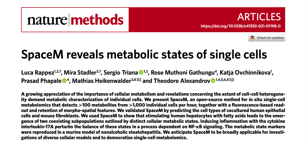
Thrilled to see a key work of @alexandrovteam out in @naturemethods! 🤩
Rappez, Stadler et al SpaceM reveals metabolic states of single cells
https://t.co/KAUY6p6LVH
1/5