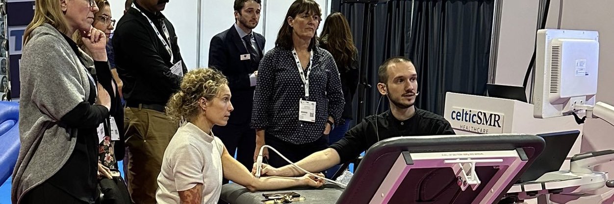
Excited to share our recent publication with Dr. Malliaropoulos in Ultrasound Journal, on MSK ultrasound in clinical practice and medical education.
Read the full article here: https://t.co/rb5lpttEnR
#sportsmed#ultrasound#medicaleducation#anatomy
Excited to share our latest publication in the JDMS, with Nabeel Alghamdi and Nikos Malliaropoulos, focusing on the transformative role of ultrasound in musculoskeletal care and patient outcomes.
Full article here: https://t.co/3iJSfmXeWT
#sportsmed#ultrasound#msk
Looking forward to presenting on ’Rehab consideration in FAI Syndrome and Hip Dysplasia’ next week at Newcastle at the ‘Early intervention in hip surgery’
This conference will provide core information on hip pathology in young adults, covering the current approaches to diagnosis and management.
As well as providing a comprehensive review of the current state of non-operative treatment and hip joint preservation surgery, the meeting will also cover the challenges associated with total hip replacement performed in a young population and specific including developmental FAI Syndrome and Hip dysplasia, Perthes disease much more.
Last Few seats available at (Only £40)
https://t.co/CaBj6lZG7n
Such a privilege to present on elbow ultrasound, focusing on anatomical variants and differential diagnoses at #SonoCon2024 in #Vancouver. Huge thanks to Audrey, Jody and the organising committee for the invite!
Key points from my lecture are in the thread below 👇
Histologically a fibrocartilage, the plica presents as a triangular hyperechoic structure in the HRJ space, positioned below the level of capitulum and radial head. Moderate vascularisation and abundant nerve endings, thickness of 1.7-2.2mm in asymptomatic individuals.
Such a privilege to present on elbow ultrasound, focusing on anatomical variants and differential diagnoses at #SonoCon2024 in #Vancouver. Huge thanks to Audrey, Jody and the organising committee for the invite!
Key points from my lecture are in the thread below 👇
👉Radiohumeral synovial plica: A fold in continuity with the capsule, blending with the CET, 77% prevalence in asymptomatic patients. The lateral fold, visible on ultrasound, appears in 5-20% of cases.
👉Accessory ossicles: Six types with a prevalence of 0.77%. Types I and II, located in the coronoid and olecranon fossae, can limit flexion/extension, often requiring surgical intervention. Differentiate from calcifications by smooth cortex and oval/circular acoustic shadowing.
👉Medial supracondylar process (prevalence 0.7-2.5%, more common in males, linked to CdLS). Occurs with or without the ligament of Struthers, or the ligament may exist on its own. Potential entrapment site for the median nerve and brachial artery, or rarely the ulnar nerve.
Off to #Vancouver for Sonography Canada Conference 2024 to present on anatomical variants of the elbow, exploring insights through #ultrasound and cadaver studies to aid differential diagnoses. Excited to connect with colleagues and share insights in the field of sonography.
A sweet moment! A special award from Prof Tsiridis at his presidential dinner
Being the only woman among amazing males this award was an empowering & strong message to all girls out there
Your presence will be seen, your voice will be heard, your hard work will be recognised!