Adhikari’s Image of the Week: Prostate Calculus. Most are asymptomatic, but large/numerous calculi may cause pelvic pain, dysuria, prostatitis or chronic infection. #POCUS
Adhikari’s Image of Week: Hemangioma! Classic appearance is a homogeneous hyperechoic mass, measuring <3cm in diameter with acoustic enhancement and sharp margins. #POCUS
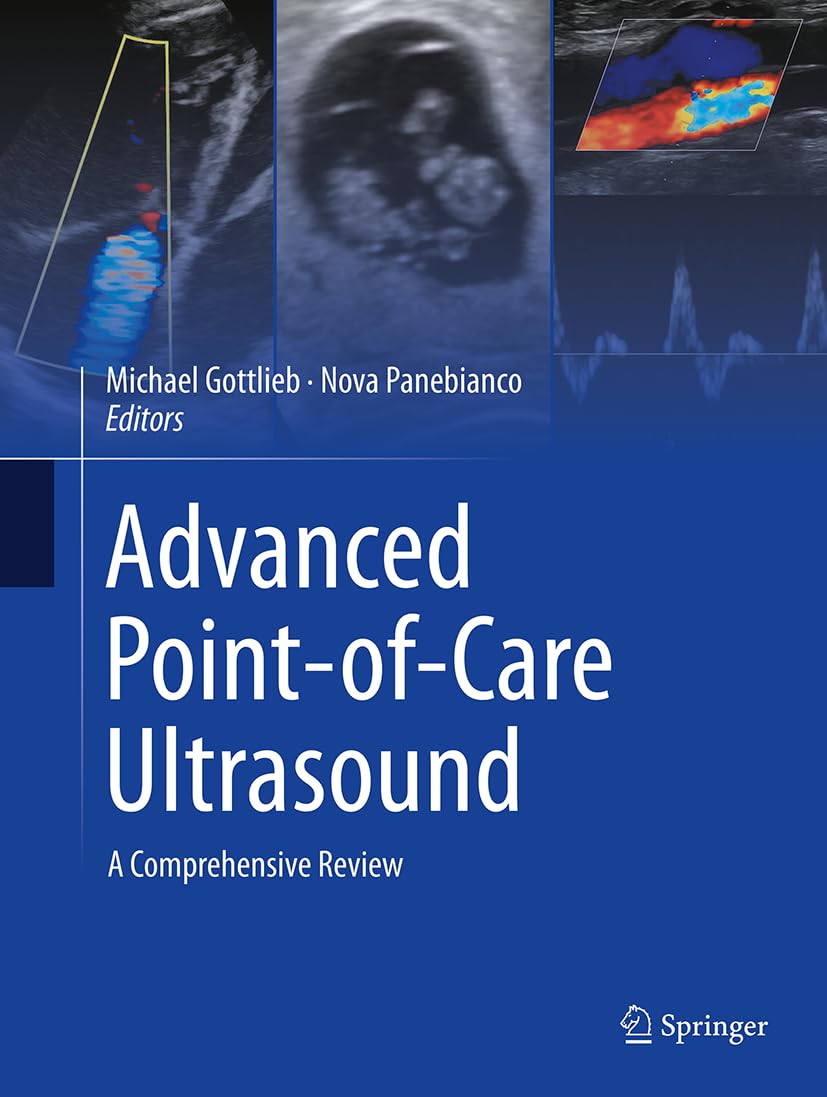
Excited to share that our new Advanced Ultrasound textbook is the #1 New Release across multiple medical specialties!!
Grateful to the numerous authors, editors, and other contributors who have made this an incredible resource for the #POCUS community
https://t.co/O9kbecsTe7
Congratulations to Dr. Nik Theyyunni @HeyDrNik who received the ACEP National Emergency Medicine Excellence in Bedside Teaching Award!🎉 @UMichiganEM@MaizeandLube
Adhikari's Image of the Week: G-tube placement confirmation. Once visualized in the stomach, color Doppler can be applied over the tube to enhance visualization by motion artifact during gentle tube oscillation. #POCUS
Nominations are now open for the #SAEM AEUS Academy awards! Nominate yourself or someone you admire for this honor. Awards will be presented at the #SAEM23 Annual Meeting. Nominate now: https://t.co/x02pBnxSro
Adhikari's Image of the Wk: Adenomyomatosis. Can be focal or diffuse. US shows wall thickening w/multiple small cystic pockets (Rokitansky-Aschoff sinuses) and comet-tail artifacts representing cholesterol aggregates. #POCUS
Adhikari's Image of the Week: US features are non-specific: Echogenic medullary pyramids, some may cast posterior shadowing. Predominantly bilateral. Distinguishing between medullary nephrocalcinosis can be difficult, as many times these conditions coexist. #POCUS