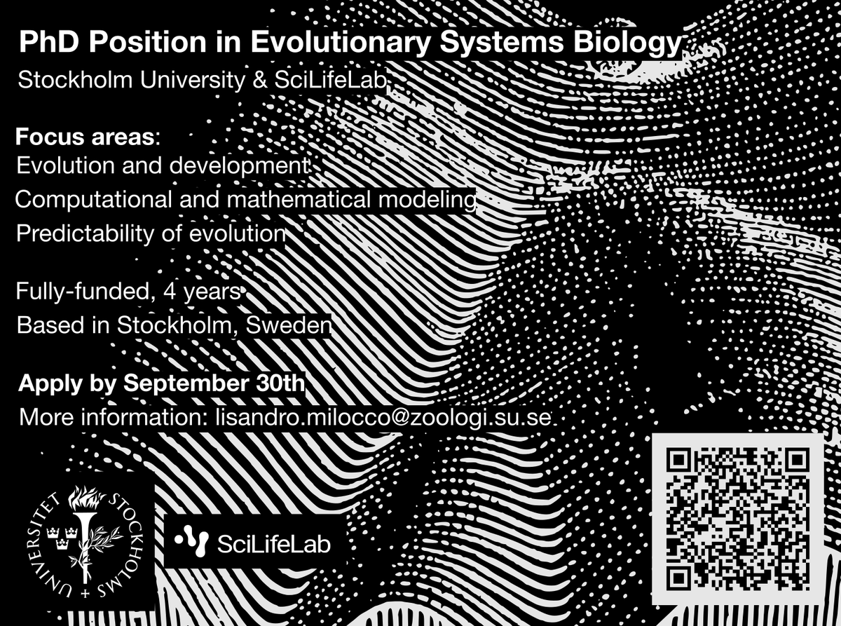
🚨 We're hiring!
PhD position in Evolutionary Systems Biology at Stockholm University & SciLifeLab
🎓 Focus: evolution, development, computational & mathematical modeling, prediction
📍 Stockholm, Sweden
🕒 4 years, fully-funded
🔍 Details: https://t.co/nIbcxSeF8T
Please share!
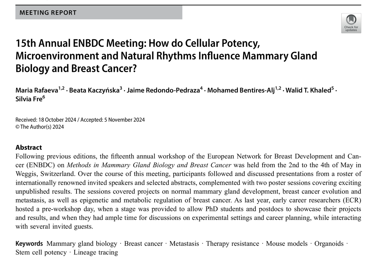
🔔Check out the meeting report from the 15th @enbdc
Workshop on Methods in Mammary Gland Biology & Breast Cancer, held in Weggis 🇨🇭 (May 2–4, 2024), for highlights from talks, poster sessions, & early career researcher workshop.
https://t.co/4FiokmC7WJ
Issue 15 is complete!
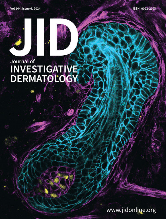
On the cover: A confocal maximum intensity projection image created with Imaris showing an E17.5 mouse wild-type mammary gland wholemount stained with epithelial cell marker EpCAM (green). See Research article by Satta et al.
https://t.co/rHL7SYTKS0
Here's the @jidjournal June cover video https://t.co/q9j0a4zocb
Mammary bud epithelium adopts hair follicle cell fate upon contact with the hair follicle dermal condensate. E13.5 mouse mammary bud epithelium expressing mGFP (cyan) was manually separated from the surrounding mammary mesenchyme after enzyme treatment, and recombined with the E16.5 back skin mesenchyme dissected from a mouse embryo ubiquitously expressing mTdtomato (magenta). The recombinant was cultured for 6 days before fixation and whole-mount immunostaining with the dermal papilla marker Sox2 (yellow). Z-stack images were captured using Leica SP8 Upright confocal microscope with an HC PL APO 20x/0.75 IMM CORR CS2 objective. For more detail, see the article “Stabilization of epithelial beta-catenin compromises mammary call fate acquisition and branching morphogenesis” by Satta et al https://t.co/K8Ndciapfa.
#dermtwitter #dermatology #dermresearch #dermscience
It’s here! The June issue of the @theJIDJournal is out now https://t.co/ca0zprQP66
Cover image:
Mammary Bud Epithelium Adopts Hair Follicle Cell Fate upon Contact with the Hair Follicle Dermal Condensate E13.5 mouse mammary bud epithelium expressing mGFP (cyan) was manually separated from the surrounding mammary mesenchyme after enzyme treatment, and recombined with the E16.5 back skin mesenchyme dissected from a mouse embryo
ubiquitously expressing mTdtomato (magenta). The recombinant was cultured for 6 days before fixation and whole-mount immunostaining with the dermal papilla marker Sox2 (yellow). Z-stack images were captured using Leica SP8 Upright confocal
microscope with an HC PL APO 20x/0.75 IMM CORR CS2 objective.
Image courtesy of Qiang Lan of Cell and Tissue Dynamics Research Program, Institute of Biotechnology, Helsinki Institute of Life Sciences (HiLIFE), University of Helsinki, Finland.
#dermatology #skinresearch #mammary #derm #dermatologist #dermresearch #dermscience
I am thrilled to share my postdoc work @MikkolaLab@HiLIFE_helsinki@BIOTECH_UH@helsinkiuni published @eLife. In this project, we tried to understand the regulation of the initial branching and pattern formation in embryonic mouse mammary gland.
https://t.co/6OrxySJOHF
Glad to see its final look. I started this project during my PhD, and thanks all the co-authors, especially @SanamPeyvandi and Prof. Curzio Rüegg @unifr, in working together to finish it.
Tumor-educated Gr1+CD11b+ cells drive breast cancer metastasis via OSM/IL6-JAK-induced cancer cell plasticity: https://t.co/Guwr82Gu3o
@sqianglan@SanamPeyvandi@unifr#Oncology
@MariaCaffarel@SanamPeyvandi@unifr Thank you Maria for your nice words and support. Your encouragement kept me motivated and made this project to be real. 🙏