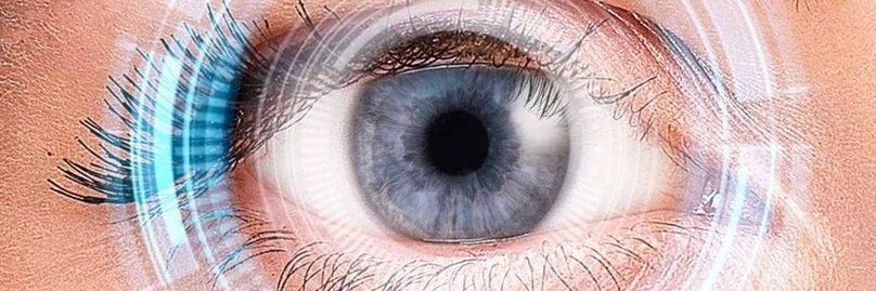
Our doctors are committed to providing quality, individualized eye care which includes lasik and cataract surgeries, routine care, and treatment of eye disease.
Ever wonder what happens during an auto-refractor test? 🏠✨
Before your LASIK or vision consultation, we use this quick test to estimate your prescription—all you have to do is stare at the little farmhouse!
#HoustonLasikandEye#LASIK#AutoRefractor#eyehealth#visiongoals
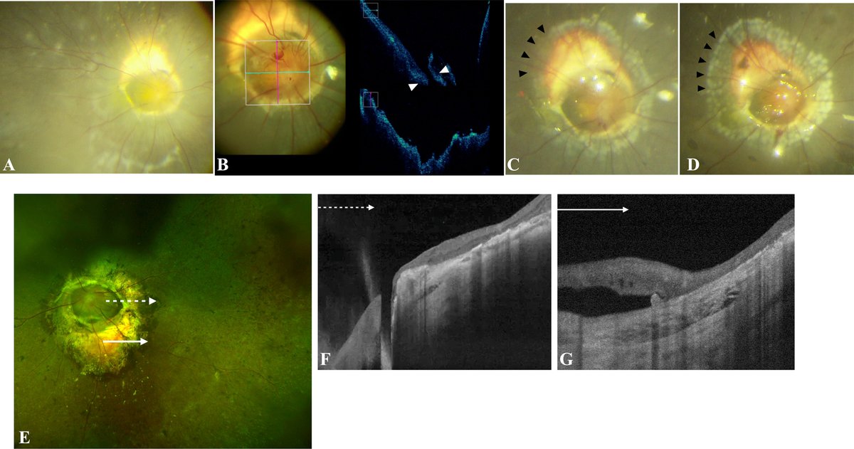
Visual Recovery After Surgery in Epiretinal Membrane Eyes with Retinal Nerve Fiber Layer Schisis and Internal Limiting Membrane Tear
https://t.co/ZmxiEReDV1
#ophthalmology#retina
A 76-year-old woman was referred for a 9 × 6 mm bi-lobed, pink-beige mass with smooth surface of the right lower lid, slowly enlarging for 8 months (A). There was no history of previous cancer. The lesion was surgically excised, revealing a dermal tumor that consisted of infiltrating sheets of mildly pleomorphic cells with oval nuclei, separated by deposits of mucin (B and C). Cells expressed cytokeratin 7, epithelial membrane antigen, GATA binding protein 3, neuron specific enolase, and synaptophysin (D). Cytokeratin 20 was negative. A mammogram was normal. A diagnosis of endocrine mucin-producing sweat gland carcinoma of eyelid was made.
https://t.co/yAHxVNuxDq
#ophthalmology
Not all eyes are the same—so why should LASIK be one-size-fits-all? 👁️✨
Ready to find out which procedure is right for you? Book your consultation today! 📲
#CustomLASIK#PRK#VisionGoals#EyeHealth#ByeByeGlasses
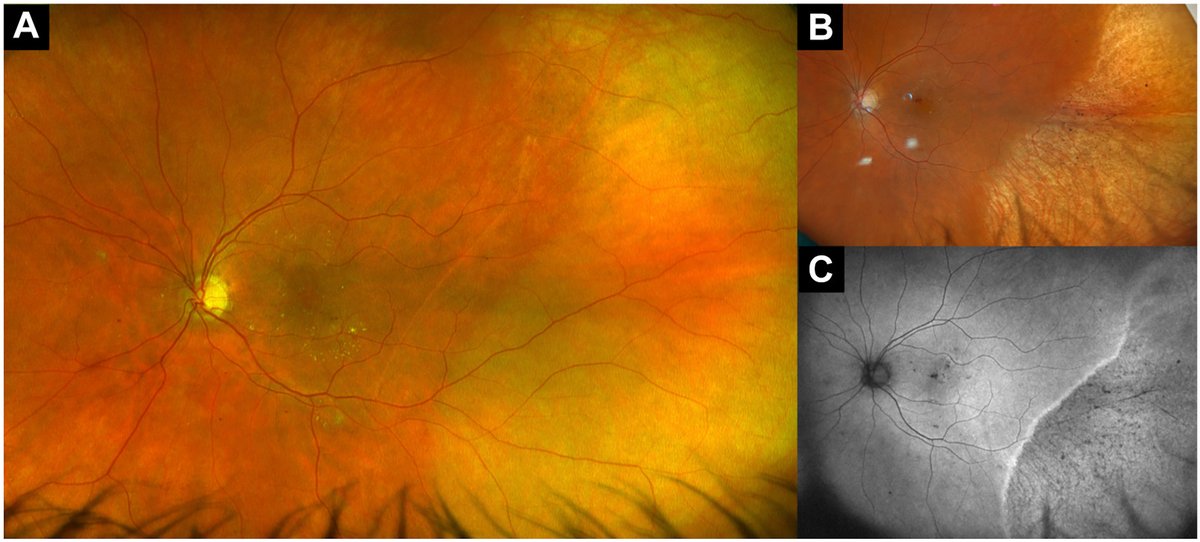
A 56-year-old woman with diabetic macular edema presented from elsewhere after subconjunctival anesthesia was complicated by inadvertent subretinal injection of 2% lidocaine. Visual acuity dropped to hand motion, and an anterior chamber tap was performed by the injecting provider to lower the intraocular pressure (IOP). The following day, vision recovered to baseline 20/30 with normal IOP, and fundus examination revealed temporal retinal whitening spanning 5 hours, sparing the macula (A). Five years later, fundus examination showed temporal pigmentary changes (B), and fundus autofluorescence demonstrated a hyperautofluorescent demarcation line with adjacent areas of speckled hypoautofluorescence (C). Subretinal injection of lidocaine can cause long-term retinal pigmentary changes, but good visual acuity if the macula is spared.
https://t.co/hHpaniPwlS
#ophthalmology #retina
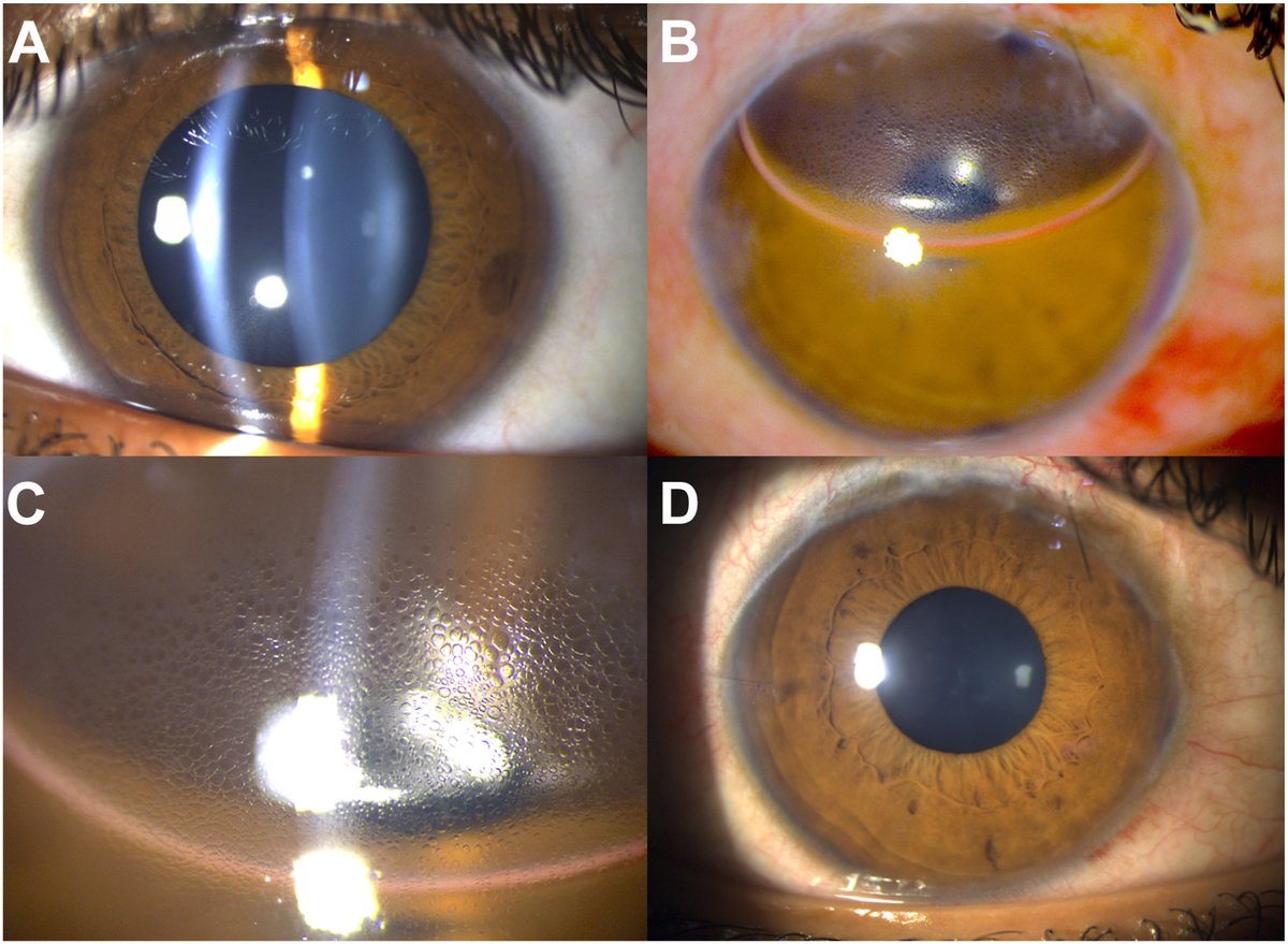
A 21-year-old man with juvenile open-angle glaucoma (A) underwent gonioscopy-assisted transluminal trabeculotomy. Intracameral air was injected at the end of surgery to pressurize the globe and prevent reflux bleed. The next day, focal corneal epithelial bullae appeared in the region over the partially resorbed air bubble (B, C). The bullae resolved by day 5 with air resorption (D), and visual acuity recovered to 6/6. While intracameral air is known to affect the endothelium, this case suggests it can also cause localized, transient epithelial microcysts at the air–cornea interface, and warrants caution in eyes with a potentially compromised endothelium.
https://t.co/x4Zep79ELI
@manchanda1995
#ophthalmology #glaucoma
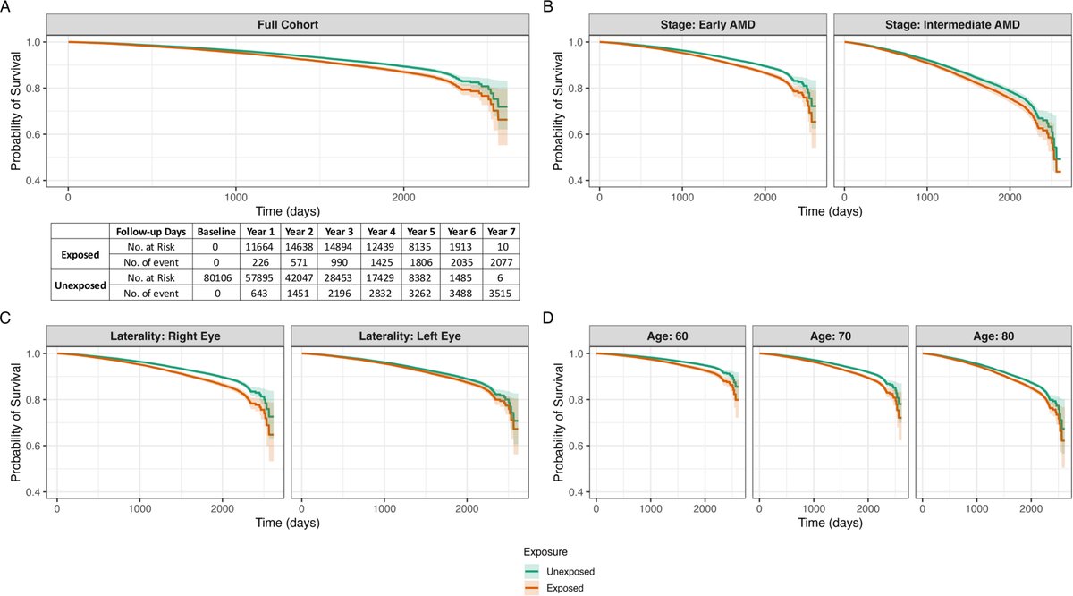
High resolution and multimodal structural phenotyping of intermediate and advanced non-neovascular age-related macular degeneration
https://t.co/5MJHnTtjix
#ophthalmology
Study Explores Reasons Behind IOL Explantation
Several key patterns arose, linked to implant material and design. Read more: https://t.co/y004xexmZJ
#cataractsurgery#cornea#optometry
Indirect ophthalmoscopy can feel awkward in the beginning. Optimizing setup, posture, and lens selection can make the #ophthalmic exam significantly easier. #YOInfo https://t.co/64C9JrFDKC
Upgrade your vision, ditch the readers 👁️⚡️
If you’ve noticed your vision shifting in your 40’s and 50’s, RLE could be your answer.
Clear vision is always in style. Schedule your consultation with Houston Lasik and Eye today!
#RLE#Presbyopia#Cataract#ClearVision#Readers
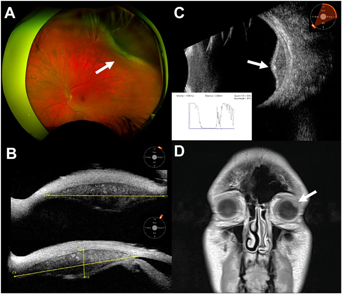
A 51-year-old man underwent brain magnetic resonance imaging that disclosed a left globe mass. Fundus imaging showed an amelanotic lesion with posterior exudative deposits, without orange pigment or subretinal fluid (A). Ultrasound biomicroscopy (B) and B-scan (C) revealed a solid ciliary body mass with marked intralesional vascularity, hyperechoic calcific deposits, and A-scan (C, subset) demonstrated low internal reflectivity. Magnetic resonance imaging showed a T1 hyperintense ciliary body mass, but was unchanged compared with magnetic resonance imaging from 10 years prior (D, 3.4 mm height). The constellation of a ciliary body mass, intrinsic vascularity, low reflectivity, increased transillumination, calcific deposits, and decade-long stability favored leiomyoma. Although definitive diagnosis would require histopathologic examination with surgical biopsy, the lesion’s indolent course supported continued surveillance.
https://t.co/CcEjynviZn
#ophthalmology #retina
Several Inflammatory Skin Diseases Associated with Increased Risk of Keratoconus
Immune dysregulation and elevated proinflammatory cytokines may contribute to the condition by disrupting collagen integrity.
https://t.co/uFRLUnmbFG
#keratoconus#cornea#optometry
Did you squint or pause to read? 🧐
Friendly reminder that your annual eye exam is just as important as any other checkup!
Give us a call to book your spot today!🗓️
#visioncare#eyes#wellness#eyedoctor#eyeexam
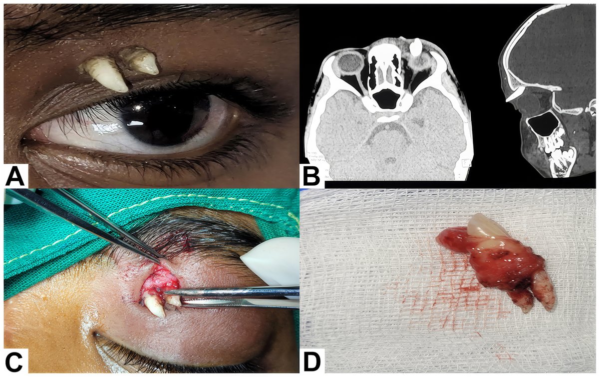
A 16-year-old boy presented with a painless swelling of the left upper eyelid for 1 month, with no history of trauma. Examination revealed 2 hard, well-defined whitish projections arising from a fullness in the medial third of the upper eyelid (A). Computed tomography demonstrated a well-defined hyperdense structure with crown–root morphology arising from the left orbital roof, extending into the preseptal space and projecting externally with 2 tooth-like projections on its surface (B). Surgical excision revealed a mature canine tooth (C, D) without teratomatous elements, consistent with an ectopic tooth of the superior orbit.
From “Ectopic Canine Tooth in the Superior Orbit” by Meghana Tanwar, DNB, FRCS (Ophth), Vedant Sharma, MBBS, Usha Kim, DO, DNB. Published by Ophthalmology online on January 16, 2026.
Read: https://t.co/LabGO5DaHD
#AAO #Ophthalmology
How does LASIK work its magic? It all starts with the corneal flap.🪄👁️
Drop your questions about the LASIK process in the comments, or DM us to book your free consultation!
#eyedoctor#lasik#fyp#eyes#clearvision