We further demonstrate that our framework works in real-world settings on a 200 MP Samsung ISOCELL HP2 prototype. For both object tracking and scene text recognition tasks, our sensor attention policy reproduces the smooth pursuit and saccadic scanpaths seen in simulations, achieving 200 MP bandwidth-efficient sensing under real-world conditions with sensor noise.
[5/6]
Should have posted about this before, but my Cleo simulator paper got published in JNeurosci!
Take a look if you want to simulate advanced opto/ephys experiments at a medium (point neuron) level of realism.
https://t.co/ykmfQC5MaK
Early iSOAR 5D movie of membrane and mitochondria dynamics over 3 hrs at 3 min intervals in the head of a zebrafish embryo 6 dpf, covering a 300 x 1000 um FOV at 193 x 193 x 550 nm resolution (0.8 TB total).
🧠 When the imaging protocol masks real biology: In a new iScience paper, Dirkx et al. address a subtle but important problem in human iPSC-derived neuronal cultures. Fluo-4 AM dye-based Ca²⁺ imaging is widely used to monitor neuronal activity, but the associated imaging medium exchange can disrupt the spontaneous synchronized network bursts that mark neuronal maturation. In other words, the workflow itself may alter the biology you want to measure.
To show that the issue comes from the imaging procedure itself, the authors used the MaxTwo HD-MEA system as a gold-standard functional reference. MaxTwo recordings showed that fresh imaging medium abolishes synchronized bursting while leaving overall firing intact, whereas re-applying each well’s own conditioned imaging medium preserves spontaneous bursting much more faithfully after dye loading.
This was more than a technical optimization. In agreement with the MaxWell HD-MEA ground truth, the team could still detect the expected hyperexcitable network phenotype in a KCNQ2-related epilepsy model with calcium imaging. A simple workflow change, but a meaningful step forward for functional phenotyping, disease modeling, and drug screening when single-cell spatial resolution matters.
👉 Read the full publication here – https://t.co/6El0oSkXon
👏 Congratulations to Nina Dirkx, Bob Asselbergh, Peter Verstraelen, Jonas Van Lent, Els De Vriendt, Vincent Timmerman, Winnok H. De Vos, and Sarah Weckhuysen from the VIB Center for Molecular Neurology, the University of Antwerp, and University Hospital Antwerp, Antwerp, Belgium.
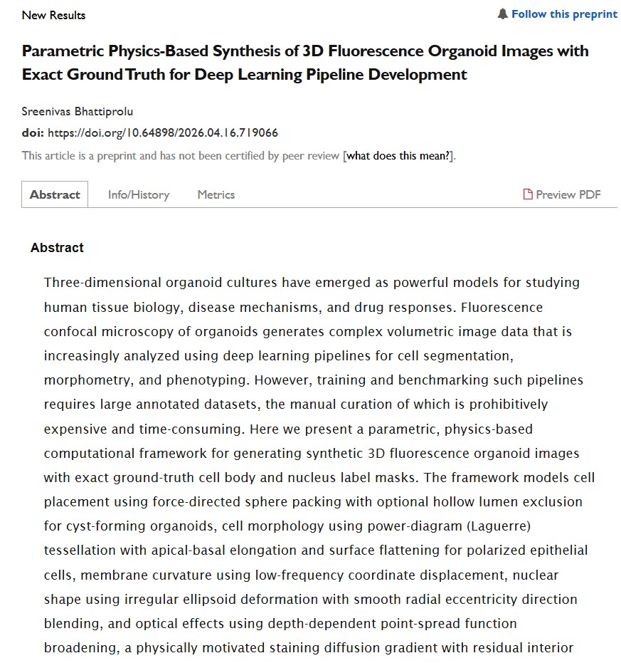
Excited to share my latest preprint on bioRxiv!
Synthetic 3D fluorescence organoid images with exact ground-truth masks for training deep learning models + testing pipelines before real data.
https://t.co/6Rt3FwcyI7
#microscopy#Biotech#DrugDiscovery
📷 Expansion microscopy turns 11 — and our comprehensive Primer is now live in @MethodsPrimers ! A decade of ExM, 1000+ papers, and still accelerating. Co-authored with the father of the field, @eboyden3 , + Ha Vo & Rong Xu. 📷 Free to read: https://t.co/3LTyIZQgpT
Preprint out arguing that we should build the techology to translate (compile) molecularly annotated connectomes into dynamics. I think this is incredibly important. https://t.co/173FDhYtUn
Wrote a review on voltage imaging at the invitation of Neurophotonics, with @UrsLucasBoehm and Zeguan Wang. This method transformed how you see neural activity in small animals, but it is still somewhat exclusive. Hope this helps break the barrier.
https://t.co/NbvBDt3eXO
Read my critical thoughts on this: "We are watching two disciplines trade their worst habits. Neuroscience is mistaking benchmarked prediction for understanding, and machine learning is mistaking mechanistic language for mechanism. ..."
Introducing #FASIM: A new microscopy method combining SIM with fluorescence anisotropy, ~100 nm resolution with 0.56% relative error—a >20-fold accuracy improvement, allowing us to map the "crowdedness" (viscosity/molecular interactions) in real-time.
https://t.co/ugDTpw5ZsJ
Aging may feel gradual… but what if it’s not?
In our paper out today, we tracked fish continuously from puberty until death.
This gave us a unique view of how aging unfolds across the adult lifespan.
🧵
Our live tissue clearing paper is out in @naturemethods! We achieved optical clearing of mammalian brain tissues without compromising normal neuronal function. Big congrats to @Shigenori774 and our wonderful collaborators! 🎉
https://t.co/joMB5odihK (1/10)
A new open-source platform has been developed to facilitate the functional phenotyping of human stem cell-derived neurons by integrating calcium imaging, optogenetic stimulation, and automated analysis into a single accessible workflow.
https://t.co/5TOTAlzKRn
First whole-brain recording of social sound processing in a vertebrate @danionella . Surprises start in the hindbrain; thalamus gates conspecific calls; male and female brains diverge downstream. Work by Jörg Henninger & me with and at the @benjulab
If you are an experienced builder/user of advanced custom microscopes, want to do more of the same with some pretty amazing hardware, and want to be a key part of the Cell Observatory Initiative, please consider applying!
🚀 Looking to hire a light-sheet microscopy scientist to help us design, build, and optimize the next-gen platform that will power mammalian connectomics at scale. If you’re excited to join a fast-paced team doing high-impact science, take a look: https://t.co/wRN2INtnwd🔬⚙️
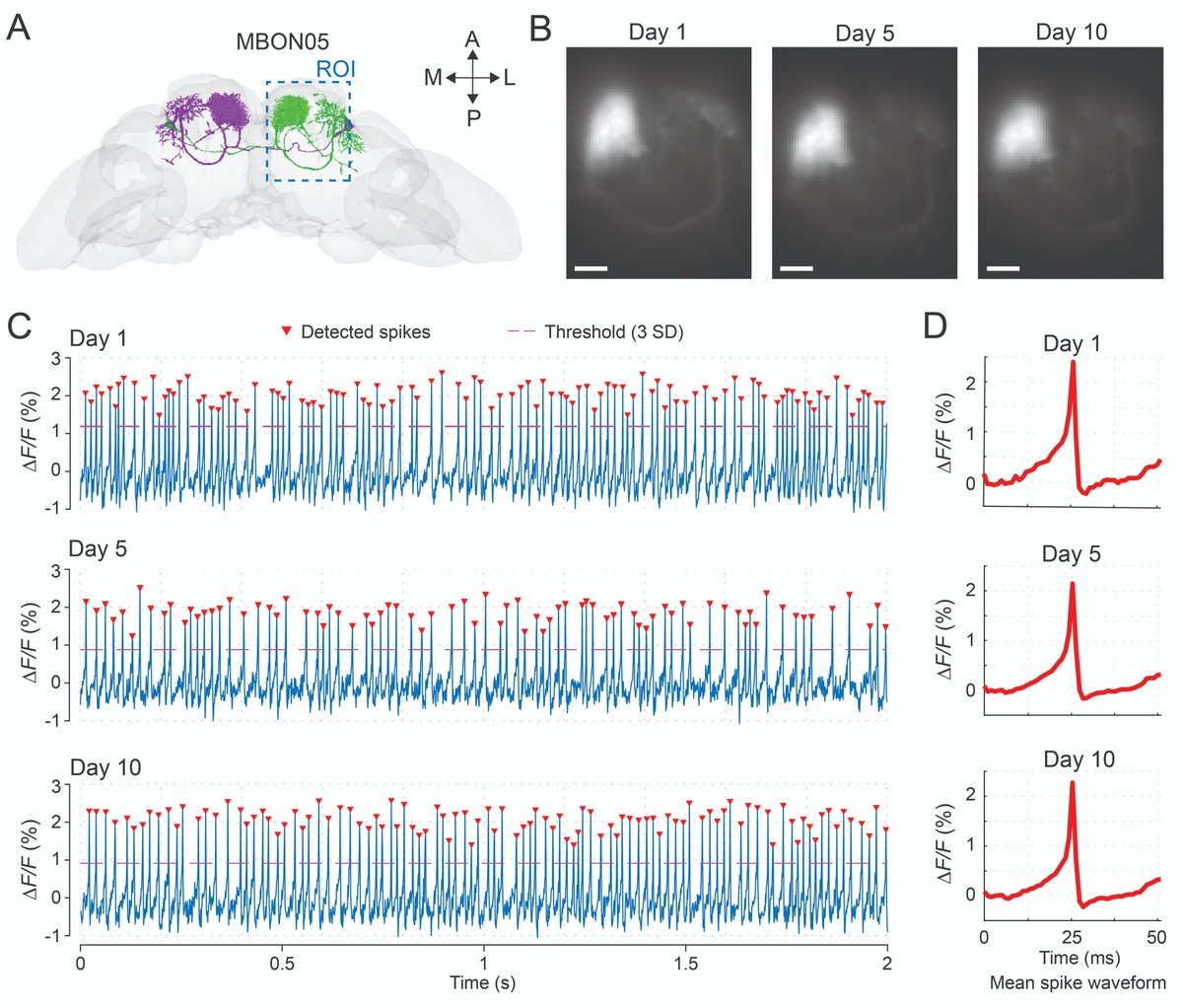
Drosophila folks! We just shared a new protocol for surgical preparation enabling long-term imaging in adult flies. It’s low-cost, simple, and highly repeatable. For demonstration, we also show 10-day voltage imaging and 1-week population calcium imaging.
https://t.co/kHaVPwi1sx
A sneak peak of a complex and technically challenging experiment that my lab developed: https://t.co/UXXUargZqM. Super proud of PhD candidate Hannah Johnson for showcasing our Whole-gut spatial genomic analysis in #zebrafish.
This video illustrates one landmark in the protocol after multiple rounds of sequential #HCR and 3D imaging in zebrafish larvae to reveal spatial expression of numerous mRNAs in the same specimen. Data from these imaging data sets are then computationally analyzed for spatial cell groups, spatially variable genes, and differentially expressed genes along 3D.
We are leveraging this systems-level SGA to uncover unappreciated mechanistic insight at the cell and tissue levels into #ENS construction. Stay tuned for our work that exploits this pipeline within various mutant and perturbation conditions. Reach out if you are interested in trying this! #fruitypebbles