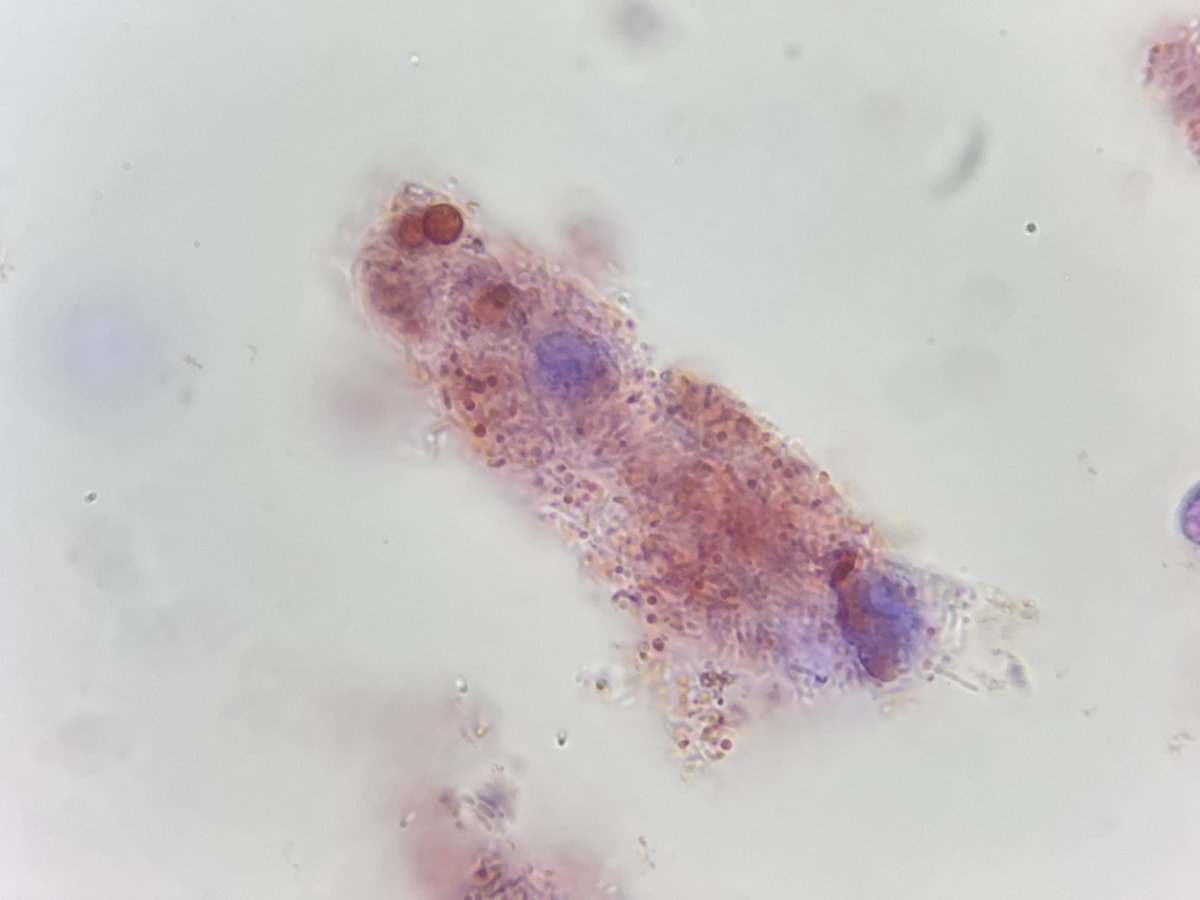
Cytoplasmic vacuolization
Morphological change indicating cell degeneration. The origin of vacuoles is dilated organelles (endoplasmic reticulum, Golgi complex, mitochondria or lysosomes) due to the entry of water.
#urinarysediment#urinemicroscopy#urinalysis
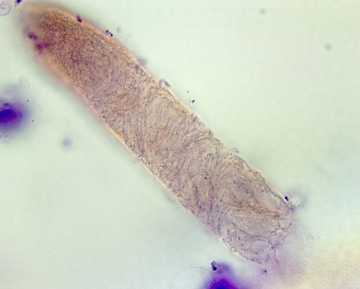
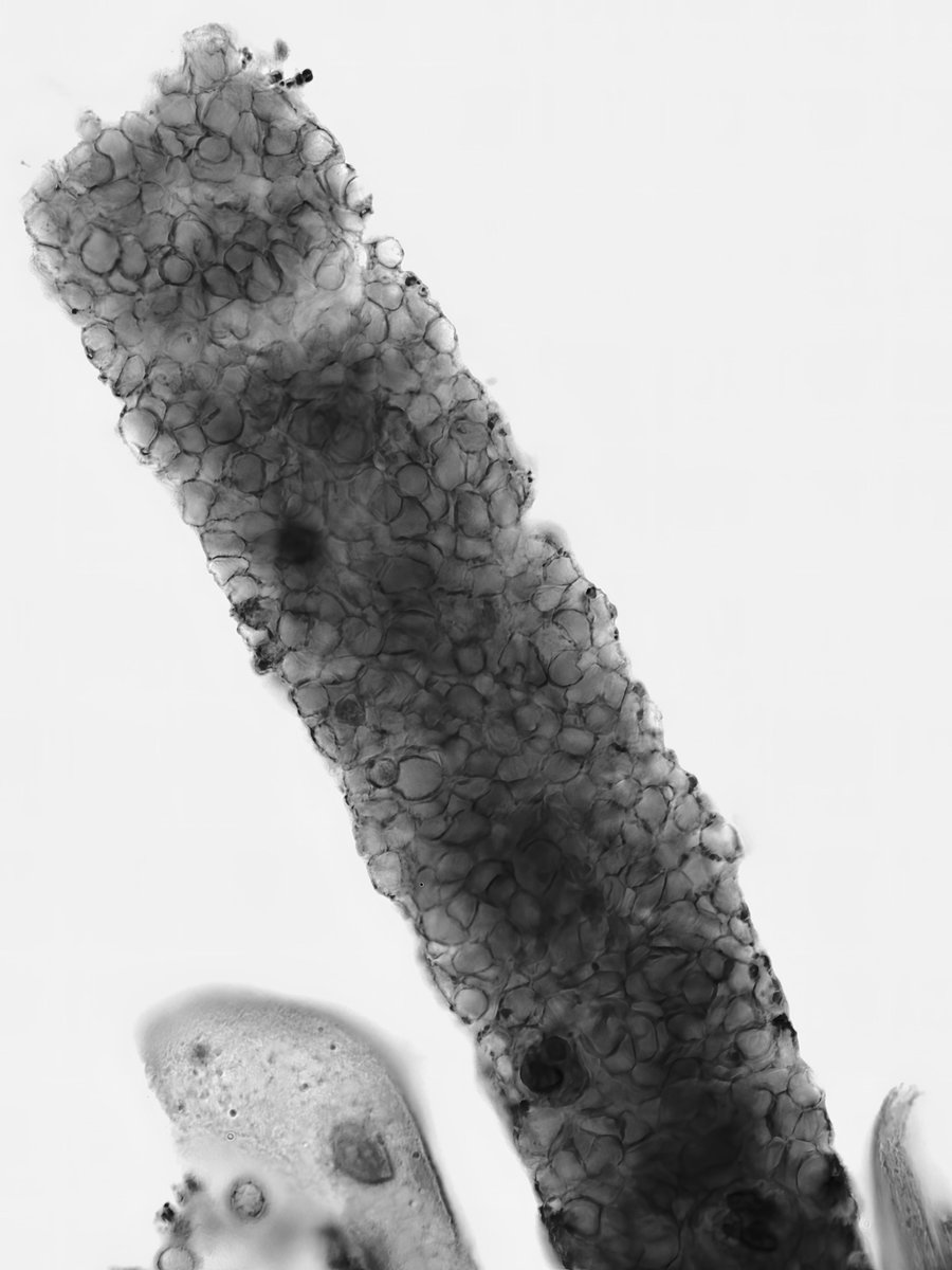
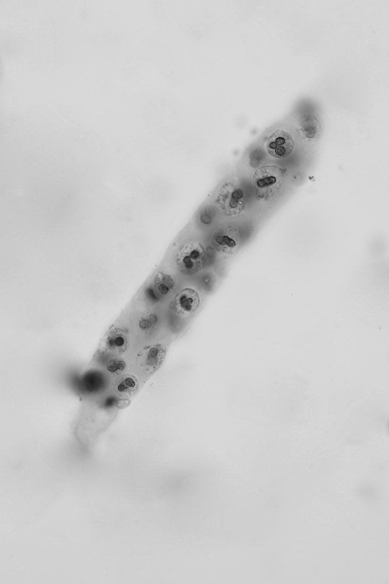
Mixed cellular cast (RBC's, WBC's, and tubular epithelial cells) with a fibrin matrix - brightfield with SM stain - converted to grayscale - #UrinarySediment#UrineMicroscopy
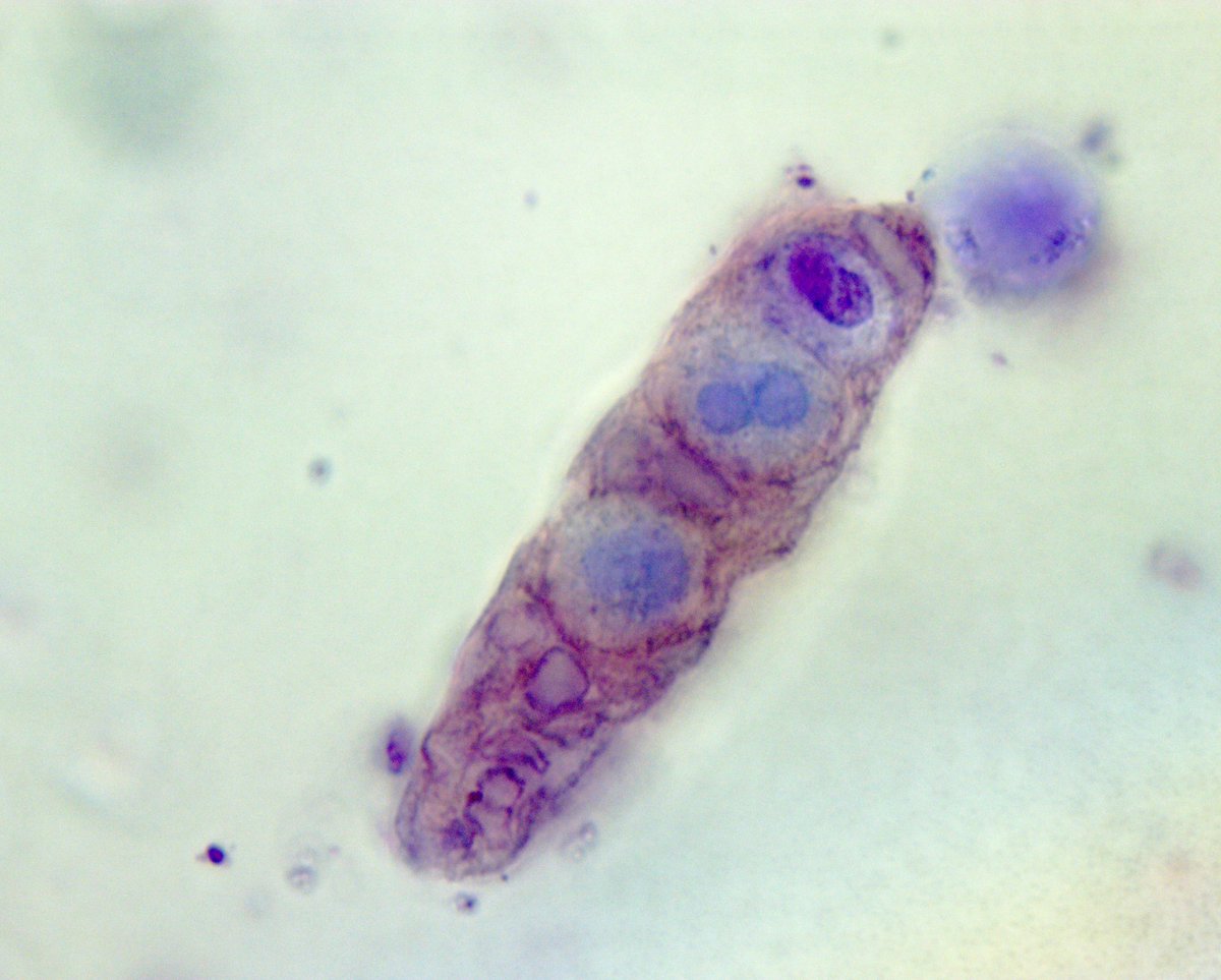
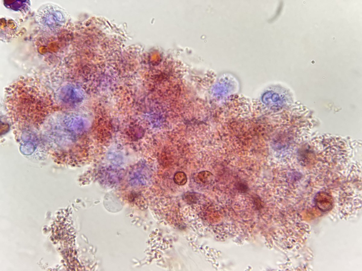
Cellular casts, unstained r difficult to characterize it. SM stain allows better visualization but not always able to distinguish WBCs from RTECs. Prescott-Brodie (PB) aka Mohty stain shows WBCs with distinct dark blue whereas RTECs look fuchsia #UrinarySediment#UrineMicroscopy
Demonstration that nucleated cells in casts from a hyperbilirubinuric specimen are RTEC. PB stain shows that cells do not turn blue (or green: yellow+blue), therefore, they are not WBC. AKI ESLD cholestasis. #UrinarySediment