Making cardiovascular medicine as simple as possible, but not simpler.
ECG| ECHO| CMR| CXR |Interventional |MedEd
Tweets are only for educational purposes.
Sepsis is a medical emergency. Early recognition and timely intervention can save lives.
The Surviving Sepsis Campaign (SSC) 2026 Guidelines provide updated recommendations for the management of sepsis and septic shock.
In this 12-page educational series, we cover:
1. Definitions, epidemiology and pathophysiology
2. Screening, recognition and initial assessment
3. Diagnostic investigations and infection identification
4. Antimicrobial therapy and source control
5. Initial resuscitation and fluid therapy
6. Vasopressors and hemodynamic management
7. Respiratory support and mechanical ventilation
8. Adjunctive therapies
9. Additional supportive management
10. Goals of care and shared decision-making
11. Transitions of care and sepsis survivorship
12. Practical clinical algorithms and key recommendations
High-yield principles:
- Recognize sepsis early.
- Give antimicrobials promptly when indicated.
- Reassess fluid responsiveness and tissue perfusion.
- Use norepinephrine as the first-line vasopressor.
- Individualize treatment and achieve timely source control.
- Plan recovery beyond hospital survival.
Remember: Sepsis management is not a single intervention. It is a coordinated process of early recognition, infection control, organ support, repeated reassessment and recovery planning.
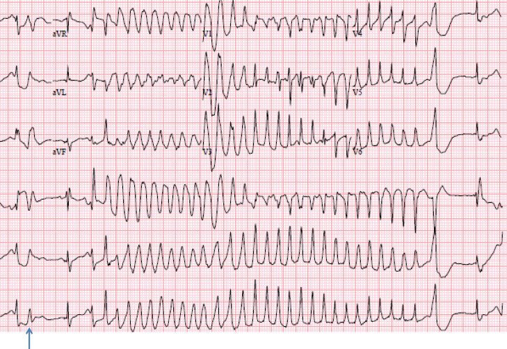
A 35-year-old patient with known WPW syndrome presents with an irregular, very rapid wide-complex tachycardia.
Which medication is appropriate for a haemodynamically stable patient?
A. Digoxin
B. Procainamide
C. Verapamil
D. Adenosine
Which of the following drugs should generally be avoided with bivalirudin because of increased bleeding risk?
A. Metoprolol
B. Atorvastatin
C. Apixaban
D. Amlodipine
COPD management goes far beyond prescribing inhalers.
The GOLD 2027 Report brings together important updates in diagnosis, classification, pharmacological treatment, exacerbation management and long-term care.
In this 12-page educational series, we cover:
1. COPD definition, risk factors and pathophysiology
2. Diagnosis and case-finding
3. Assessment and ABE classification
4. Stable COPD and risk reduction
5. Pharmacological management
6. Pulmonary rehabilitation and non-pharmacological care
7. Oxygen therapy and advanced COPD
8. Exacerbation diagnosis and assessment
9. Acute exacerbation treatment and hospital management
10. Comorbidities and multimorbidity
11. Artificial intelligence and emerging technologies
12. Practical clinical algorithms and key GOLD 2027 updates
Key message: Confirm the diagnosis, characterize the patient, individualize treatment, prevent exacerbations and reassess regularly.
14 Features to Analyze on Every ECG
1. Standardization (calibration) and technical quality
2. Heart rate(s): atrial and ventricular if not the same
3. Rhythm/AV conduction
4. PR (AV) interval
5. QRS interval (width)
6. QT/QTc intervals
7. QRS axis
8. P waves (width, amplitude, shape)
9. QRS voltages: normal, high or low
10. R wave progression in chest leads
11. Q waves (normal vs. abnormal)
12. ST segments
13. T waves
14. U waves
ECG From Scratch #74: Multifocal Atrial Tachycardia (MAT)
An irregular, rapid rhythm is not always atrial fibrillation.
Think of MAT when you see:
- At least 3 distinct non-sinus P-wave morphologies.
- Atrial rate ≥100 bpm.
- Variable PR intervals.
- An irregular ventricular rhythm.
MAT is commonly associated with severe chronic lung disease, particularly COPD.
Can you distinguish MAT from AF on a 12-lead ECG?
Which of the following is NOT a typical ECG feature supporting ventricular tachycardia?
A. Fusion beats
B. AV dissociation
C. Capture beats
D. Termination by carotid sinus massage
Correct answer: D. Immediate unsynchronised defibrillation.
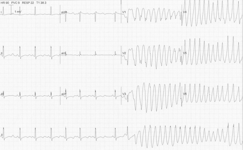
This is pulseless ventricular tachycardia (pVT), a shockable cardiac-arrest rhythm.
The priority is:
1️⃣ Start high-quality CPR.
2️⃣ Deliver an immediate unsynchronised shock (defibrillation).
3️⃣ Resume CPR immediately for 2 minutes; minimise interruptions.
4️⃣ Give epinephrine 1 mg IV/IO every 3-5 minutes according to the resuscitation algorithm.
5️⃣ For persistent/refractory VF/pVT, give amiodarone 300 mg IV/IO, followed by 150 mg if needed, or lidocaine as an alternative.
Note:
- Amiodarone and lidocaine are adjuncts for refractory shockable arrest, not substitutes for the first shock.
- Synchronized cardioversion is used for selected tachyarrhythmias with a pulse, not pulseless VT.
Clinical pearl: Always distinguish VT with a pulse from pulseless VT. The presence or absence of a pulse changes the treatment.
A patient in the ICU suddenly becomes unresponsive and pulseless. The cardiac monitor shows ventricular tachycardia.
What is the most appropriate immediate treatment?
A. Amiodarone 300 mg IV
B. Lidocaine 1.5 mg/kg IV
C. Synchronized cardioversion
D. Immediate unsynchronised defibrillation
Answer: C. No specific treatment is required
This patient has asymptomatic bradycardia with a resting heart rate of 48 bpm.
In an otherwise stable patient without symptoms or evidence of haemodynamic compromise, immediate treatment is generally unnecessary.
A 65-year-old patient is found to have a resting heart rate of 48 beats/min during a routine examination. He has no dizziness, syncope, fatigue or exercise intolerance.
What is the most appropriate management?
A. Immediate atropine administration
B. Permanent pacemaker implantation
C. No specific treatment is required
D. Start isoprenaline
ECG interpretation:
1. Regular wide-complex rhythm at approx 94 bpm.
2. Sinus bradycardia at approx 56 bpm.
3. P waves march independently through the QRS complexes.
4. AV dissociation.
5. LBBB-like morphology, reflecting ventricular activation from the idioventricular rhythm.
6. ST-segment elevation in anterolateral leads, suggesting acute myocardial injury.
Diagnosis: Accelerated idioventricular rhythm (AIVR) with AV dissociation.
Clinical pearl: AIVR typically occurs at 50–110 bpm and is often associated with reperfusion, although it can occur in other settings.
Do not mistake its wide QRS complexes for ventricular tachycardia or assume that the LBBB-like pattern represents true bundle branch block.
In this patient, chest pain and ST elevation demand urgent evaluation for acute coronary occlusion.
Answer: B. Prolonged QT interval
Torsades de pointes (TdP) is a form of polymorphic ventricular tachycardia classically associated with prolonged ventricular repolarization (prolonged QT interval).
Which ECG abnormality is most strongly associated with torsades de pointes?
A. ST-segment depression
B. Prolonged QT interval
C. Narrow QRS complex
D. Short PR interval
Answer: D. Third-degree AV block (complete heart block).
Neonatal lupus is an autoimmune condition caused by transplacental passage of maternal anti-Ro/SSA and/or anti-La/SSB antibodies.
These antibodies can damage the fetal conduction system, particularly the AV node, resulting in congenital complete heart block.
High-yield clinical clues:
- Annular erythematous skin lesions, often around the eyes and scalp.
- Maternal history of SLE or Sjögren syndrome (although the mother may be asymptomatic).
- Congenital complete AV block.
- Risk of fetal bradycardia, hydrops and neonatal pacemaker requirement.
Remember: Third-degree AV block is the classic and most serious cardiac manifestation of neonatal lupus.
Which type of heart block is most commonly associated with neonatal lupus?
A. First-degree AV block
B. Mobitz type 1
C. Mobitz type 2
D. Third-degree AV block
I make this point for every single #HCM talk. We wrote about this at @ACCinTouch.org
In patients with HCM , MR and LVOT Signal on #Echofirst can be quite confusing.
In addition to double checking the SBP with MR jet to confirm LVOT gradient/following tricks can be useful
➡️MR signal velocity is always higher because it gives the LV-to-LA gradient
• LVOT signal velocity is lower
➡️ HOCM MR envelope is wider, begins after the onset of ejection, and terminates at the end of IVR
•LVOT signal is narrower and spans the ejection time
➡️ HOCM MR envelope Curved initially but then straightens out (perpendicular to baseline) by midsystole
•LVOT signal is classic late peaking, dagger shaped, and continues to show rightward curvature in mid-late systole
Differentiating the HOCM MR Signal From Other Causes of MR
➡️Most other MR signals (except mitral valve [MV] prolapse) have onset at the isovolumetric contraction (IVC) phase and last until isovolumetric relaxation (IVR; i.e., pansystolic MR).
➡️ HOCM MR begins after IVC during the ejection phase but persists until IVR. This gives the HOCM MR signal its characteristic initial curved appearance, which is different from most other MR signals.
🔗 https://t.co/9EemADlUSg
confusing LVOT Doppler signal w mitral regurgitation due to SAM in hypertrophic CM?
double check if it’s really the LVOT signal by estimating LVOT gradient with SBP/peak MR jet
& see if this matches measured LVOT (4v2) gradient
1.Peak MR signal(velocity) gives LVP-to–LAP gradient (4V2)
2.LVSP estimated by LV-LA gradient + LAP (~10-20mm Hg)
3.LVOT gradient estimated by LVSP – systolic BP #echofirst @echo_batman
Cardiology learning just got more interactive.
✳ CARDIA is a free, browser-based 3D cardiac learning platform that takes you from basic heart anatomy to advanced interventional cardiology.
Explore:
- Interactive 3D cardiac anatomy
- ECG and electrophysiology
- Echocardiography, cardiac CT and MRI
- Coronary angiography and IVUS
- Hemodynamics and pressure-volume loops
- Coronary physiology (FFR, iFR, CFR, IMR)
- Cardiovascular pharmacology
Change a parameter and see how the physiology responds.
No installation. No account. Completely free.
A useful resource for medical students, residents, cardiologists and anyone interested in cardiovascular medicine.
Explore CARDIA: https://t.co/v3jeDQpoMz
RADIAL ARTERY ACCESS: TECHNIQUE MATTERS
Radial access is the preferred approach for coronary angiography and PCI in many clinical settings, particularly ACS, because it reduces bleeding and vascular complications compared with femoral access.
Key steps for successful access:
1. Palpate the radial pulse and prepare the wrist.
2. Infiltrate local anaesthetic.
3. Puncture at approximately 30-45°.
4. Introduce a 0.018-inch guidewire gently. Never force resistance.
5. Advance the sheath carefully over the wire.
6. Administer an appropriate antispasm cocktail and procedural anticoagulation.
7. Flush and secure the sheath before catheter introduction.
Practical pearls:
- Ultrasound guidance can improve first-pass success, particularly in difficult access.
- Use an appropriately sized hydrophilic sheath.
- Recognize and manage radial artery spasm early.
- Assess for bleeding, haematoma and radial artery occlusion after the procedure.
Successful radial access is not simply about entering the artery. It is about achieving access safely while preserving the artery for future use.