The global leader in miniature mulitiphoton technologies (m2PM, m3PM), offering high-resolution in vivo multiphoton imaging that advances brain research.
🧠 Thank You, SfN 2026! What an Incredible Week!
What an inspiring few days at SfN 2026 in Washington, DC!
A big thank you to everyone who visited TRANSVISTA Booth #1704 — from researchers exploring miniature multiphoton imaging for the first time to long-time collaborators sharing their latest discoveries and experiences with our technology.
We truly valued every conversation, scientific discussion, and opportunity to connect with the neuroscience community. It was especially rewarding to see the enthusiasm around miniature 2-photon & 3-photon imaging and how these technologies are helping researchers push the boundaries of in vivo neuroscience.
This year, we were also delighted to have our affiliated company, CSR Biotech, joining us at the booth. Together, we enjoyed connecting with the research community and showcasing our technologies and capabilities.
A special thank you to the scientists who participated in our Meet the Scientists sessions and generously shared their research and insights. Your contributions made the discussions even more meaningful!
SfN 2026 may be over, but the conversations and collaborations continue.
Thank you for being part of our journey. We look forward to seeing what we can discover together next!
TRANSVISTA — Keep Moving Freely.
#SfN2026 #Neuroscience #MiniatureMicroscopy #TwoPhotonMicroscopy #InVivoImaging #mini2p
🧠 Day 3 at SfN 2026 — More Conversations, More Discoveries!
The past two days have been filled with insightful discussions and inspiring exchanges during our 𝐌𝐞𝐞𝐭 𝐭𝐡𝐞 𝐒𝐜𝐢𝐞𝐧𝐭𝐢𝐬𝐭𝐬 sessions!
We've enjoyed hearing researchers share their work, discussing real-world imaging challenges, and exploring new possibilities in miniature multiphoton imaging.
And there's still more to come!
🎤 Join us for today's two Meet the Scientists sessions:
🕑 2:00–2:30 PM
🕒 3:00–3:30 PM
Come hear directly from fellow researchers, exchange ideas, and discover new perspectives on miniature 2-Photon imaging.
📍 TRANSVISTA Booth #1704
Walter E. Washington Convention Center
Haven't joined us yet? There's still time! Stop by, meet our team, and be part of the conversation.
We look forward to seeing you today!
#SfN26 #MeetTheScientists #Neuroscience #MiniatureMicroscopy #TwoPhotonMicroscopy #InVivoImaging #mini2p
🧠 Day 2 at SfN 2026 — The Conversations Continue!
What a great start to SfN 2026! It was wonderful meeting researchers, reconnecting with familiar faces, and sharing ideas about the future of miniature multiphoton imaging.
We're back for Day 2 and would love to see you!
🔬 Stop by TRANSVISTA Booth #1704 to:
- Explore our miniature 2P & 3P imaging solutions
- See how we're helping researchers push the boundaries of in vivo imaging
- Meet our team and exchange ideas
🎁 And yes, we have some special little gifts waiting for you! Come say hello and pick up a little something from the TRANSVISTA team.
📍 Walter E. Washington Convention Center
📅 November 16 | SfN 2026
📌 Booth #1704
See you on the show floor!
#SfN26 #Neuroscience #TwoPhotonMicroscopy #Mini2P #InVivoImaging
🚀 Tomorrow, We're Bringing Miniature 2P Imaging to SfN 2026!
The wait is almost over! SfN 2026 kicks off tomorrow, and the TRANSVISTA team is ready to welcome you in Washington, DC.
Visit us at Booth #1704 to:
🔬 𝐄𝐱𝐩𝐞𝐫𝐢𝐞𝐧𝐜𝐞 𝐋𝐢𝐯𝐞 𝐃𝐞𝐦𝐨𝐬
See our miniature two-photon imaging technology in action.
🧠 𝐌𝐞𝐞𝐭 𝐭𝐡𝐞 𝐒𝐜𝐢𝐞𝐧𝐭𝐢𝐬𝐭𝐬
Hear firsthand experiences from researchers using TRANSVISTA's SUPERNOVA systems. Join our sessions on November 15–17.
📌 𝐄𝐱𝐩𝐥𝐨𝐫𝐞 𝐎𝐮𝐫 𝐑𝐞𝐬𝐞𝐚𝐫𝐜𝐡
Discover our poster on long-working-distance miniature two-photon microscopy for cross-species deep-brain imaging.
📍 SfN 2026 | Booth #1704
📅 November 15–18, 2026
📌 Walter E. Washington Convention Center, Washington, DC
Whether you're exploring new imaging technologies or looking for solutions for your next experiment, we'd love to connect!
Safe travels, and we look forward to seeing you tomorrow at Booth #1704!
#SfN26 #Neuroscience #MiniatureMicroscopy #TwoPhotonMicroscopy #InVivoImaging #Mini2P
🎤 𝐌𝐞𝐞𝐭 𝐎𝐮𝐫 𝐒𝐜𝐢𝐞𝐧𝐭𝐢𝐟𝐢𝐜 𝐔𝐬𝐞𝐫𝐬 𝐚𝐭 #SfN26!
Curious how researchers are using TRANSVISTA's miniature 2-photon imaging technologies in their experiments?
Join us at Booth #1704 for firsthand insights and experiences from TRANSVISTA users. Hear how they apply our imaging technologies in their research, exchange ideas, and explore new possibilities for neuroscience discovery.
📅 November 15–17
🕒 Multiple 30-minute sessions throughout the day
Stay tuned for more details about our speakers!
📍 𝐖𝐚𝐥𝐭𝐞𝐫 𝐄. 𝐖𝐚𝐬𝐡𝐢𝐧𝐠𝐭𝐨𝐧 𝐂𝐨𝐧𝐯𝐞𝐧𝐭𝐢𝐨𝐧 𝐂𝐞𝐧𝐭𝐞𝐫 | 𝐁𝐨𝐨𝐭𝐡 #1704
We look forward to seeing you there!
Explore TRANSVISTA: https://t.co/f491R4CFS1
#SfN26 #Neuroscience #MiniatureTwoPhoton #InVivoImaging #DrivingDiscovery
🔬 𝐑𝐞𝐝𝐞𝐟𝐢𝐧𝐢𝐧𝐠 𝐕𝐞𝐫𝐬𝐚𝐭𝐢𝐥𝐢𝐭𝐲. 𝐃𝐫𝐢𝐯𝐢𝐧𝐠 𝐃𝐢𝐬𝐜𝐨𝐯𝐞𝐫𝐲.
Every research question is different. So are the imaging needs.
From different lasers and probes to diverse animal models and applications, TRANSVISTA's miniature 2P & 3P imaging technologies offer versatile solutions for 𝘪𝘯 𝘷𝘪𝘷𝘰 neuroscience research.
But our commitment goes beyond the microscope. From consultation and training to ongoing application support, we work alongside researchers to help turn imaging possibilities into scientific discoveries.
📍 Meet us at SfN 2026 | Booth #1704
📅 November 15–18 | Washington, DC
Explore our imaging solutions at https://t.co/f491R4CFS1 or contact us at [email protected] to discuss your research needs.
#TRANSVISTA #SfN26 #Neuroscience #MiniatureTwoPhoton #InVivoImaging
🔊 Save the Date | SfN 2026
TRANSVISTA is heading to Neuroscience 2026!
Join us in Washington, DC, from November 15–18 to explore advanced in vivo imaging solutions for freely moving animals and discover new possibilities in neuroscience research.
𝐈𝐧𝐭𝐞𝐫𝐞𝐬𝐭𝐞𝐝 𝐢𝐧 𝐥𝐞𝐚𝐫𝐧𝐢𝐧𝐠 𝐦𝐨𝐫𝐞?
🌐 Explore our miniature 2P & 3P imaging technologies at https://t.co/f491R4DdHz
🤝 Connect with our team at [email protected] to discuss your research needs or schedule a demo.
📍 Visit us at Booth #1704 and discover new possibilities for your neuroscience research!
We look forward to connecting with you at SfN 2026!
#SfN2026 #Neuroscience #InVivoImaging #MiniatureTwoPhoton #Miniature2P
𝐒𝐜𝐢𝐞𝐧𝐜𝐞 𝐀𝐝𝐯𝐚𝐧𝐜𝐞𝐬 | 𝐌𝐢𝐜𝐫𝐨𝐠𝐥𝐢𝐚: 𝐓𝐡𝐞 𝐁𝐫𝐚𝐢𝐧'𝐬 "𝐒𝐥𝐞𝐞𝐩 𝐒𝐭𝐚𝐛𝐢𝐥𝐢𝐳𝐞𝐫𝐬"?
Why do we get sleepier the longer we stay awake?
Sleep pressure builds during wakefulness, but exactly how the brain senses it, and turns it into stable sleep, is still not well understood.
A new study published in Science Advances, led by teams from Academician Heping Cheng at Peking University and Professor Xiaochun Gu at Southeast University, offers a new answer.
The study, "Microglia stabilize sleep homeostasis via adenosine A₃ receptor signaling," reveals a surprising role for microglia in regulating sleep.
🔬 𝐌𝐢𝐜𝐫𝐨𝐠𝐥𝐢𝐚 𝐬𝐞𝐧𝐬𝐞 𝐬𝐥𝐞𝐞𝐩 𝐩𝐫𝐞𝐬𝐬𝐮𝐫𝐞
Using miniature two-photon microscopy in freely behaving mice, researchers tracked calcium activity and morphological changes alongside animal behavior, EEG/EMG, and levels of extracellular adenosine.
Microglial calcium activity closely tracked brain and behavioral states—it was found to be higher during wakefulness, lower during NREM sleep, and shifting rapidly at state transitions.
More strikingly, prolonged sleep deprivation increased microglial activity, which then gradually returned to baseline during recovery sleep. This suggests microglia actively respond to sleep pressure, rather than simply following changes in behavior.
🧩 𝐀𝐝𝐞𝐧𝐨𝐬𝐢𝐧𝐞 𝐀₃ 𝐫𝐞𝐜𝐞𝐩𝐭𝐨𝐫: 𝐭𝐡𝐞 𝐦𝐢𝐬𝐬𝐢𝐧𝐠 𝐥𝐢𝐧𝐤
Adenosine is a well-established signal of sleep pressure and its extracellular levels shifted across the sleep-wake cycle in step with microglial activity.
Further experiments identified the adenosine A₃ receptor (A₃R) as a key pathway driving microglial calcium dynamics.
When A₃R was selectively deleted in microglia, both state-dependent calcium activity and morphological changes were weakened. These mice also transitioned more frequently between wakefulness and NREM sleep and showed increased sleep fragmentation—without any significant change in total sleep time.
🧠 𝐅𝐫𝐨𝐦 𝐢𝐦𝐦𝐮𝐧𝐞 𝐬𝐮𝐫𝐯𝐞𝐢𝐥𝐥𝐚𝐧𝐜𝐞 𝐭𝐨 𝐬𝐥𝐞𝐞𝐩 𝐫𝐞𝐠𝐮𝐥𝐚𝐭𝐢𝐨𝐧
Together, these findings support a model in which microglia "read" sleep-pressure signals through A₃R, then dynamically adjust their own activity and morphology to help stabilize sleep-wake states.
In other words, microglia may act as a built-in stabilizer for sleep homeostasis.
🔬 𝐖𝐡𝐲 𝐢𝐦𝐚𝐠𝐢𝐧𝐠 𝐭𝐞𝐜𝐡𝐧𝐨𝐥𝐨𝐠𝐲 𝐦𝐚𝐭𝐭𝐞𝐫𝐬
Miniature two-photon microscopy makes it possible to observe cellular dynamics at high resolution during natural behavior and throughout sleep-wake cycles in freely moving animals.
The imaging technology behind this research was developed by Academician Heping Cheng's lab and brought to market as commercial imaging systems by TRANSVISTA.
Read the full article: DOI: 10.1126/sciadv.aef3957
Know more about TRANSVISTA: https://t.co/f491R4CFS1
Figure from Zhao et al., Science Advances (2026). CC BY 4.0.
#ScienceAdvances #Neuroscience #SleepResearch #Microglia #Miniature2P
A retrosplenial neuron firing near a wall could mean two very different things. Either "there's a boundary to my left," or "that's the north wall." Different coordinate systems... and the brain switches between them constantly.
A new study in PNAS localizes that conversion, not to a region, but to a subregion and a specific projection pathway.
Dr. Miao's group at Peking University imaged the 𝗿𝗲𝘁𝗿𝗼𝘀𝗽𝗹𝗲𝗻𝗶𝗮𝗹 𝗰𝗼𝗿���𝗲𝘅 (𝗥𝗦𝗖) 𝗶𝗻 𝗳𝗿𝗲𝗲𝗹𝘆 𝗺𝗼𝘃𝗶𝗻𝗴 𝗺𝗶𝗰𝗲. TRANSVISTA is proud this work was done on our miniaturized two-photon platform.
𝗞𝗲𝘆 𝗳𝗶𝗻𝗱𝗶𝗻𝗴𝘀:
1️⃣ The transformation runs front to back. Anterior RSC is dominated by egocentric boundary coding. Posterior RSC shows enhanced allocentric representation, and only posterior cells fire selectively for a particular wall.
2️⃣ Posterior RSC encodes sparsely but integrates densely. Fewer tuned neurons, yet more conjunctive cell types than chance predicts. A GLM shows why: head-direction information converges onto posterior boundary cells.
3️⃣ The projection to entorhinal cortex is curated, not relayed. RSC→MEC neurons are enriched for those conjunctive cells. RSC forwards the cells that have already done the transformation.
𝗪𝗵𝘆 𝘁𝗵𝗶𝘀 𝘀𝘁𝘂𝗱𝘆 𝗻𝗲𝗲𝗱𝗲𝗱 𝗮 𝗺𝗶𝗻𝗶𝗮𝘁𝘂𝗿𝗶𝘇𝗲𝗱 𝘁𝘄��-𝗽𝗵𝗼𝘁𝗼𝗻 𝘀𝘆𝘀𝘁𝗲𝗺: Boundary coding is defined by how an animal moves through its environment. Head-fixation removes the variable under study. Imaging on TRANSVISTA's mTPM ran at 600 × 512, ~9 Hz, 920 nm excitation, through a GRIN lens and prism. Behavior was captured on a 30 Hz infrared camera, every stream locked by hardware timestamps... which is what makes "this cell fires when the animal faces this wall" quantifiable rather than an inference.
Full paper linked here: https://t.co/N1JuWKMIgu
Know more about TRANSVISTA: https://t.co/f491R4DdHz
#Neuroscience #TwoPhotonImaging #InVivoImaging #SpatialNavigation
🧠 A head direction cell fires when an animal faces a particular way. It's one of the cleanest results in systems neuroscience, and cells have long been classified in binary terms: a neuron is a head direction cell or it isn't.
A recent study in Nature Communications challenges this view. Lui et al. recorded the same entorhinal cells as mice explored the same arena twice: once freely, and once head-fixed on a self-driving cart. Surprisingly, roughly half of the head direction cells recorded during free movement stopped being tuned on the cart. Meanwhile, a larger group of previously untuned cells took over the job.
☑️Key Findings:
1. Cells trade the job back and forth. Of ~4,500 cells recorded in a cue-rich arena, 3.9% were tuned only while freely moving, 10.6% only on the cart, and 3.2% in both conditions. Put the mouse back on its feet and the old assignment returns. Nothing is lost; it's handed off.
2. The code on the cart is better, not worse. Being carried around should blur a directionally tuned signal built partly from self-motion. Instead, tuning became sharper, more stable, and easier for a decoder to read.
3.Only one group behaves like a classic attractor. Cells tuned in both conditions maintained their temporal relationships, while the two swing groups lost that structure when they switched. This suggests two coding schemes running side by side.
4. Sensory cues drive it. Movement doesn't. Remove visual and odor cues from the walls and the effect disappears. Change the cart's speed and nothing happens. Whatever the cortex is responding to, it isn't the motion.
5. It has to be learned, and takes about one session. The first time a mouse rides the cart, the new map assembles block by block. Tuning sharpens and decoding error drops. By the second session, it is already established. Newly recruited cells also develop a spatial habit, firing when the animal faces the nearest wall.
There is no version of this experiment that works without a microscope small enough to sit on a mouse's head. The team used a miniaturized two-photon microscope (mTPM V2 with LF headpiece) from TRANSVISTA (formerly Transcend Vivoscope), exciting GCaMP6f at 920 nm and recording at 9.5 Hz, 600 × 512 pixels, with a 534 × 455 µm FOV. Reaching the MEC required an implanted microprism, while the re-attachable headpiece enabled recordings across multiple sessions and paradigms.
Behavior and imaging ran on the same clock through GINKGO-mTPM software, with heading extracted from overhead infrared video using DeepLabCut.
And here is the detail that makes the comparison honest: the cart carried mice bodily around a real arena instead of pinning them to a treadmill in a virtual one. Vestibular feedback intact. Real walls, real smells. Head fixed, world still moving.
Read the full article here: https://t.co/5KjTf5kVCR
Know more about TRANSVISTA: https://t.co/f491R4CFS1
#Neuroscience #Miniature2P #BrainImaging #FreelyMoving #SpatialNavigation
That's a wrap on FENS 2026! 👋
What an inspiring week it has been!
From meaningful conversations at our booth to insightful discussions during the poster session, #FENS2026 gave us the opportunity to connect with researchers, collaborators, partners, and friends from around the world.
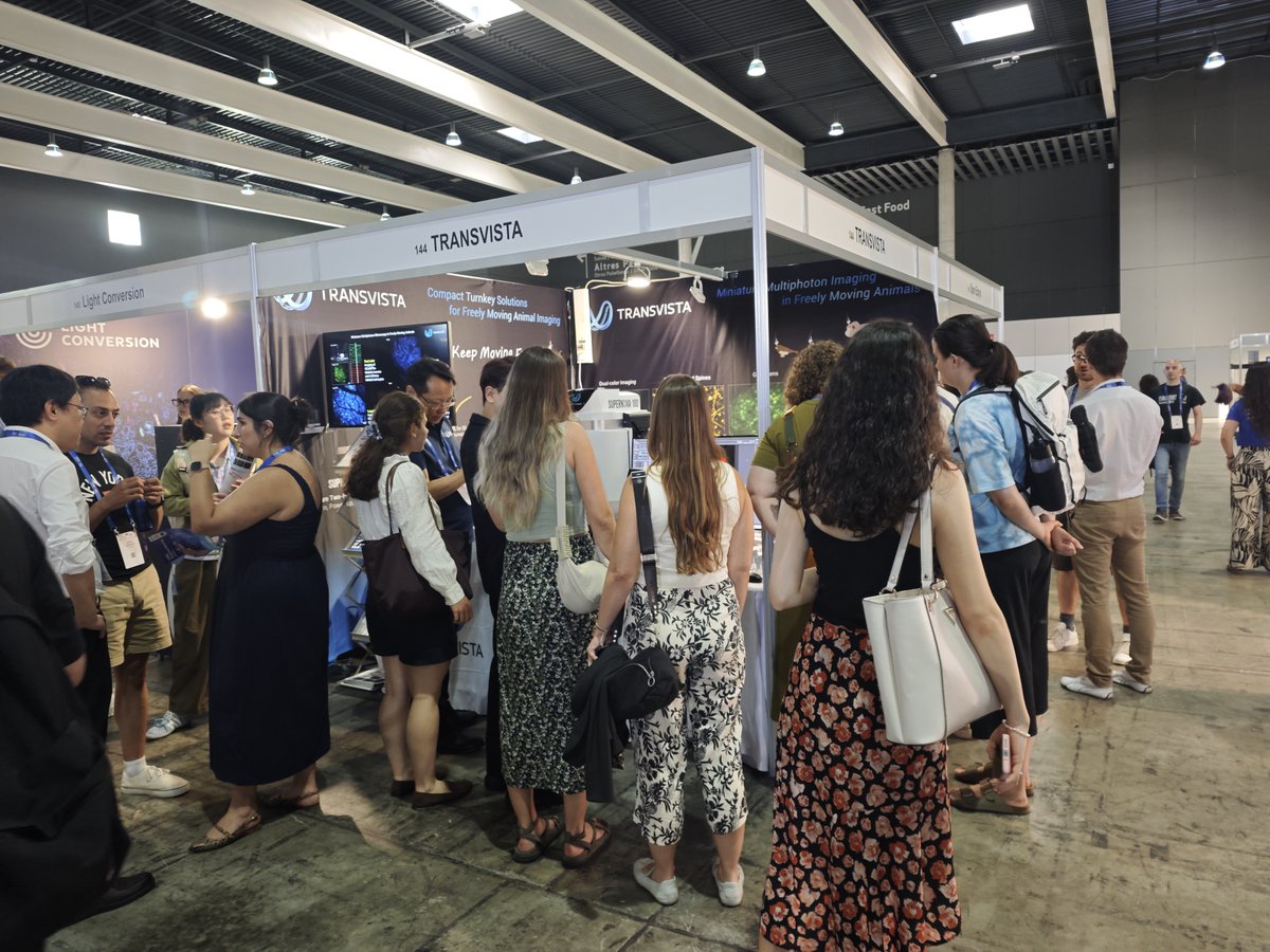
Thank you to everyone who stopped by Booth #144, shared your research, asked thoughtful questions, and exchanged ideas with our team. Every conversation reminds us why we're passionate about developing imaging technologies that help advance neuroscience research.
A special thank you to the @FENSorg organizers for bringing together such a vibrant scientific community, and to everyone who made this week so memorable.
Until we meet again—we look forward to seeing you at the next conference!
Learn more about TRANSVISTA: https://t.co/f491R4DdHz
#FENS2026 #Neuroscience #TwoPhotonMicroscopy #InVivoImaging #MiniatureMicroscopy #BrainResearch
It's the final day of FENS 2026!
Before the meeting comes to a close, we'd love to invite you to our poster presentation this morning:
📄 Poster No. PS07-10AM-008
𝗔 𝗟𝗼𝗻𝗴-𝗪𝗼𝗿𝗸𝗶𝗻𝗴-𝗗𝗶𝘀𝘁𝗮𝗻𝗰𝗲 𝗠𝗶𝗻𝗶𝗮𝘁𝘂𝗿𝗲 𝗧𝘄𝗼-𝗣𝗵𝗼𝘁𝗼𝗻 𝗠𝗶𝗰𝗿𝗼𝘀𝗰𝗼𝗽𝗲 𝗘𝗻𝗮𝗯𝗹𝗲𝘀 𝗖𝗿𝗼𝘀𝘀-𝗦𝗽𝗲𝗰𝗶𝗲𝘀 𝗜𝗺𝗮𝗴𝗶𝗻𝗴 𝗳𝗿𝗼𝗺 𝗥𝗼𝗱𝗲𝗻𝘁𝘀 𝘁𝗼 𝗡𝗼𝗻𝗵𝘂𝗺𝗮𝗻 𝗣𝗿𝗶𝗺𝗮𝘁𝗲𝘀
🗓️ Today | 11:30 AM–1:00 PM
🎤 Presented by Runlong Wu, PhD
This work introduces a long-working-distance miniature two-photon microscope that enables neuronal-resolution imaging across multiple animal models—from freely moving mice to naturally behaving marmosets and head-unrestrained macaques. We're looking forward to discussing the technology, the science behind it, and its potential applications with researchers.
And if you haven't had a chance to visit us yet, there's still time to stop by Booth #144 before FENS wraps up!
Thank you to everyone who has visited our booth over the past few days—we've truly enjoyed the inspiring conversations and the opportunity to connect with the neuroscience community.
See you soon!
#FENS2026 #Neuroscience #TwoPhotonMicroscopy #InVivoImaging #MiniatureMicroscopy #BrainResearch @FENSorg
Thank you for an amazing first day at FENS 2026!
It was wonderful to reconnect with familiar faces and meet many new researchers from around the world. We truly appreciate all the inspiring conversations and the interest in our miniature two-photon imaging technologies.
We'll be back at Booth #144 tomorrow, with another opportunity to Meet the Scientists from 2:30–3:30 PM. If you'd like to discuss your research, explore our technologies, or see a live demo, we'd be delighted to welcome you.
See you on Day 2!
#FENS2026 #Neuroscience #TwoPhotonMicroscopy #InVivoImaging #MiniatureMicroscopy #BrainResearch @FENSorg
See you tomorrow at FENS Forum 2026! 👋
The wait is almost over—we're looking forward to meeting the neuroscience community in Barcelona!
If you're attending FENS, be sure to stop by Booth #144 to explore our latest innovations in miniature two-photon imaging and in vivo imaging technologies. Our team will be on site throughout the meeting to answer questions, demonstrate our technologies, and discuss your research.
𝗔𝘁 𝗼𝘂𝗿 𝗯𝗼𝗼𝘁𝗵, 𝘆𝗼𝘂 𝗰𝗮𝗻 𝗲𝗻𝗷𝗼𝘆:
🔹 Live demos of our latest imaging technologies
🔹 Meet the Scientists sessions (July 8–9 | 2:30–3:30 PM)
🔹 A cute booth giveaway waiting for you! 🐭
In addition, don't miss our poster presentation in the official FENS Poster Area:
📄 𝗔 𝗟𝗼𝗻𝗴-𝗪𝗼𝗿𝗸𝗶𝗻𝗴-𝗗𝗶𝘀𝘁𝗮𝗻𝗰𝗲 𝗠𝗶𝗻𝗶𝗮𝘁𝘂𝗿𝗲 𝗧𝘄𝗼-𝗣𝗵𝗼𝘁𝗼𝗻 𝗠𝗶𝗰𝗿𝗼𝘀𝗰𝗼𝗽𝗲 𝗘𝗻𝗮𝗯𝗹𝗲𝘀 𝗖𝗿𝗼𝘀𝘀-𝗦𝗽𝗲����𝗶𝗲𝘀 𝗜𝗺𝗮𝗴𝗶𝗻𝗴 𝗳𝗿𝗼𝗺 𝗥𝗼𝗱𝗲𝗻𝘁𝘀 𝘁𝗼 𝗡𝗼𝗻𝗵𝘂𝗺𝗮𝗻 𝗣𝗿𝗶𝗺𝗮𝘁𝗲𝘀 (𝗣𝗼𝘀𝘁𝗲𝗿 𝗡𝗼. 𝗣𝗦𝟬𝟳-𝟭𝟬𝗔𝗠-𝟬𝟬𝟴)
📅 July 10 | 11:30 AM–1:00 PM
🎤 Presented by Runlong Wu, PhD
Whether you'd like to explore our technologies at the booth or discuss our latest research at the poster session, we'd love to connect.
📍 Booth #144
📅 FENS Forum 2026 | July 7–10 | Barcelona
See you soon!
#FENS2026 #Neuroscience #TwoPhotonMicroscopy #InVivoImaging #MiniatureMicroscopy #BrainResearch @FENSorg
We're excited to share that the 𝗦𝗨𝗣𝗘𝗥𝗩𝗜𝗦𝗜𝗢𝗡 𝟳𝟴𝟬 will also be on display at our booth during #FENS2026.
The 𝗦𝗨𝗣𝗘𝗥𝗩𝗜𝗦𝗜𝗢𝗡 𝟳𝟴𝟬 is an in vivo two-photon imaging system that enables 𝗻𝗼𝗻-𝗶𝗻𝘃𝗮𝘀𝗶𝘃𝗲, 𝗹𝗮𝗯𝗲𝗹-𝗳𝗿𝗲𝗲, 𝗿𝗲𝗮𝗹-𝘁𝗶𝗺𝗲 imaging of skin at subcellular resolution. It supports research in areas such as skin physiology, determatology diagnosis, and drug evaluation.
If you're interested in expanding in vivo imaging beyond the brain, stop by our booth to learn more about the 𝗦𝗨𝗣𝗘𝗥𝗩𝗜𝗦𝗜𝗢𝗡 𝟳𝟴𝟬 and discuss its research applications with our team.
📍 Booth 144
We look forward to seeing you at FENS 2026!
Learn more about us: https://t.co/f491R4DdHz
#FENS2026 #Neuroscience #InVivoImaging #MultiphotonMicroscopy #SkinImaging #Neurotechnology
We're excited to share that our work will be presented at #FENS2026:
𝗔 𝗟𝗼𝗻𝗴-𝗪𝗼𝗿𝗸𝗶𝗻𝗴-𝗗𝗶𝘀𝘁𝗮𝗻𝗰𝗲 𝗠𝗶𝗻𝗶𝗮𝘁𝘂𝗿𝗲 𝗧𝘄𝗼-𝗣𝗵𝗼𝘁𝗼𝗻 𝗠𝗶𝗰𝗿𝗼𝘀𝗰𝗼𝗽𝗲 𝗘𝗻𝗮𝗯𝗹𝗲𝘀 𝗖𝗿𝗼𝘀𝘀-𝗦𝗽𝗲𝗰𝗶𝗲𝘀 𝗜����𝗮𝗴𝗶𝗻𝗴 𝗳𝗿𝗼𝗺 𝗥𝗼𝗱𝗲𝗻𝘁𝘀 𝘁𝗼 𝗡𝗼𝗻-𝗛𝘂𝗺𝗮𝗻 𝗣𝗿𝗶𝗺𝗮𝘁𝗲𝘀 (𝗔𝗯𝘀𝘁𝗿𝗮𝗰𝘁 𝗡𝗼. 𝟮𝟯𝟯𝟳)
Current miniature two-photon microscopes have significantly advanced in vivo imaging in freely behaving animals, but their limited working distance can restrict applications across species and experimental paradigms.
In this work, we developed a long-working-distance miniature two-photon microscope featuring 𝗶𝗻𝘁𝗲𝗿𝗰𝗵𝗮𝗻𝗴𝗲𝗮𝗯𝗹𝗲 𝗼𝗯𝗷𝗲𝗰𝘁𝗶𝘃𝗲𝘀, enabling 𝗵𝗶𝗴𝗵-𝗿𝗲𝘀𝗼𝗹𝘂𝘁𝗶𝗼𝗻 𝗶𝗺𝗮𝗴𝗶𝗻𝗴 𝗮𝗰𝗿𝗼𝘀𝘀 𝗺𝘂𝗹𝘁𝗶𝗽𝗹𝗲 𝗮𝗻𝗶𝗺𝗮𝗹 𝗺𝗼𝗱𝗲𝗹𝘀—𝗳𝗿𝗼𝗺 𝗳𝗿𝗲𝗲𝗹𝘆 𝗺𝗼𝘃𝗶𝗻𝗴 𝗺𝗶𝗰𝗲 𝗮𝗻𝗱 𝗻𝗮𝘁𝘂𝗿𝗮𝗹𝗹𝘆 𝗯𝗲𝗵𝗮𝘃𝗶𝗻𝗴 𝗺𝗮𝗿𝗺𝗼𝘀𝗲𝘁𝘀 𝘁𝗼 𝗵𝗲𝗮𝗱-𝘂𝗻𝗿𝗲𝘀𝘁𝗿𝗮𝗶𝗻𝗲𝗱 𝗺𝗮𝗰𝗮𝗾𝘂𝗲𝘀.
The system supports deep brain imaging, flexible imaging configurations, and neuronal-resolution recordings across species, providing new opportunities for comparative studies of brain function.
📍 Poster No. PS07-10AM-008
📅 July 10, 2026 | 11:30–13:00
🎤 Runlong Wu, PhD
The ability to perform neuronal-resolution imaging across rodents and non-human primates may help bridge experimental models and enable new comparative studies of brain function.
Learn more about our technology: https://t.co/03rUSW8lVy
#FENS2026 #Neuroscience #TwoPhotonMicroscopy #MiniatureMicroscopy #InVivoImaging #Neurotechnology #NonHumanPrimates @FENSorg
We are proud to share that the groundbreaking work on “multicolor miniature two-photon microscopy”, developed by Prof. Runlong Wu and the team led by Prof. Heping Cheng and Prof. Aimin Wang, has been selected as one of the “2025 China Optics Top 10 Social Impact Events (Light10)” and was honored at Light Conference 2026.
This recognition highlights the growing impact of multicolor miniature two-photon imaging in advancing neuroscience and biomedical research. By enabling high-resolution, multicolor deep brain imaging in freely behaving animals, this technology allows researchers to observe cellular and subcellular dynamics under more physiologically relevant conditions.
This recognition reflects the tremendous potential of miniature multiphoton imaging technologies to transform neuroscience research. At TRANSVISTA, we are committed to translating these innovations into practical tools that help researchers explore neural activity with unprecedented freedom, resolution, and depth.
Know more about us: https://t.co/f491R4CFS1
#LightConference2026 #Light10 #Neuroscience #BiomedicalImaging #TwoPhotonMicroscopy #BrainResearch #FreelyMoving
🎉 A significant milestone for dermatological imaging in China.
The first Chinese academic atlas dedicated to the clinical application of two-photon microscopy in dermatology, 𝗔𝘁𝗹𝗮𝘀 𝗼𝗳 𝗧𝘄𝗼-𝗣𝗵𝗼𝘁𝗼𝗻 𝗠𝗶𝗰𝗿𝗼𝘀𝗰𝗼𝗽𝘆 𝗳𝗼𝗿 𝗦𝗸𝗶𝗻 𝗗𝗶𝘀𝗲𝗮𝘀𝗲𝘀, was officially released at the 31st Annual Meeting of the Chinese Society of Dermatology.
Edited by Prof. Yong Cui and academically reviewed by Prof. Rusong Meng, the atlas brings together multicenter clinical experience and provides a comprehensive reference for the use of 𝘁𝘄𝗼-𝗽𝗵𝗼𝘁𝗼𝗻 𝗺𝗶𝗰𝗿𝗼𝘀𝗰𝗼𝗽𝘆 𝗶𝗻 𝗻𝗼𝗻-𝗶𝗻𝘃𝗮𝘀𝗶𝘃𝗲 𝘀𝗸𝗶𝗻 𝗶𝗺𝗮𝗴𝗶𝗻𝗴. Covering 𝟯𝟰 𝗰𝗼𝗺𝗺𝗼𝗻 𝗱𝗲𝗿𝗺𝗮𝘁𝗼𝗹𝗼𝗴𝗶𝗰𝗮𝗹 𝗰𝗼𝗻𝗱𝗶𝘁𝗶𝗼𝗻𝘀 𝗮𝗻𝗱 𝟳𝟮 𝗶𝗻 𝘃𝗶𝘃𝗼 𝗶𝗺𝗮𝗴𝗶𝗻𝗴 𝗰𝗮𝘀𝗲𝘀, it highlights a new diagnostic paradigm that integrates morphology, tissue composition, and functional information.
At TRANSVISTA, we are proud to support the advancement of this field through our 𝗦𝗨𝗣𝗘𝗥𝗩𝗜𝗦𝗜𝗢𝗡 𝟳𝟴𝟬 𝘁𝘄𝗼-𝗽𝗵𝗼𝘁𝗼𝗻 𝘀𝗸𝗶𝗻 𝗶𝗺𝗮𝗴𝗶𝗻𝗴 𝗽𝗹𝗮𝘁𝗳𝗼𝗿𝗺, helping researchers and clinicians explore 𝗵𝗶𝗴𝗵-𝗿𝗲𝘀𝗼𝗹𝘂𝘁𝗶𝗼𝗻, 𝗹𝗮𝗯𝗲𝗹-𝗳𝗿𝗲𝗲, 𝗶𝗻 𝘃𝗶𝘃𝗼 𝘀𝗸𝗶𝗻 𝗶𝗺𝗮𝗴𝗶𝗻𝗴 and accelerate the translation of cutting-edge optical technologies into clinical practice.
Congratulations to all authors, editors, and contributors on this landmark publication. We look forward to seeing two-photon imaging continue to expand its impact in dermatology and beyond.
Know more about SUPERVISION 780: https://t.co/a0UZQdK2gA
News source: Umer Technology
#TwoPhotonMicroscopy #Dermatology #MedicalImaging #SkinImaging #BiomedicalImaging #SUPERVISION780 #TRANSVISTA
Sleep disturbances are common in chronic stress, depression, and neurodegenerative disorders; however, the neural mechanisms linking stress to disrupted sleep remain poorly understood. A groundbreaking study published in the journal Neuron investigated the effects of chronic stress on sleep.
Dr. Hailong Dong, Dr. Guangchao Zhao and their team demonstrated that chronic stress increases the excitability of ventral tegmental area (VTA) dopamine neurons, leading to excessive dopamine release in the mPFC, which subsequently disrupts sleep spindle generation and stabilizes non-rapid eye movement (NREM) sleep.
𝗞𝗲𝘆 𝗙𝗶𝗻𝗱𝗶𝗻𝗴𝘀:
1️⃣ Fragmented Sleep: Chronic stress increased total NREM sleep duration but resulted in fragmented sleep and frequent transitions between sleep and wakefulness, resembling hypersomnia and poor sleep quality.
2️⃣ Impaired Sleep Spindles: Sleep spindles, which are essential for sleep stability and memory, showed reduced density and duration, disrupted morphology, and decreased coupling with slow-wave activity, indicating a significant impact on deep NREM sleep.
3️⃣ VTA Dopamine Neuron Hyperactivity: Chronic stress led to increased activity of VTA dopamine neurons, which was linked to disrupted sleep spindle continuity and increased sleep fragmentation.
4️⃣ Excessive Dopamine Release: Chronic stress raised dopamine levels in the medial prefrontal cortex (mPFC), affecting glutamatergic neurons. Activating the VTA to mPFC pathway replicated sleep issues, while inhibiting it improved sleep structure.
5️⃣ Therapeutic Strategies: Combining D1 and D2 receptor blockade showed significant recovery from sleep fragmentation. Inhibiting stress-induced HCN channels in VTA dopamine neurons restored sleep-related abnormalities.
𝗔𝗽𝗽𝗹𝗶𝗰𝗮𝘁𝗶𝗼𝗻 𝗼𝗳 𝗠𝗶𝗻𝗶𝗮𝘁𝘂𝗿𝗲 𝗧𝘄𝗼-𝗣𝗵𝗼𝘁𝗼𝗻 𝗜𝗺𝗮𝗴𝗶𝗻𝗴:
One of the most innovative aspects of the study was 𝘁𝗵𝗲 𝘂𝘀𝗲 𝗼𝗳 𝗳𝗮𝘀𝘁, 𝗵𝗶𝗴𝗵-𝗿𝗲𝘀𝗼𝗹𝘂𝘁𝗶𝗼𝗻 𝗺𝗶𝗻𝗶𝗮𝘁𝘂𝗿𝗲 𝘁𝘄𝗼-𝗽𝗵𝗼𝘁𝗼𝗻 𝗺𝗶𝗰𝗿𝗼𝘀𝗰𝗼𝗽𝘆 (𝗙𝗛𝗜𝗥𝗠-𝗧𝗣𝗠) 𝗶𝗻 𝗳𝗿𝗲𝗲𝗹𝘆 𝗺𝗼𝘃𝗶𝗻𝗴 𝗺𝗶𝗰𝗲. This technique enabled researchers to monitor dopamine dynamics in the mPFC using a GRIN lens while simultaneously recording EEG and EMG signals during natural sleep. Through this combined approach, the researchers demonstrated that elevated dopamine pulses disrupt spindle continuity after CSDS.
Article DOI: 10.1016/j.neuron.2026.04.037
Know more about us: https://t.co/f491R4DdHz
#Neuroscience #TwoPhoton #InVivoImaging #BrainImaging #FreelyMoving