De vez en cuando pónganse en el lugar del otro y entiendan que lo que para ustedes no tiene ninguna importancia al otro le puede doler un montón y eso no significa que esté exagerando... 🙌
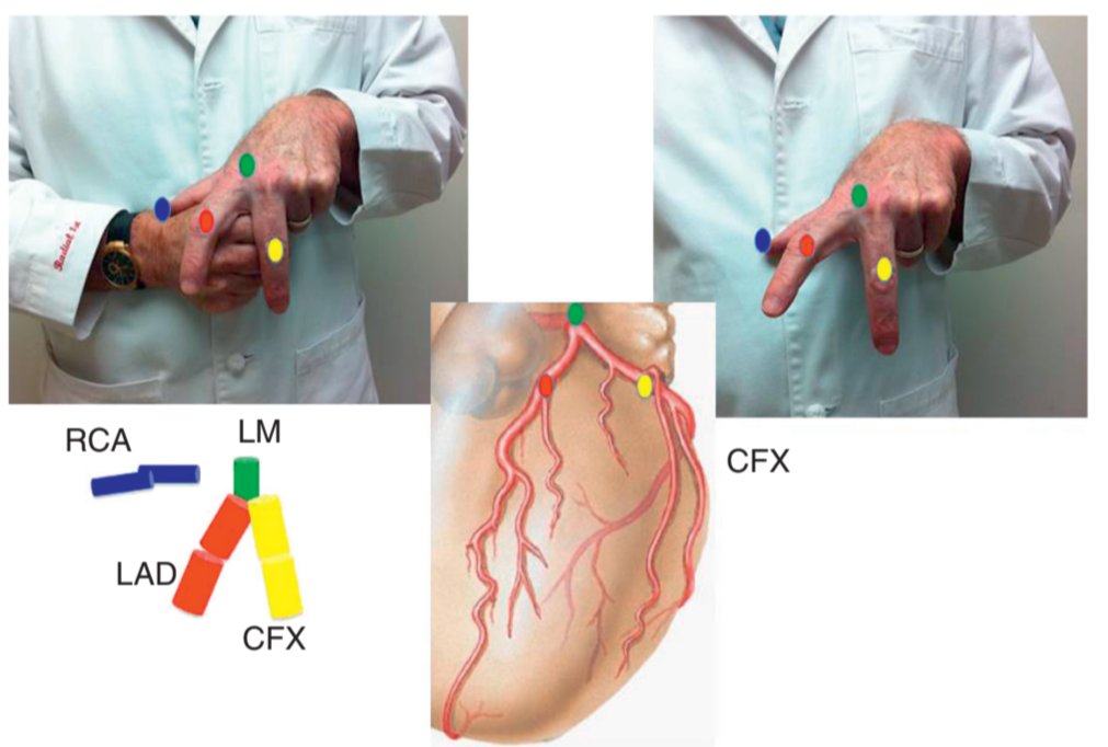
LEARN THE CORONARY ARTERIES WITH YOUR HAND
A simple fist and fingers model can make coronary anatomy much easier to remember.
Think of the closed fist as the heart:
🟥 LAD: index finger, running down the anterior interventricular groove
🟨 LCx: middle finger, wrapping around the left side in the AV groove
🟦 RCA: thumb, beginning superiorly and continuing in the AV groove
🟩 LM: short segment before dividing into the LAD and LCx
The PDA arises distally and runs in the posterior interventricular groove. Its origin depends on coronary dominance.
This simple 3D mental model helps connect coronary anatomy with angiographic projections before you even enter the cath lab.
Reference: Kern M. Cath Lab Digest. 2011;19(8).
Echo From Scratch | Post #23
PSAX at the Aortic Valve Level
The parasternal short-axis view at the aortic valve level is one of the most useful views for understanding the relationship between the four cardiac valves.
In this plane, the aortic valve sits centrally and should show its characteristic Y-shaped configuration in diastole.
What should you identify?
1️⃣ Aortic valve and all 3 cusps
2️⃣ RVOT
3️⃣ Pulmonary artery
4️⃣ Right atrium
5️⃣ Left atrium
6️⃣ Tricuspid valve
7️⃣ Pulmonary valve
⭕Aortic valve
In diastole, the three cusps meet in the classic Y-shaped pattern:
R = right coronary cusp
L = left coronary cusp
N = noncoronary cusp
In systole, the cusps open and the central orifice becomes much more circular.
ECG From Scratch #33: Left Atrial Abnormality
If right atrial abnormality tends to make the P wave taller, left atrial abnormality tends to make it wider.
(Why)
Because the left atrium normally depolarizes after the right atrium. Delayed or prolonged left atrial activation can therefore increase the total duration of atrial depolarization.
The classic ECG clue
P-wave duration ≥0.12 s (120 ms)
That is at least 3 small boxes.
The P wave may also become:
Broad and notched or humped
The second hump reflects delayed left atrial activation.
This pattern was historically called P mitrale, particularly because it was associated with mitral valve disease. The broader term left atrial abnormality (LAA) is preferable because a broad P wave does not necessarily prove anatomical left atrial enlargement.
Look at V1
🔵Lead V1 can provide another important clue.
A normal P wave in V1 is often biphasic:
small positive component ➡️ negative component
With LAA, the terminal negative component may become:
≥1 mm deep
and/or
>40 ms wide
Why?
The right atrium lies more anteriorly, while the left atrium is positioned posteriorly.
So the initial positive component reflects right atrial activation, while the prominent terminal negative component reflects delayed left atrial activation directed posteriorly, away from V1.
Where can LAA appear?
Common settings include:
- Mitral stenosis or regurgitation
- Aortic stenosis or regurgitation
- Hypertensive heart disease
- Dilated, hypertrophic, or restrictive cardiomyopathy
- Coronary artery disease
An important caveat
A broad or notched P wave does not prove anatomical left atrial enlargement.
A patient may have a broad P wave because of delayed atrial conduction even when imaging does not show an enlarged left atrium.