Foot and mouth disease
Here’s a complete visual and structured overview of Foot-and-Mouth Disease (FMD) in cattle, including its etiology, clinical signs, treatment, prevention, and control. I’ve also prepared an educational infographic you can use for teaching or revision.
---
🦠 Etiology
- Cause: Foot-and-Mouth Disease Virus (FMDV), genus Aphthovirus, family Picornaviridae
- Serotypes: Seven (A, O, C, SAT1, SAT2, SAT3, Asia1) – important because immunity is serotype-specific
- Transmission:
- Direct contact with infected animals (saliva, milk, feces, urine)
- Airborne spread (up to several kilometers)
- Contaminated feed, water, equipment, clothing, vehicles
- Survives for long periods in the environment, making control difficult
---
🐄 Clinical Signs in Cattle
- Incubation period: 2–14 days
- Key signs:
- Fever, depression, anorexia
- Excessive salivation, drooling
- Vesicles/blisters in mouth, tongue, lips, dental pad → rupture → painful ulcers
- Lesions on feet → lameness, reluctance to move
- Vesicles on teats → mastitis, reduced milk yield
- Sudden drop in productivity (milk, weight gain)
---
💉 Treatment
- No specific antiviral treatment (supportive care only)
- Measures:
- Pain relief and anti-inflammatories
- Antibiotics to prevent secondary bacterial infections
- Soft feed and good nursing care
- Note: Recovery is possible, but animals remain debilitated for weeks.
---
🛡️ Prevention & Control
- Vaccination: Inactivated FMD vaccines available; must match circulating serotype
- Biosecurity: Restrict movement of animals, vehicles, and people
- Quarantine: Isolate infected herds immediately
- Disinfection: Clean and disinfect equipment, housing, and clothing
- Surveillance: Laboratory confirmation (RT-PCR, ELISA) essential for outbreak management
- Policy: In Ethiopia, FMD is a major transboundary disease, requiring national and regional coordination
#FMD #veterinary
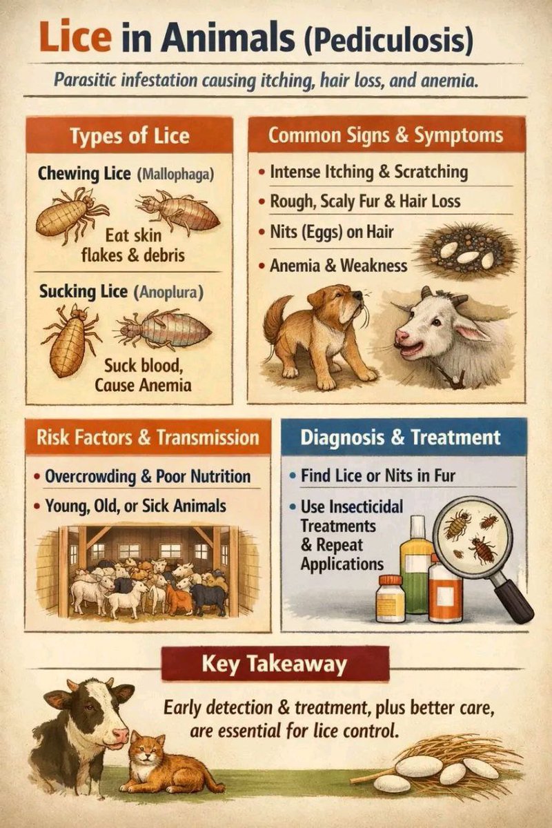
Lice in animals are small, wingless, host-specific ectoparasites that cause intense itching (pruritus), hair loss (alopecia), and skin irritation, primarily affecting animals in close contact. They include sucking lice (blood-feeders) and biting lice (skin debris-feeders), which thrive in winter and can cause anemia, poor coat condition, and reduced production in livestock.
Key Aspects of Animal Lice:
Types: Sucking lice (Anoplura, mammals only) and Chewing/Biting lice (Mallophaga, birds and mammals).
Signs: Intense itching, hair loss, restlessness, scabby/thickened skin, and weight loss or low milk yield.
Transmission: Spread through direct contact between animals or contaminated bedding/grooming tools.
Seasonality: Infestations often increase during winter months.
Treatment: Insecticides, sprays, shampoos, and pour-on products (e.g., fipronil, selamectin, pyrethroids).
Host Specificity: Lice from one animal species (e.g., dog lice) rarely infest humans or other animal species.
Diagnosis and Management:
Lice and their eggs (nits) are visible to the naked eye, often found in matted areas, around ears, neck, and shoulders. Treatment often requires repeating 2–3 weeks later to kill newly hatched nymphs. Improving nutrition, reducing stress, and avoiding overcrowding help prevent outbreaks.
Lice in animals are small, wingless, host-specific ectoparasites that cause intense itching (pruritus), hair loss (alopecia), and skin irritation, primarily affecting animals in close contact. They include sucking lice (blood-feeders) and biting lice (skin debris-feeders), which thrive in winter and can cause anemia, poor coat condition, and reduced production in livestock.
Key Aspects of Animal Lice:
Types: Sucking lice (Anoplura, mammals only) and Chewing/Biting lice (Mallophaga, birds and mammals).
Signs: Intense itching, hair loss, restlessness, scabby/thickened skin, and weight loss or low milk yield.
Transmission: Spread through direct contact between animals or contaminated bedding/grooming tools.
Seasonality: Infestations often increase during winter months.
Treatment: Insecticides, sprays, shampoos, and pour-on products (e.g., fipronil, selamectin, pyrethroids).
Host Specificity: Lice from one animal species (e.g., dog lice) rarely infest humans or other animal species.
Diagnosis and Management:
Lice and their eggs (nits) are visible to the naked eye, often found in matted areas, around ears, neck, and shoulders. Treatment often requires repeating 2–3 weeks later to kill newly hatched nymphs. Improving nutrition, reducing stress, and avoiding overcrowding help prevent outbreaks.
Lice in animals are small, wingless, host-specific ectoparasites that cause intense itching (pruritus), hair loss (alopecia), and skin irritation, primarily affecting animals in close contact. They include sucking lice (blood-feeders) and biting lice (skin debris-feeders), which thrive in winter and can cause anemia, poor coat condition, and reduced production in livestock.
Key Aspects of Animal Lice:
Types: Sucking lice (Anoplura, mammals only) and Chewing/Biting lice (Mallophaga, birds and mammals).
Signs: Intense itching, hair loss, restlessness, scabby/thickened skin, and weight loss or low milk yield.
Transmission: Spread through direct contact between animals or contaminated bedding/grooming tools.
Seasonality: Infestations often increase during winter months.
Treatment: Insecticides, sprays, shampoos, and pour-on products (e.g., fipronil, selamectin, pyrethroids).
Host Specificity: Lice from one animal species (e.g., dog lice) rarely infest humans or other animal species.
Diagnosis and Management:
Lice and their eggs (nits) are visible to the naked eye, often found in matted areas, around ears, neck, and shoulders. Treatment often requires repeating 2–3 weeks later to kill newly hatched nymphs. Improving nutrition, reducing stress, and avoiding overcrowding help prevent outbreaks.
Lice in animals are small, wingless, host-specific ectoparasites that cause intense itching (pruritus), hair loss (alopecia), and skin irritation, primarily affecting animals in close contact. They include sucking lice (blood-feeders) and biting lice (skin debris-feeders), which thrive in winter and can cause anemia, poor coat condition, and reduced production in livestock.
Key Aspects of Animal Lice:
Types: Sucking lice (Anoplura, mammals only) and Chewing/Biting lice (Mallophaga, birds and mammals).
Signs: Intense itching, hair loss, restlessness, scabby/thickened skin, and weight loss or low milk yield.
Transmission: Spread through direct contact between animals or contaminated bedding/grooming tools.
Seasonality: Infestations often increase during winter months.
Treatment: Insecticides, sprays, shampoos, and pour-on products (e.g., fipronil, selamectin, pyrethroids).
Host Specificity: Lice from one animal species (e.g., dog lice) rarely infest humans or other animal species.
Diagnosis and Management:
Lice and their eggs (nits) are visible to the naked eye, often found in matted areas, around ears, neck, and shoulders. Treatment often requires repeating 2–3 weeks later to kill newly hatched nymphs. Improving nutrition, reducing stress, and avoiding overcrowding help prevent outbreaks.
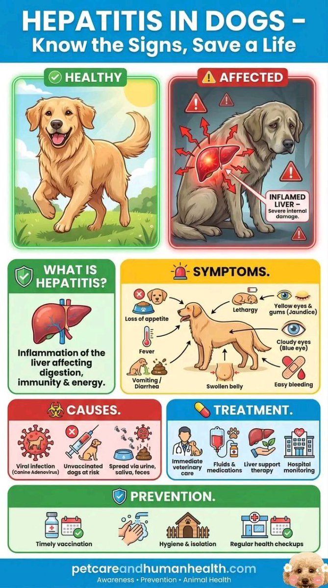
Hepatitis in dogs
Types of Hepatitis in Dogs
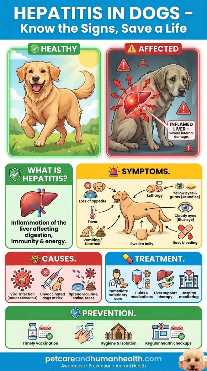
There are two types of hepatitis commonly seen in dogs, infectious canine hepatitis and canine chronic hepatitis. Chronic is defined as an infection that has been causing damage for some time (at least a few weeks), whereas acute hepatitis can manifest over only a few days.
Infectious Canine Hepatitis
Infectious canine hepatitis is an acute contagious disease in dogs caused by the canine adenovirus 1. The virus targets the infected pet's liver, lungs, kidneys, spleen, lining of blood vessels and occasionally other organs. Signs of infectious canine hepatitis can vary widely from slight fever to death.
Canine Chronic Hepatitis
Canine chronic hepatitis is a condition that is associated with infectious canine hepatitis. Breeds of dogs that are predisposed to the disease include Skye Terriers, Springer Spaniels, Beagles, West Highland White Terriers, Cocker Spaniels, Maltese, Labrador Retrievers, Doberman Pinschers, Bedlington Terriers, Standard Poodles and Chihuahuas.
Symptoms
Infectious canine hepatitis can cause a variety of signs and symptoms. If your dog is suffering from infectious hepatitis you may notice one or more of the following symptoms:
Watery discharge from eyes and nose
Congestion
Slight fever
Deficiency of blood clotting
Blindness
Loss of appetite
Thirst
Enlarged tonsils
Yellow, jaundiced look to ears, gums and skin
Eye inflammation
Severe depression
Abdominal pain (occasional)
Vomiting (occasional)
Bruised or reddened nose and mouth
Spontaneous bleeding
Severe cases:
Red dots on skin
Bruised or reddened nose and mouth
Swelling (neck, head, lymph nodes)
Seizures
Death
Treatment
Depending on the results of the biopsy, your vet may recommend treating the disease with a broad-spectrum antibiotic, anti-inflammatory or immunosuppressive medication.
Sometimes, a painful spasm can be associated with cornea clouding in the eye. Your vet can prescribe eye ointment to alleviate your pup's pain. If your dog is experiencing corneal clouding, it is essential to protect your pup's eye from bright light.
Treatment options can range from intravenous fluid therapy to hospitalization. Your dog will require blood work on a regular basis for monitoring purposes.
Preventing Hepatitis in Dogs
A mandatory vaccine is the most widely used and important preventive measure for infectious canine hepatitis. Your dog will typically receive this vaccine along with their canine distemper vaccinations.
The hepatitis vaccine is typically given to puppies at about 7 to 9 weeks of age, with the first booster between 11 and 13 weeks, after which they’ll be protected.
To remain protected against this serious condition, your dog will need to keep receiving the booster shots throughout their life - with another one at 15 months, then each year to keep the infection away.
#veterinary #veterinarymedicine #dogsofinstagram #doglover #dog #doglife #HepatitisAwareness #HepatitisB
Hepatitis in dogs
Types of Hepatitis in Dogs
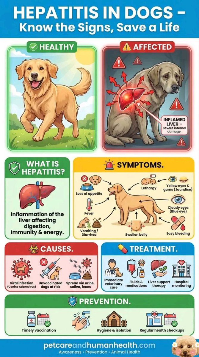
There are two types of hepatitis commonly seen in dogs, infectious canine hepatitis and canine chronic hepatitis. Chronic is defined as an infection that has been causing damage for some time (at least a few weeks), whereas acute hepatitis can manifest over only a few days.
Infectious Canine Hepatitis
Infectious canine hepatitis is an acute contagious disease in dogs caused by the canine adenovirus 1. The virus targets the infected pet's liver, lungs, kidneys, spleen, lining of blood vessels and occasionally other organs. Signs of infectious canine hepatitis can vary widely from slight fever to death.
Canine Chronic Hepatitis
Canine chronic hepatitis is a condition that is associated with infectious canine hepatitis. Breeds of dogs that are predisposed to the disease include Skye Terriers, Springer Spaniels, Beagles, West Highland White Terriers, Cocker Spaniels, Maltese, Labrador Retrievers, Doberman Pinschers, Bedlington Terriers, Standard Poodles and Chihuahuas.
Symptoms
Infectious canine hepatitis can cause a variety of signs and symptoms. If your dog is suffering from infectious hepatitis you may notice one or more of the following symptoms:
Watery discharge from eyes and nose
Congestion
Slight fever
Deficiency of blood clotting
Blindness
Loss of appetite
Thirst
Enlarged tonsils
Yellow, jaundiced look to ears, gums and skin
Eye inflammation
Severe depression
Abdominal pain (occasional)
Vomiting (occasional)
Bruised or reddened nose and mouth
Spontaneous bleeding
Severe cases:
Red dots on skin
Bruised or reddened nose and mouth
Swelling (neck, head, lymph nodes)
Seizures
Death
Treatment
Depending on the results of the biopsy, your vet may recommend treating the disease with a broad-spectrum antibiotic, anti-inflammatory or immunosuppressive medication.
Sometimes, a painful spasm can be associated with cornea clouding in the eye. Your vet can prescribe eye ointment to alleviate your pup's pain. If your dog is experiencing corneal clouding, it is essential to protect your pup's eye from bright light.
Treatment options can range from intravenous fluid therapy to hospitalization. Your dog will require blood work on a regular basis for monitoring purposes.
Preventing Hepatitis in Dogs
A mandatory vaccine is the most widely used and important preventive measure for infectious canine hepatitis. Your dog will typically receive this vaccine along with their canine distemper vaccinations.
The hepatitis vaccine is typically given to puppies at about 7 to 9 weeks of age, with the first booster between 11 and 13 weeks, after which they’ll be protected.
To remain protected against this serious condition, your dog will need to keep receiving the booster shots throughout their life - with another one at 15 months, then each year to keep the infection away.
#veterinary #veterinarymedicine #dogsofinstagram #doglover #dog #doglife #HepatitisAwareness #HepatitisB
Hepatitis in dogs
Types of Hepatitis in Dogs
There are two types of hepatitis commonly seen in dogs, infectious canine hepatitis and canine chronic hepatitis. Chronic is defined as an infection that has been causing damage for some time (at least a few weeks), whereas acute hepatitis can manifest over only a few days.
Infectious Canine Hepatitis
Infectious canine hepatitis is an acute contagious disease in dogs caused by the canine adenovirus 1. The virus targets the infected pet's liver, lungs, kidneys, spleen, lining of blood vessels and occasionally other organs. Signs of infectious canine hepatitis can vary widely from slight fever to death.
Canine Chronic Hepatitis
Canine chronic hepatitis is a condition that is associated with infectious canine hepatitis. Breeds of dogs that are predisposed to the disease include Skye Terriers, Springer Spaniels, Beagles, West Highland White Terriers, Cocker Spaniels, Maltese, Labrador Retrievers, Doberman Pinschers, Bedlington Terriers, Standard Poodles and Chihuahuas.
Symptoms
Infectious canine hepatitis can cause a variety of signs and symptoms. If your dog is suffering from infectious hepatitis you may notice one or more of the following symptoms:
Watery discharge from eyes and nose
Congestion
Slight fever
Deficiency of blood clotting
Blindness
Loss of appetite
Thirst
Enlarged tonsils
Yellow, jaundiced look to ears, gums and skin
Eye inflammation
Severe depression
Abdominal pain (occasional)
Vomiting (occasional)
Bruised or reddened nose and mouth
Spontaneous bleeding
Severe cases:
Red dots on skin
Bruised or reddened nose and mouth
Swelling (neck, head, lymph nodes)
Seizures
Death
Treatment
Depending on the results of the biopsy, your vet may recommend treating the disease with a broad-spectrum antibiotic, anti-inflammatory or immunosuppressive medication.
Sometimes, a painful spasm can be associated with cornea clouding in the eye. Your vet can prescribe eye ointment to alleviate your pup's pain. If your dog is experiencing corneal clouding, it is essential to protect your pup's eye from bright light.
Treatment options can range from intravenous fluid therapy to hospitalization. Your dog will require blood work on a regular basis for monitoring purposes.
Preventing Hepatitis in Dogs
A mandatory vaccine is the most widely used and important preventive measure for infectious canine hepatitis. Your dog will typically receive this vaccine along with their canine distemper vaccinations.
The hepatitis vaccine is typically given to puppies at about 7 to 9 weeks of age, with the first booster between 11 and 13 weeks, after which they’ll be protected.
To remain protected against this serious condition, your dog will need to keep receiving the booster shots throughout their life - with another one at 15 months, then each year to keep the infection away.
#veterinary #veterinarymedicine #dogsofinstagram #doglover #dog #doglife #HepatitisAwareness #HepatitisB
Hepatitis in dogs
Types of Hepatitis in Dogs
There are two types of hepatitis commonly seen in dogs, infectious canine hepatitis and canine chronic hepatitis. Chronic is defined as an infection that has been causing damage for some time (at least a few weeks), whereas acute hepatitis can manifest over only a few days.
Infectious Canine Hepatitis
Infectious canine hepatitis is an acute contagious disease in dogs caused by the canine adenovirus 1. The virus targets the infected pet's liver, lungs, kidneys, spleen, lining of blood vessels and occasionally other organs. Signs of infectious canine hepatitis can vary widely from slight fever to death.
Canine Chronic Hepatitis
Canine chronic hepatitis is a condition that is associated with infectious canine hepatitis. Breeds of dogs that are predisposed to the disease include Skye Terriers, Springer Spaniels, Beagles, West Highland White Terriers, Cocker Spaniels, Maltese, Labrador Retrievers, Doberman Pinschers, Bedlington Terriers, Standard Poodles and Chihuahuas.
Symptoms
Infectious canine hepatitis can cause a variety of signs and symptoms. If your dog is suffering from infectious hepatitis you may notice one or more of the following symptoms:
Watery discharge from eyes and nose
Congestion
Slight fever
Deficiency of blood clotting
Blindness
Loss of appetite
Thirst
Enlarged tonsils
Yellow, jaundiced look to ears, gums and skin
Eye inflammation
Severe depression
Abdominal pain (occasional)
Vomiting (occasional)
Bruised or reddened nose and mouth
Spontaneous bleeding
Severe cases:
Red dots on skin
Bruised or reddened nose and mouth
Swelling (neck, head, lymph nodes)
Seizures
Death
Treatment
Depending on the results of the biopsy, your vet may recommend treating the disease with a broad-spectrum antibiotic, anti-inflammatory or immunosuppressive medication.
Sometimes, a painful spasm can be associated with cornea clouding in the eye. Your vet can prescribe eye ointment to alleviate your pup's pain. If your dog is experiencing corneal clouding, it is essential to protect your pup's eye from bright light.
Treatment options can range from intravenous fluid therapy to hospitalization. Your dog will require blood work on a regular basis for monitoring purposes.
Preventing Hepatitis in Dogs
A mandatory vaccine is the most widely used and important preventive measure for infectious canine hepatitis. Your dog will typically receive this vaccine along with their canine distemper vaccinations.
The hepatitis vaccine is typically given to puppies at about 7 to 9 weeks of age, with the first booster between 11 and 13 weeks, after which they’ll be protected.
To remain protected against this serious condition, your dog will need to keep receiving the booster shots throughout their life - with another one at 15 months, then each year to keep the infection away.
#veterinary #veterinarymedicine #dogsofinstagram #doglover #dog #doglife #HepatitisAwareness #HepatitisB
Anatomy of blood supply of cow udder
Anatomy of blood supply of cow udder.
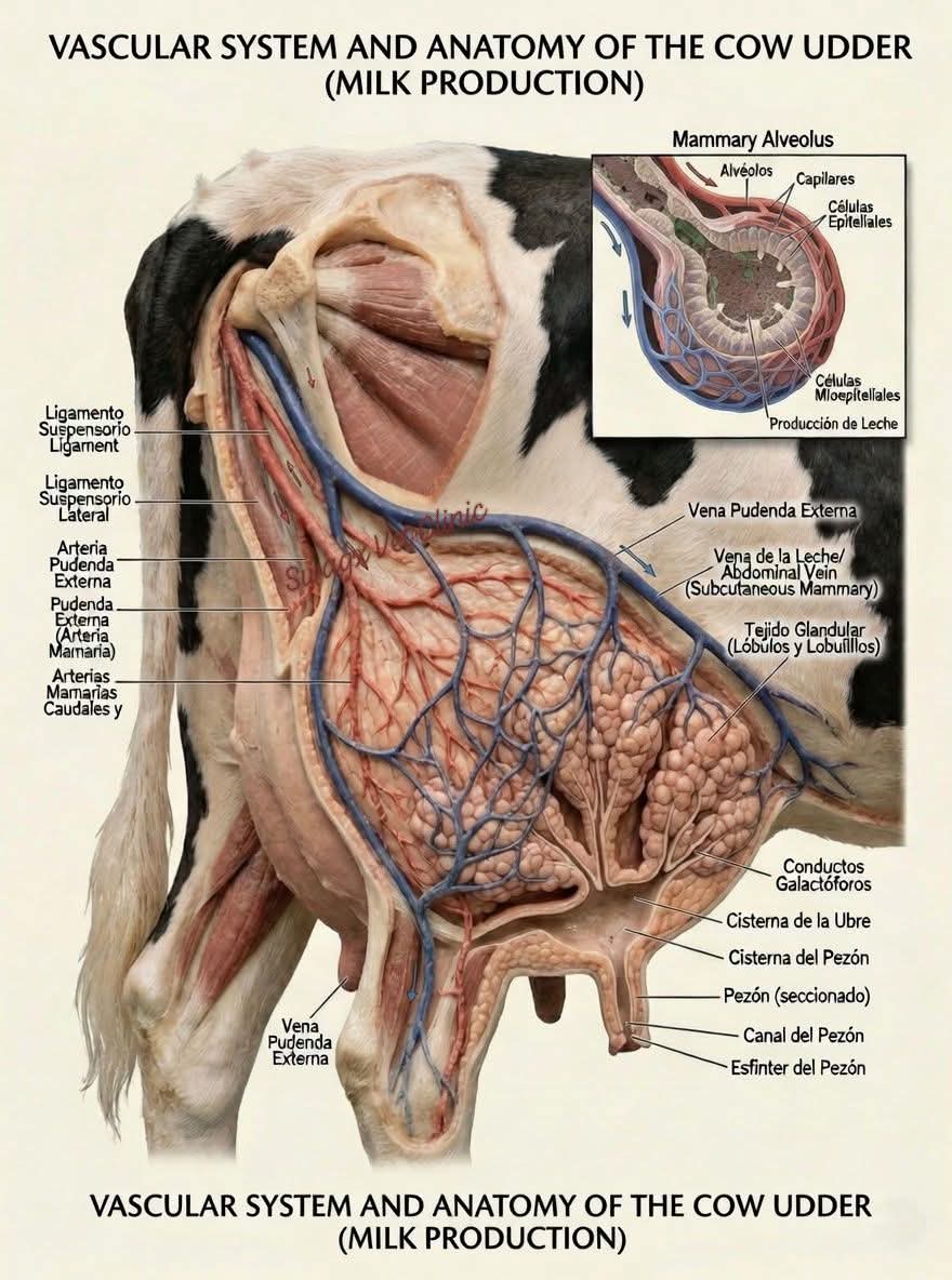
The cow's udder receives a massive blood supply, requiring 400–500 liters of blood flow to produce 1 liter of milk, primarily via the external pudendal arteries (left and right), which branch into cranial and caudal mammary arteries. These arteries enter via the inguinal canal, forming sigmoid flexures for flexibility.
Arterial Supply
External Pudendal Artery: The main supply, originating from the abdominal aorta via the external iliac artery. It passes through the inguinal canal and splits into cranial and caudal mammary arteries, forming a, sigmoid flexure which allows for expansion.
Perineal Artery: Provides a minor contribution to the posterior dorsal part of the udder.
Capillary Beds: Extensive networks surround each alveolus for nutrient exchange.
Venous Drainage
Subcutaneous Abdominal Vein ("Milk Vein"): Responsible for carrying approximately 90% of the blood away from the udder, running along the ventral abdomen.
External Pudendal Vein: Drains blood back through the inguinal canal.
Perineal Vein: Drains the posterior part of the udder.
Venous Ring: A base structure where all veins contribute, located at the top of the udder.
Functional Anatomy
Independence: Each half (left and right) of the udder has its own separate vascular system, though small connections exist.
Adaptation: Sigmoid flexures in the main vessels allow them to stretch as the udder fills.
Blood-Milk Barrier: Nutrients are transferred from capillaries to alveoli, where they are transformed into milk.
Reproductive cycle of cattle
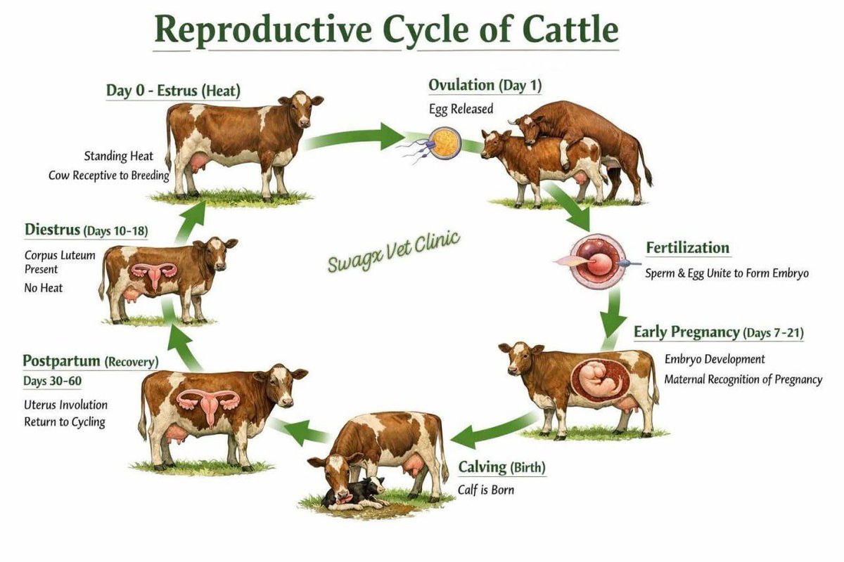
The cow's reproductive cycle (estrous cycle) lasts an average of 21 days (range 18–24) and is a continuous, polyestrous process of four stages—proestrus, estrus (heat), metestrus, and diestrus. Key hormonal drivers are estrogen (driving heat behavior) and progesterone (maintaining pregnancy), aimed at preparing the uterus and maturing a follicle for ovulation.
NADIS +4
Key Stages of the 21-Day Cycle
Proestrus (Days 17–21): The Corpus Luteum (CL) regresses, progesterone drops, and a new dominant follicle grows rapidly, producing high estrogen.
Estrus (Day 0/Heat): High estrogen levels cause standing heat (behavioral signs, restlessness, standing to be mounted). Ovulation occurs ~12–30 hours after the start of standing heat.
Metestrus (Days 1–5): Ovulation occurs and the Corpus Luteum (CL) (CL) begins to form at the site of the ruptured follicle.
Diestrus (Days 5–17): The CL is fully functional, producing high levels of progesterone to prepare the uterus for pregnancy. If no embryo is present, the uterus releases prostaglandin
, causing the CL to regress and starting a new cycle.
Key Reproductive Features
Polyestrous: Cows cycle continuously year-round until pregnant.
Follicular Waves: 2 to 3 waves of follicles grow during each cycle.
Pregnancy: If fertilized, the embryo prevents CL regression, maintaining high progesterone.
Puberty: Occurs between 10–12 months of age.
Reproductive cycle of cattle
The cow's reproductive cycle (estrous cycle) lasts an average of 21 days (range 18–24) and is a continuous, polyestrous process of four stages—proestrus, estrus (heat), metestrus, and diestrus. Key hormonal drivers are estrogen (driving heat behavior) and progesterone (maintaining pregnancy), aimed at preparing the uterus and maturing a follicle for ovulation.
NADIS +4
Key Stages of the 21-Day Cycle
Proestrus (Days 17–21): The Corpus Luteum (CL) regresses, progesterone drops, and a new dominant follicle grows rapidly, producing high estrogen.
Estrus (Day 0/Heat): High estrogen levels cause standing heat (behavioral signs, restlessness, standing to be mounted). Ovulation occurs ~12–30 hours after the start of standing heat.
Metestrus (Days 1–5): Ovulation occurs and the Corpus Luteum (CL) (CL) begins to form at the site of the ruptured follicle.
Diestrus (Days 5–17): The CL is fully functional, producing high levels of progesterone to prepare the uterus for pregnancy. If no embryo is present, the uterus releases prostaglandin
, causing the CL to regress and starting a new cycle.
Key Reproductive Features
Polyestrous: Cows cycle continuously year-round until pregnant.
Follicular Waves: 2 to 3 waves of follicles grow during each cycle.
Pregnancy: If fertilized, the embryo prevents CL regression, maintaining high progesterone.
Puberty: Occurs between 10–12 months of age.