Uzun yıllardır sürdürdüğümüz çabalarımız nihayet karşılık buldu. Süresiz nafaka uygulaması, Anayasa Mahkemesi tarafından iptal edildi.
Şimdi sıra; aileyi güçlendiren, kadın ve erkeği güvence altına alan yeni bir düzenlemede.
İnternette "bulunamayan" kitap yoktur, sadece yanlış arama yöntemi vardır.
📌 Kaydedin, lazım olur.
Aradığınız herhangi bir kitabı, makaleyi veya kaynağı saniyeler içinde doğrudan cihazınıza indirmek için bu basit formülü kullanabilirsiniz:
❌ Eski Yöntem: Kitap Adı + "oku" veya "indir" yazıp reklam dolu sitelerde kaybolmak.
✅ Yeni Yöntem: Kitap Adı + "filetype:pdf" veya Kitap Adı + "doctype:pdf"
Örnek Uygulama:
1. Google'a gir.
2. "Babil'in En Zengin Adamı doctype:pdf" yaz.
3. Çıkan sonuçlarda PDF yazan dosya bağlantısına tıkla.
Bu yöntem sadece kitaplar için değil; sunumlar için .ppt, tablolar için .xls formatlarında da hayat kurtarır.
...yani "filetype:ppt" veya "filetype:xls" aratabilirsiniz.
ℹ️ Naçizane tavsiyem kitapları doğrudan satın almak ancak bazen kitapları bulamayabilir veya bütçe ayıramayabiliriz. Bu yöntem dar zamanlar için...
📌 Kaydedin, bir gün lazım olabilir.
🤔 Sizin bildiğiniz ve "oha abi!!! çok iyiymiş" dediğiniz bir life hack var mı? Yorum yazın. 👇
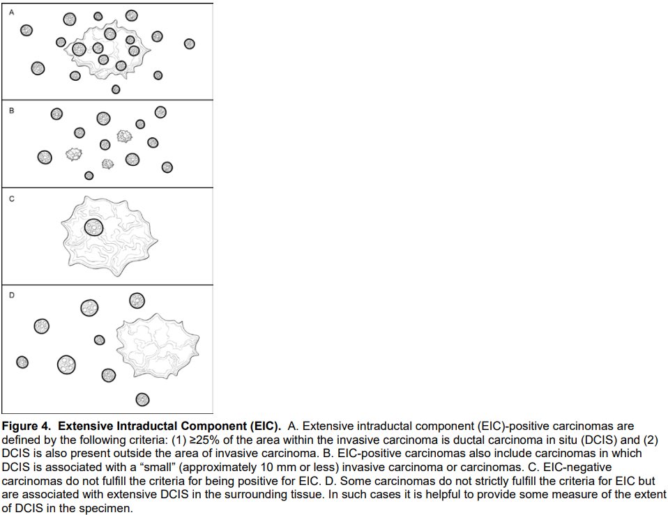
@LaurenERosenMD@Pathologists There are two cases for which you can call EIC. A) DCIS is >=25% of tumor area and present outside (as in example case in original tweet; B) extensive DCIS is associated with pT1a/b invasive carcinoma.
✨Inflammatory Rhabdomyoblastic Tumor (IRMT)
(with emphasis on biological behavior and diagnostic pitfalls) ✨
A rare, newly defined tumor that sits right in the middle between rhabdomyoma and rhabdomyosarcoma – and can be mistaken for ILMS, IMT, or even pleomorphic RMS if you are not paying attention. 👀
📚 Definition
Soft tissue neoplasm with skeletal muscle differentiation, exuberant lymphohistiocytic inflammatory infiltrate, characteristic near‑haploidy, and intermediate malignant potential (borderline, rarely metastasizing).
Clinicopathologic, immunohistochemical, and genetic studies combining cases previously labeled as “inflammatory leiomyosarcoma” (ILMS) and “histiocyte‑rich rhabdomyoblastic tumor” (HRRMT) have shown that they represent a single entity, now proposed under the term IRMT; in the current WHO (2020), however, these tumors ainda constam sob o rótulo de ILMS, e IRMT é a nomenclatura em transição.
📊 Epidemiology
Young to middle‑aged adults (mean ~40–50 years).
Marked male predominance.
Occasional association with neurofibromatosis type 1 (NF1), both germline and somatic.
📍 Sites
Deep soft tissues of thigh and other extremities.
Trunk, retroperitoneum/pelvis, abdominal wall.
Rare cases in head and neck region and hypopharyngeal/submucosal sites of the upper aerodigestive tract.
🧬 Pathogenesis
Typical genomic profile: near‑haploidy with extensive LOH and retained disomy of chromosomes 5/22 (often also 18/20/21), sometimes followed by whole‑genome duplication (pseudodiploidy/hyperdiploidy).
Recurrent NF1 mutations; TP53 often subclonal, potentially marking progression.
During progression to RMS: additional gains/losses involving 9p21 (CDKN2A/CDKN2B) and other oncogene/tumor suppressor loci.
A minor subset of otherwise typical IRMTs does not show the classic near‑haploid pattern; these cases are currently interpreted as genetic variants within the IRMT spectrum, without clear evidence of more aggressive clinical behavior.
🩺 Clinical Features
Slowly growing mass present for months/years, usually painless.
Median size ~4–7 cm, but can exceed 10 cm in long‑standing tumors.
“Pure” IRMT: indolent course; IRMT with progression to RMS: aggressive behavior, with lung/bone/soft tissue metastases in a substantial proportion of patients.
🧪 Laboratory Diagnosis
Diagnosis relies on histopathology + IHC + ideally genomic profiling.
No specific serum biomarkers.
Near‑haploidy/LOH can be demonstrated by SNP‑array, OncoScan, or NGS panels with copy‑number analysis.
🔬 Histopathology
Well‑circumscribed, often encapsulated mass, with:
Fibrous capsule and peripheral lymphoid aggregates.
Spindle to epithelioid tumor cells with “glassy” eosinophilic cytoplasm, lacking cross‑striations.
Hyperchromatic, sometimes bizarre nuclei, but low mitotic activity in typical cases (usually ≤1/10 HPF).
Dense background of histiocytes (including foamy and Touton‑like cells), lymphocytes, and plasma cells, often obscuring the neoplastic cells.
Calcifications, hyalinized vessels, and “old lesion” features consistent with slow growth.
In RMS progression:
First pattern – nodules of small, monotonous rhabdomyoblastic cells with reduced inflammation and low‑to‑moderate mitotic activity.
Second pattern – highly cellular spindle/epithelioid sarcoma with marked atypia, frequent mitoses, and coagulative necrosis, often requiring IHC to recognize skeletal muscle differentiation.
🧫 Immunohistochemistry
Hallmark: skeletal muscle phenotype in a rich lymphohistiocytic background.
Positive:
Diffuse desmin.
Strong PAX7 in most cases (>50% of tumor cells).
MyoD1 and myogenin: positive but usually focal/scant (sometimes negative for one of them, especially myogenin).
CD163 highlights background histiocytes and xanthoma‑cell aggregates.
Variable:
SMA may be focally positive.
Negative (typically):
H‑caldesmon (h‑CD) – strong argument against true smooth muscle differentiation.
ALK, ROS1, CD21, CD23, CD34, S100, SOX10, pancytokeratins, MDM2 (no amplification).
RMS arising in IRMT can retain the same IHC profile in early overgrowth areas; in frankly high‑grade areas, desmin/myogenin/MyoD1 may be more limited, and broad IHC plus molecular workup are often needed.
🧩 Differential Diagnosis (pitfalls!)
Key “red flags” against conventional IRMT: widely infiltrative growth, high mitotic index (>3–4 mitoses/10 HPF), Ki‑67 >10%, and large confluent necrotic areas – nesses cenários, pensar primeiro em RMS ou outro sarcoma de alto grau, não em IRMT típico.
Most relevant entities:
Spindle cell and pleomorphic rhabdomyosarcoma (including MYOD1‑mutated and fusion‑positive subsets).
Other malignant tumors with skeletal muscle immunophenotype (dedifferentiated liposarcoma or melanoma with heterologous rhabdomyoblastic differentiation).
Superficial CD34‑positive fibroblastic tumor.
Inflammatory myofibroblastic tumor (IMT, ALK+).
Follicular dendritic cell sarcoma.
For RMS arising in IRMT:
If a conventional IRMT component is present at the periphery, the diagnosis is straightforward.
If no typical IRMT area is identifiable and you see an “inflammatory RMS”, a confident label of “RMS arising in IRMT” really requires demonstration of the IRMT‑type genomic profile (near‑haploidy with 5/22 disomy).
📉 Prognosis
“Pure” IRMT: intermediate malignancy – dozens of reported cases with no metastasis, with only very rare exceptions (e.g., intra‑abdominal spread); local recurrence is also uncommon when completely excised.
RMS arising in IRMT: high rates of metastasis, recurrence, and disease‑related death, behaving like high‑risk RMS; nonetheless, it remains a small subset of all IRMT/RMS.
💊 Treatment
Complete surgical excision with negative margins is the treatment of choice and is generally sufficient for IRMT without malignant transformation.
Chemo/radiotherapy follow RMS/high‑grade soft tissue sarcoma protocols only when a rhabdomyosarcomatous component or metastatic disease is present.
🧠 Take-Home Messages @Notas_Patologia
Think IRMT when you see a deep soft tissue mass in a young/middle‑aged man, well‑circumscribed, with xanthomatous inflammatory background and desmin/PAX7+, H‑caldesmon–.
ILMS and HRRMT are now best regarded as the same spectrum, unified under IRMT, even though the current WHO still lists them under “inflammatory leiomyosarcoma”.
Classic near‑haploidy with disomy of 5/22 is highly suggestive, but a minority of typical cases lacks this pattern and is still managed within the IRMT spectrum.
Marked infiltrative growth, high mitotic index, and extensive necrosis point away from conventional IRMT and toward RMS or another high‑grade sarcoma.
For “inflammatory” RMS without obvious IRMT areas, calling it “RMS arising in IRMT” requires genomic evidence of the IRMT‑type near‑haploid profile.
Selected References:
Cloutier JM et al. Modern Pathology 2021;34:758–769.
Boham SK, Chen S. Human Pathology Reports 2022;28:300645.
Odate T et al. Modern Pathology 2024;37:100359.
Dehner CA et al. Modern Pathology 2023;36:100131.
Michal M. Surgical Pathology Clinics 2023; IRMT review.
WHO Classification of Soft Tissue and Bone Tumours, 5th ed.
PathologyOutlines: https://t.co/yteSms2T6w
Disclaimer
This text is an educational summary for healthcare professionals and students. It does not replace full pathology reports, local guidelines, or individualized clinical decision-making.
#MedicalEducation #NotasDePatologia #SoftTissuePathology #Sarcoma #MolecularPathology
🖋️ Case and slide coloration courtesy of Dr. Alexandre Carneiro (@AmcarneiroMD), as part of an academic partnership project.