It’s #TechnicalTuesday and we’re looking at segmentation.
In their @BiologyOpen paper, @SaraRolfe, et al. present MEMOS, a deep learning-enabled tool for segmentation of 3D imaging data from mouse embryos implemented as an extension of the @3DSlicerApp.
https://t.co/cePOCB49gJ
4/5) The multimodality-capable (3DE, CT, CMR) tricuspid valve modeling tools we created for this study are now available open-source @3DSlicerApp#SlicerHeart#SlicerSALT
[2/2] Trevor is an academic “triple threat” in clinical imaging, translational research, and education, demonstrated by his selection as the 2021 Clinical Fellow Teacher of the Year among ALL fellows at CHOP. While we hope we can keep him at CHOP, he will thrive wherever he goes.
This is a game changer. Segment 100+ structures in any whole-body CT image in 2 minutes using TotalSegmentator in @3dslicerapp
All free, open-source software. Runs on any computer, no GPU is required. See more information at https://t.co/oH1vjz3KoJ
As featured in @NVIDIAGTC, @ProjectMONAI provides a catalytic ML framework for Christian Herz and other @JolleyLab members to derive clinically relevant insights from 3D images of children's hearts. @3DSlicerApp#SlicerHeart https://t.co/TdRqpV1lMQ
[1/2] Congratulations to Stephen Ching et al. on the application of virtual and physical simulation to plan optimal TEER access and catheter path in normal and congenitally abnormal cardiac anatomy. https://t.co/Yy4fgoWIt9
After annular curve creation and valve leaflet segmentation, the new #SlicerHeart Leaflet Analysis, Valve Quantification, & Valve Papillary Analysis modules make full valve quantification easy in @3DSlicerApp. Visit the Jolley Lab on YouTube to see more! https://t.co/2G48t7QnSt
Two new #SlicerHeart modules, Valve Annulus Analysis and Valve Segmentation, facilitate the creation of annular curves and valve leaflet segmentations in @3DSlicerApp. For a complete overview, visit the JolleyLab on YouTube. https://t.co/9lP30Mg6yL
With the new #SlicerHeart Echo Volume Render module, volume rendering in @3DSlicerApp is simple and powerful. In addition to improved ROI, customization tools include smoothing, thresholding, and more. Full video available on the Jolley Lab’s YouTube! https://t.co/JKwKmDxdUf
Congratulations to Trevor Williams, @alanacianciulli, et al. on their demonstration of how volume rendering & quantitative modeling can inform complex truncal valve repair. @3DSlicerApp#SlicerHeart https://t.co/ctiKr4OL3V
Join us for @SlicerMorph online office hours the 4th Tuesday of each month, 11am PT.
On 9/27 I'll be giving an overview of the exciting new features now available in @3DSlicerApp v5.0.3 (https://t.co/2Xmf7Xbco3). A great time to update your app if you haven't in a while!
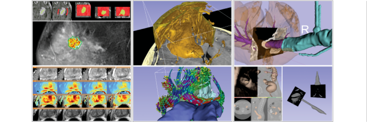
SlicerHeart extension is released with 20+ modules for cardiac 4D ultrasound visualization, heart valve analysis, device placement simulation, etc. along with an open-access overview paper.
We are thrilled to publish our description of #SlicerHeart, a open source, cardiac-focused image processing extension for @3DSlicerApp that has now been downloaded over 44,000 times. https://t.co/yCkjB0OCmm
Super-high-resolution CT @hip_ct of heart valve leaflets from the Human Organ Atlas (https://t.co/RPUGcHWV9D). Visualized in @3dslicerapp. See video tutorial for data import and rendering setup at https://t.co/vL3Jbq38lF
Hi. It`s a pleasure to share this video that shows the left female sacroiliac articulation from a different approach.
CT(3 planes)+SEGMENTATION+3D VISION+TRANSPARENCY
Segmentation with @3DSlicerApp. Thanks¡
Happy #FossilFriday ¡
In this short video I show how the Majungasaurus skull can be completely disarticulated.
Cretaceous Period 70 to 66 mya.
Complex anatomy become easy anatomy ¡
Scale 1/1
All from open access ¡
Thanks to :
@3dSlicerAPP@Morphosource@Witmerlab
Ductal stenting has become an alternative to surgical shunt palliation. Tomorrow, Dr. Mudit Gupta will preview an evolving 3D modeling tool for procedural planning of PDA stenting at @SCAI#SCAI2022. @3DSlicerApp#SlicerHeart



![JolleyLab's tweet photo. [1/3] The #SlicerHeart extension for @3DSlicerApp has now been downloaded more than 50,000 times! Thank you to all who have contributed, especially @lassoan, @_ChristianHerz_, @cs_pinter, @sunderlandkyl, Steve Pieper, Gabor Fichtinger, & Ron Kikinis. https://t.co/MIos1bQMUr](https://pbs.twimg.com/media/FrsNiszXwAEA6Xj.jpg)





