Have you been cutting corners when it comes to coronal anatomy?
Do you just say a lesion is in the inferior frontal region & hope no one asks for details?
Turn the corner when it come to coronal anatomy!
Here’s an easy way to remember this frontal anatomy you NEED to know!
Only three things to remember:
➡️Gyrus Rectus is above the nose. Remember this bc gyrus rectus means straight & it’s straight like your nose is straight!
➡️Lateral orbital gyrus is along the medial roof of the, well, orbit!
➡️Inferior, middle, and superior frontal gyri are arranged like a hand fan in the coronal plane above the gyrus rectus & lateral orbital gyrus. Remember Frontal and Fan both start w/F!
Now when it comes to coronal anatomy, hopefully you’ve cornered the market!
Members of the Fleischner Society have compiled and just released an updated Glossary of Terms for Thoracic Imaging that replaces previous glossaries published in 1984, 1996, and 2008. https://t.co/5BrYc5Tt4g
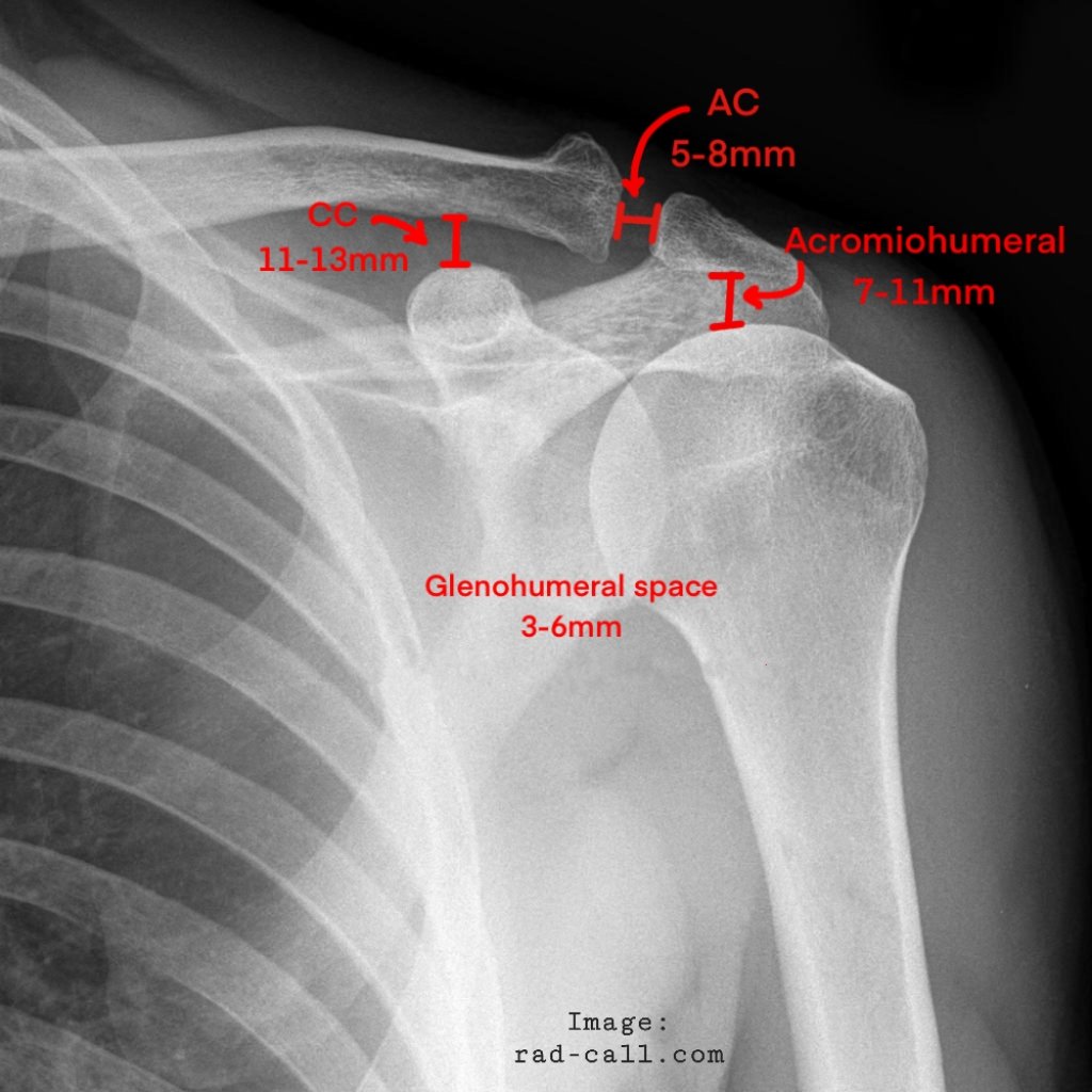
NORMAL SHOULDER MEASUREMENTS on AP view:
●GHJ space <6 mm.
●ACJ should be no greater than 7 mm.
●Coracoclavicular distance no greater than 13 mm
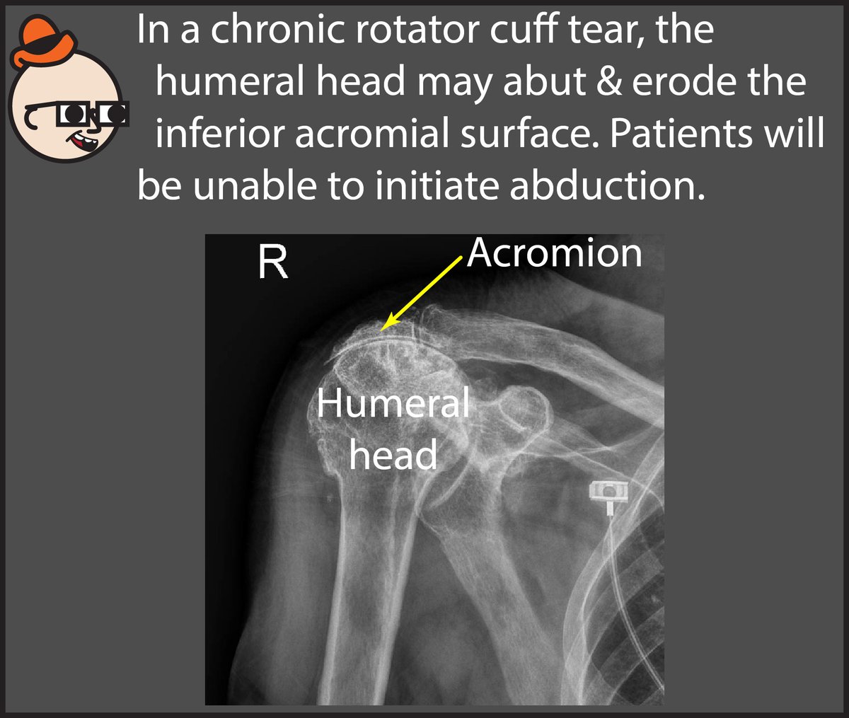
●AHD (Acromiohumeral distance) <7 mm is highly suggestive of a large rotator cuff tear.
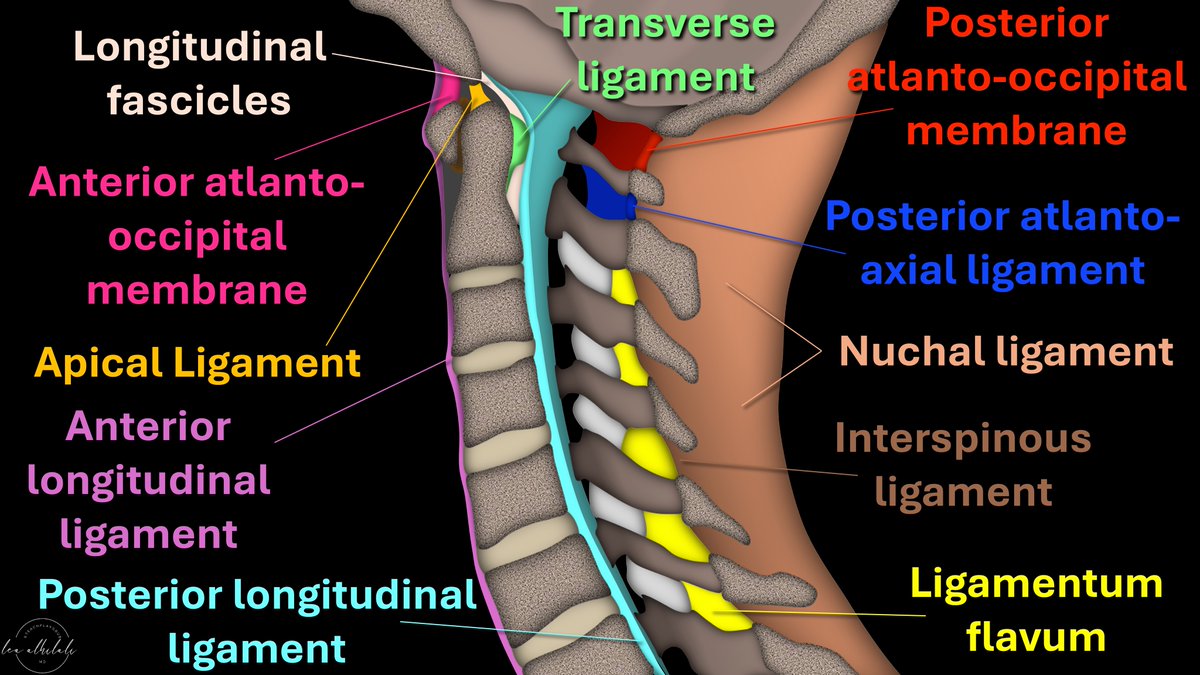
Afraid to stick your neck out when it comes to naming cervical spine ligaments?
Know the anterior longitudinal ligament (ALL) & that’s ALL?
Is the nuchal ligament a NEW CALL for you?
How many ligaments do YOU know?
Here’s are the ligaments to know & how you can remember them
➡️ALL:
🔸As it’s name implies, it runs ALL the way down the spine from the occiput to the sacrum
🔸It’s the ANTERIOR longitudinal ligament so it is ANTERIOR to the vertebral bodies
➡️PLL:
🔸PLL stands for POSTERIOR longitudinal ligament, so it’s posterior to vertebral bodies
🔸I call it “PARTIALLY longitudinal ligament” bc it only runs from C2 inferiorly
🔸Above C2 it’s the tectorial membrane.
🔸I remember this bc it has a different name but is TECT-nically part of the PLL
➡️Cranio-cervical ligaments:
🔸In the sagittal plane C2 looks like a mountain peak
🔸Ligaments here are arranged how you climb a mountain!
▶️AT Last you reach the ANTERIOR mountain (first is ANTERIOR ATLanto-occipital membrane)
▶️You hike up the APEX (next is APICAL ligament)
▶️Finally, you have to TRAVERSE the LONG way down (TRANSVERSE & LONGitudinal fibers)
➡️Posterior ligaments:
▶️Ligamentum flavum:
🔸I remember this bc Flavum & Facets both start w/F = Flavum is by the Facets
🔸F is also for FAR FIVE, so ligamentum flavum is only on the lowest/farthest five cervical vertebral bodies
🔸Above this, membranes are named for what they connect (posterior atlanto-occipital & posterior atlanto-axial membrane)
▶️Nuchal ligament:
🔸This is where we see the nuchal fold & is the equivalent of the supraspinous ligament from the occiput to C7.
Now you know the major cervical ligaments you can see on sagittal images!
Hopefully, now you will be ready when injury to these structures occurs in your neck of the woods!
Happy to share that I've been awarded an R01 from @NIH_NINDS !! We will be developing multidimensional connectome based models and quantitative pain assessments in individuals with SCI and neuropathic pain. @TJUHNeurosurg@ResearchAtJeff
Do you know your facial skeletal anatomy well enough to report trauma CT in a surgically relevant and precise manner?
#Radres check out our chapter written with @mhanaokaMD@drsuyash
https://t.co/RAOOHrOX0z
10 tips for radiology residents to get the most out of residency
I have made my fair share of mistakes during residency. I was lucky to have wise mentors, people who taught us by example. Tips to help you make the most out of your residency:
1. Be the person who picks out the toughest and most complex cases in the department to report: if there is one thing you implement from this post, let it be this. You will get sufficient exposure to the regular bread and butter radiology cases. Given a choice between reporting an easy and difficult case, always choose the latter. It might be an MRI for a brain tumor, CT complex craniofacial trauma, CECT for a laryngeal malignancy, an HRCT thorax with 10 different things going on in the lungs, complex congenital heart disease CT, the dreaded ICU radiograph, an MRCP of a hepatobiliary malignancy, a complex gynaec ultrasound, a foot MRI of a complex pathology, etc. It will be difficult initially but it is absolutely worth it in the long run. You get a very deep sense of professional satisfaction from handling complex cases well, whether diagnostic or interventional.
2. Develop rapport with referring physicians and surgeons. Be friends with them. You will learn more from a 10 min talk over coffee / tea with a top physician / surgeon than you will by spending 2 hours with any radiology textbook. Get to know the things they want you to mention in the report. Take follow ups of your cases from them.
3. Be approachable. Be the radiologist who the physicians and surgeons know they can trust to give an opinion on the complex and emergency cases for which they come to the department to discuss. You will also learn a hell of a lot.
4. Learn to report radiographs. Ignore radiographs at your own peril, master the art of reporting them well.
5. Seek to master all organ systems, all modalities: unless you are a subspecialist working in a tertiary orthopaedic / neuro / gastro hospital, you need to be an excellent general radiologist. Read up and keep reporting all organ system cases. You owe it to your patients.
6. Respect your craft. Read, report, work hard, be on time. Good old fashioned stuff, never goes out of fashion.
7. Respect the machines. Learn to optimise ultrasound, MRI, CT protocols. Learn optimum patient positioning for radiographs.
8. Never stop reading and learning. Don't stop reading even after you finish residency. A senior consultant at one of my previous hospitals read 1 radiology article from a journal every single day. Done day in, day out for more than a decade leads to phenomenal compounding. He also embodied a lot of the tips mentioned here. No surprise he was the most sought after radiologist in the hospital whose opinion was considered the final word.
9. Go above and beyond the call of duty. Spend as much time in the department as you reasonably can and see as many cases as you can, read reports of seniors and consultants. Might not be possible for everyone and am not advocating stressing yourself out, that is a recipe for burnout. But every little bit goes a long way. I did this in Grant Medical College as an assistant professor when my wife would be on her cardiology residency on call in Sion hospital. The physicians and surgeons would know more often than not I would be available in the department in the evenings for a quick opinion on in-patient and emergency cases. Again, you learn a lot!
10. Always keep in mind the patient behind the image. What is an 'interesting' or 'beautiful' case for you is horrible for the patient.
Review of Perilymphatic Disease:
Pulmonary lymphatics & the “lymphatic pattern” on imaging.
[1/5]
…from Dr. Jeffrey Galvin’s talk at #STR2021 & condensed by @MikeHarowiczMD.
#radres#Radtwitter#MedTwitter#FOAMrad#FOAMmed


![thoracicrad's tweet photo. Review of Perilymphatic Disease:
Pulmonary lymphatics & the “lymphatic pattern” on imaging.
[1/5]
…from Dr. Jeffrey Galvin’s talk at #STR2021 & condensed by @MikeHarowiczMD.
#radres #Radtwitter #MedTwitter #FOAMrad #FOAMmed https://t.co/e7i9oLrsLS](https://pbs.twimg.com/media/GC3A0VGaoAERBiI.jpg)
![thoracicrad's tweet photo. Review of Perilymphatic Disease:
Pulmonary lymphatics & the “lymphatic pattern” on imaging.
[1/5]
…from Dr. Jeffrey Galvin’s talk at #STR2021 & condensed by @MikeHarowiczMD.
#radres #Radtwitter #MedTwitter #FOAMrad #FOAMmed https://t.co/e7i9oLrsLS](https://pbs.twimg.com/media/GC3A0VBaoAExJzh.jpg)






![thoracicrad's tweet photo. Review of Perilymphatic Disease:
Pulmonary lymphatics & the “lymphatic pattern” on imaging.
[1/5]
…from Dr. Jeffrey Galvin’s talk at #STR2021 & condensed by @MikeHarowiczMD.
#radres #Radtwitter #MedTwitter #FOAMrad #FOAMmed https://t.co/e7i9oLrsLS](https://pbs.twimg.com/media/GC3A0XIagAEPLK4.jpg)


