Physeal bar: diagnosis and treatment
Patient with a history of Salter-Harris type IV fracture in the distal tibia, which on radiological control shows a focal closure of the physis in its central region.
A physeal bar results from an injury or infection in an unfused physis, consisting of a bone bridge that crosses the growth plate and causes an alteration in growth and/or a deformity. The lower extremities, especially the distal tibial physis, are the most susceptible due to its wavy contour (Kump's protuberance).
Mapping of the physeal bar can help in the treatment. Resection of the bar is considered if <50% of the physis is affected.
To assist the surgeon in the procedure, a CT scan can be used with a biopsy needle from the previously determined cortical window to the center of the bone fusion, thereby facilitating its subsequent resection and finally interposing fat or soft tissue at the surgical site to prevent the bar from reforming.
Cierre de un excelente Congreso, con temas novedosos, otras disciplinas que se incorpora a la Medicina del Fútbol y en general a la Medicina Deportiva @AEMEF_
Plantar plate tear: ultrasound diagnosis
A 23-year-old patient with plantar pain at the level of the second metatarsophalangeal joint for two weeks after playing football continuously.
Physical examination reveals pain with mobility of the second metatarsophalangeal joint, predominantly in the plantar region. A claw toe morphology is associated with an overload of the extensor brevis and longus muscles of the fingers.
An ultrasound is performed, which shows an interruption in the distal third of the plantar plate associated with a cartilage interface sign. A dynamic study shows herniation of the flexor tendon through the defect.
Treatment is performed with 1 cc of PRP infiltration in the plate defect and intra-articular on two occasions separated by seven days associated with crutches discharge, with pain resolution after 10 days and progressive return to sports activity.
Ultrasound diagnosis of anterior talofibular ligament tear
Patient suffering from ankle twisting, presenting lateral pain and functional impotence that prevent him from continuing with sports activity.
The most frequent injury in an ankle sprain is the tear of the anterior talofibular ligament, which consists of two bundles (upper and lower) that can be easily visualized in the ultrasound study and that are generally associated with the cephalic extension of the rupture affecting the joint capsule of the tibiotalar joint.
The diagnosis can be made both in a static study and in a dynamic examination by inverting the ankle or “milking” the tibiotalar intra-articular fluid, as seen at the beginning of this video.
In high-performance athletes, PRP infiltration around the ligament can be combined with physiotherapy treatment to promote healing, and it is associated with intra-articular injection to reduce secondary arthritis caused by joint instability.
Another case of pubalgia on US.
When the condition is centered in the midline, some refer to these injuries as a midline rectus abdominis-adductor longus pubic plate tear. Where the injury appears to be centered/starts at the midline and extends to the sides with involvement of the common aponeurosis, usually bilaterally asymmetric, more extensive on one side.
This is a classic example, with a defect of the tenoperiosteal interface of the common aponeurosis RA/AL communicating on both sides crossing the midline, more extensive on the right.
If you want to avoid confusion with terminology, the description is the most important thing.
#mskrad
Parosteal osteosarcoma: Diagnosis
A 43-year-old patient with gonalgia of 3 months' evolution associated with a sensation of a posterior mass.
An X-ray was performed, which showed a dense lobulated ovoid lesion attached to the posterior cortex of the distal metaphysis of the femur.
A CT study was initially completed, where penetration of the cortex by the lesion was observed, and an MRI assessment was subsequently performed, which showed a peripheral mass with hypointense T1 and T2 (the presence of high signal inside suggests a high-grade tumor) and ruled out bone marrow invasion.
A biopsy was performed under CT control for histological confirmation, and the result was parosteal osteosarcoma. This tumor corresponds to 4% of osteosarcomas, occurs mainly in the 3-4 decades, and its characteristic location is the posterior region of the femur. Because they tend to be low-grade lesions, they are usually treated, as in this case, with a surgical resection, with an excellent survival prognosis.
✍🏻 Nuevo ‘protocolo de comunicación de emergencias’ en estadios de fútbol profesional para la temporada futbolística 2024/2025.
🤝 Este documento surge del acuerdo de @LaLigaCorp, Oficina Nacional de Deportes (OND), @rfef y @AEMEF_.
https://t.co/qLSuPTk5J3
Sural nerve neuritis: Diagnosis
Patient with pain and paresthesia in the lateral ankle and posterolateral foot region for two months after performing radiofrequency of the sural nerve.
An ultrasound study was performed, which showed a sudden change in the caliber of the sural nerve in the distal third of the ankle (there is an increase in the thickness of the connective tissue (epineurium) surrounding its five neural fascicles) associated with inflammatory changes / perineural fibrosis.
Patellar chondral fracture: diagnosis and treatment
A 14-year-old patient with gonalgia and repeated joint effusion for three weeks, with a slight sensation of joint blockage.
An MRI was performed, which showed a chondral defect in the internal patellar facet associated with an intra-articular chondral fragment in the anterior recess of the intercondylar space.
Chondral fracture is a complication of patellofemoral instability. It is more frequent between 9 and 15 years of age and has traditionally been considered an indication of early surgery. MRI is the technique of choice for its diagnosis, with a sensitivity and specificity of around 90% for chondral injuries.
Treatment consists of fixing the fragment with headless compression screws or bioabsorbable pins.
(Chondral Injury in Patellofemoral Instability. Cartilage. 2014 Jul; 5(3): 136–144. doi: 10.1177/1947603514530142)
El @valenciacf e INDIBA integran la tecnología K-Laser para mejorar la preparación, la recuperación y el rendimiento de los jugadores
➡️ INDIBA & K-Laser están comprometidos en acompañar al Club y al departamento médico con el apoyo de la mejor tecnología.
#ADNVCF 🦇
Painful interfascial fibrosis: treatment
Amateur soccer player with a history of fibrillar tear in the posterolateral myofascial junction of the right rectus femoris muscle three months ago.
Ten minutes after starting physical activity, he feels muscle overload, which prevents him from continuing with the exercise. This is due to the presence of interfascial fibrosis that “fixes” the rectus femoris and vastus intermedius muscles.
It was decided to treat using EPI (Percutaneous intratissues electrolysis), which consists of the ultrasound-guided application of a galvanic current through an acupuncture needle. This breaks the intermuscular adhesions to recover the normal mobility of both muscles and thus eliminates the pain that prevents playing soccer.
The video shows the moment when the adhesion is finally broken, and only the area of fibrosis is moved medially.
Painful interfascial fibrosis: diagnosis
Amateur soccer player with a history of fibrillar tear in the posterolateral myofascial junction of the right rectus femoris muscle three months ago. He has a sensation of muscle overload ten minutes after starting physical activity, which prevents him from continuing the exercise.
Fibrosis is the final result of the healing of a muscle tear to maintain the integrity of the tissue (reparative stage), which is progressively replaced by muscle tissue (regenerative stage) between two and three months to leave no sequelae after this period.
A late complication of healing is the formation of interfascial fibrosis that “fixes” two muscles, producing an abnormal synchronous movement between them that causes a continuous overload that prevents physical exercise. This is more frequent between the internal gastrocnemius and soleus muscles and the rectus femoris and vastus intermedius muscles.
In the present case, heterogeneous interfascial fibrosis is seen on MRI in the posterolateral location of the rectus femoris muscle. Ultrasound is the technique of choice for diagnosing painful fibrosis, firstly because, through the dynamic study, the abnormal movement of the right rectus femoris muscle can be observed in an isometric contraction compared to the normal contralateral side. Secondly, persistent peripheral vascularization in color Doppler at three months is shown, and finally, alteration of tissue elasticity is identified in EUS.
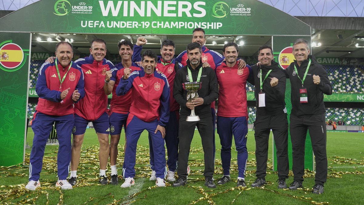
Campeones de Europa Sub-19! Tremendamente orgulloso de haber formado parte de este equipo y este grupo de trabajo. Gracias a todos los jugadores y compañeros del staff, por su implicación y dedicación, especialmente a @MarcosChenaSin que ha realizado un trabajo 🔝@SEFutbol 🇪🇦⚽️😍
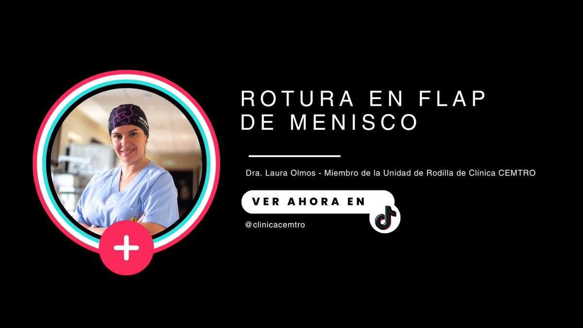
💥🦵🏻La rotura en #FLAP de #menisco es una de las variedades de las roturas meniscales
👉🏻El fragmento del menisco dañado está parcialmente unido al menisco pero puede moverse dentro de la #articulación provocando dolor y bloqueos de la #rodilla
https://t.co/F3DVidwgnI