The mitral valve is more than just two leaflets. It's a complete apparatus, and every part must work together to prevent mitral regurgitation (MR).
Key structures include:
◻️ Two leaflets: the anterior leaflet (A1-A3) and posterior leaflet (P1-P3)
◻️ Mitral annulus: the ring that supports the valve
◻️Chordae tendineae: fibrous strings that anchor the leaflets
◻️Two papillary muscles: anterolateral and posteromedial
◻️ The surrounding left atrium and left ventricle
A few high-yield facts:
- The anterior leaflet is larger, while the posterior leaflet has a broader attachment to the annulus.
- The posterior leaflet is divided into three scallops (P1, P2, P3). The matching regions of the anterior leaflet are A1, A2, and A3.
- During systole, the annulus normally contracts, helping the leaflets close tightly. If the annulus dilates, the valve may leak.
- Rupture of the chordae tendineae can cause leaflet prolapse or a flail leaflet, leading to acute mitral regurgitation.
- The posteromedial papillary muscle has a single coronary blood supply, making it more vulnerable to ischemia and rupture after myocardial infarction than the anterolateral papillary muscle, which has dual blood supply.
Understanding this anatomy makes it easier to recognize why mitral regurgitation can result from problems with the leaflets, annulus, chordae, papillary muscles, or even left ventricular remodeling.
Reference:
Dey J, Shrivastava S, Mustaqueem A. Echocardiographic Guidance in Percutaneous Approaches to Primary and Secondary Mitral Regurgitation. 2024.
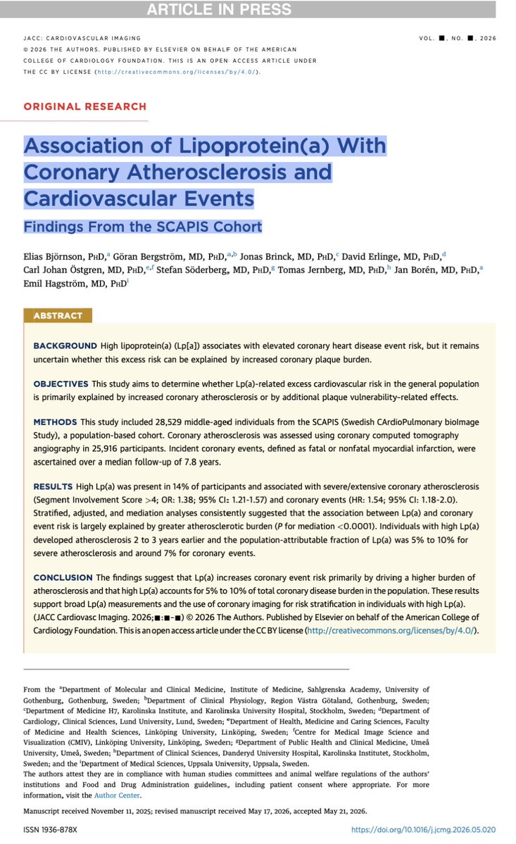
# Lp(a) Doesn't Cause Events. It Builds Plaque.
For years we've known that elevated **Lipoprotein(a)** increases cardiovascular risk.
The unanswered question was **how**.
A new *JACC: Cardiovascular Imaging* study from the **SCAPIS** cohort suggests the answer is surprisingly simple.
Among more than **28,000 individuals**, elevated Lp(a) was associated with:
- more extensive coronary atherosclerosis
- more obstructive CAD
- earlier plaque development
- higher rates of coronary events.
But here's the key finding:
Once the analysis accounted for **coronary plaque burden**, most of the excess risk associated with Lp(a) disappeared. Mediation analyses showed that the relationship between Lp(a) and events is largely explained by **more plaque—not necessarily more vulnerable plaque.**
## My Take
This paper reinforces a concept that preventive cardiology is gradually embracing:
**Risk factors matter because they create disease.**
Disease is what causes events.
That's why coronary imaging is becoming indispensable.
The question is no longer:
*"Does this patient have high Lp(a)?"*
It's:
**"What has high Lp(a) already done to this patient's arteries?"**
## Where PCCT Fits
If plaque burden is the real mediator of risk, then the next step is obvious.
Move beyond detecting stenosis.
Start quantifying plaque biology.
This is exactly where **Photon-Counting CT** has the potential to redefine preventive cardiology.
**Treat the plaque. Not the biomarker.**
#LpA #CardiacCT #PhotonCountingCT #PCCT #Atherosclerosis #PreventiveCardiology #TreatThePlaque #CardiovascularImaging
🧵 NEFROPROTECCIÓN EN 2026: lo que TODO internista debe saber.
La ERC ya no se trata solo controlando la presión arterial.
Los nuevos ensayos y KDIGO 2024 han cambiado paradigma. Estas son las claves que más impacto tienen en la práctica clínica. 👇🧵
👩🏻⚕️☕️Hoy hablaremos de:
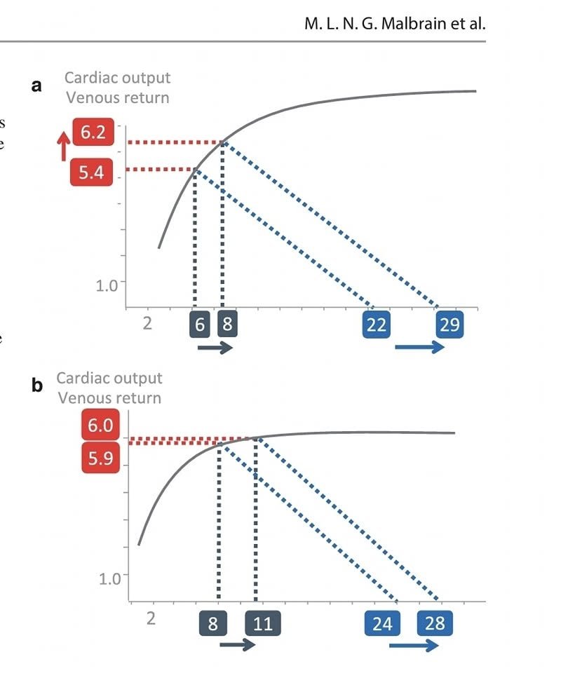
📍💧 Dinámica de los líquidos durante la reanimación...De Frank–Starling a la reevaluación de Guyton🚿
💧La administración de líquidos en el paciente crítico no debe interpretarse como una simple corrección de la “hipovolemia”, sino como una intervención hemodinámica transitoria, cuyo objetivo es aumentar el flujo sanguíneo y mejorar la perfusión tisular sin producir congestión venosa, edema intersticial ni lesión orgánica.🌀
💧Deben utilizarse cuando existe hipoperfusión, el gasto cardiaco🫀 puede aumentar con precarga y el sistema cardiovascular todavía tolera ese incremento. ➡️La reanimación moderna no persigue volumen: persigue flujo efectivo con la menor presión venosa posible.
1️⃣. Frank–Starling: Los líquidos solo funcionan si el corazón puede utilizarlos🫀💧
🌀La curva de Frank–Starling describe la relación entre el aumento de la precarga ventricular y el volumen sistólico:
💧En la porción ascendente, un incremento de la precarga puede aumentar el volumen sistólico.
💧En la zona plana, el incremento de precarga eleva principalmente las presiones de llenado, sin aumentar significativamente el gasto cardiaco.
☝🏻🤓Cuando el ventrículo derecho alcanza su límite de distensión, más líquido puede producir dilatación, aumento de la presión auricular derecha, desplazamiento septal, insuficiencia tricuspídea y disminución del llenado izquierdo.
☝🏻🤓Por tanto, precarga baja no equivale automáticamente a necesidad de líquidos, y precarga-dependencia tampoco significa que administrarlos sea beneficioso.
🚿Un paciente puede ser respondedor a volumen y, simultáneamente, presentar congestión venosa, disfunción ventricular derecha o edema pulmonar. Esta coexistencia ha sido documentada en pacientes críticos y obliga a separar los conceptos de respuesta hemodinámica y tolerancia al volumen.
💧Se considera respuesta a líquidos cuando una maniobra que incrementa la precarga produce un aumento significativo habitualmente alrededor de 10–15% del volumen sistólico o gasto cardiaco.🫀
🤨Sin embargo, la respuesta es:
🔺️Dinámica.
🔺️Dependiente de la contractilidad y poscarga.
🔺️Susceptible de cambiar después de vasopresores, ventilación, analgesia o modificación del ritmo cardiaco.
🔺️Transitoria durante la evolución del choque.
☝🏻🤓La evidencia reciente insiste en que la responsividad no debe considerarse un atributo fijo o binario del paciente, sino una condición cambiante que requiere reevaluaciones sucesivas.
2️⃣ Guyton: el gasto cardiaco depende tanto del corazón como del retorno venoso.
☝🏻🤓Guyton complementa a Frank–Starling al señalar que el gasto cardiaco no está determinado exclusivamente por la capacidad de bombeo, sino por la interacción entre:
▪️1. La función cardiaca.
▪️2. La capacidad del sistema vascular para devolver sangre al corazón.
🫀En estado estable:
✴️Gasto cardiaco = Retorno venoso
El retorno venoso puede expresarse como:
RV=Pmsf-PAD/RrV
●Pmsf: Presión media de llenado sistémico;
●PAD: Presión auricular derecha, clínicamente aproximada por la PVC;
●RRV: Resistencia al retorno venoso.
🫀El gradiente efectivo que impulsa la sangre hacia elLa presión auricular derecha no es solo una medida de “llenado”; representa también la presión de oposición o contrapresión al retorno venoso.
3️⃣Volumen estresado y no estresado
💧El volumen sanguíneo se distribuye funcionalmente en:
➡️Volumen no estresado
Llena los vasos sin ejercer una tensión relevante sobre su pared y genera poca presión de retroceso.
➡️Volumen estresado
Distiende el sistema vascular, genera la presión media de llenado sistémico y constituye el volumen funcional que impulsa el retorno venoso.🫀
🚿Los líquidos pueden aumentar el retorno venoso al incrementar el volumen estresado y desplazar la curva de retorno venoso hacia la derecha. No obstante, una fracción importante del líquido administrado puede: ■Permanecer como volumen no estresado.
■Filtrarse al intersticio.
■Elevar la presión venosa. 👇🏽
Miocardiopatía hipertrófica: claves del nuevo consenso EJHF 2026 para el diagnóstico y el tratamiento.
La miocardiopatía hipertrófica es la enfermedad cardíaca genética más frecuente, con una prevalencia estimada en torno a 1 de cada 500 personas, y una de las principales causas de insuficiencia cardíaca, arritmias y muerte súbita tanto en jóvenes como en adultos mayores. Durante décadas se entendió como una entidad de abordaje esencialmente sintomático, pero la irrupción de los inhibidores de la miosina cardíaca y la maduración de la estratificación del riesgo han transformado el escenario.
https://t.co/GPSOJuKMgw
Tratamiento de la Estenosis Mitral. 🫀💥
🔶️La EM sigue siendo una enfermedad predominantemente reumática, aunque la forma degenerativa por calcificación del anillo mitral está aumentando con el envejecimiento poblacional. El tratamiento depende de identificar correctamente su etiología, ya que las estrategias son muy diferentes.⤵
✔️ El ecocardiograma es la piedra angular.
✔️Criterios de grave: área valvular mitral ≤ 1.5 cm², gradiente medio ≥10 mmHg.
✔️La valvuloplastia mitral percutánea sigue siendo el tratamiento de elección (score de Wilkins para seleccionar candidatos).
✔️cirugía para: anatomía desfavorable para valvuloplastia, insuficiencia mitral significativa, válvula muy calcificada, fracaso previo de la valvuloplastia, necesidad de otra cirugía cardíaca concomitante.
✔️Anticoagulación: se recomienda AVK (Los ACOD aún no cuentan con evidencia suficiente para reemplazar a los AVK).
📄🆓️⤵️ @ESC_Journals 👌🏻💯
https://t.co/A9zkpASXS4
https://t.co/323G76axNh
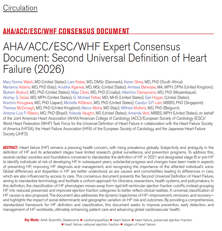
Building on the First Definition of Heart Failure, the Second Definition refines HF staging with sharper stage criteria, emphasizing early detection and individualized risk reduction and introduces the universal classification of HF causes, with explicit acknowledgment of geographic variation in HF risk and outcomes. https://t.co/YDuE4stb5s
This updated document provides a unified, internationally harmonized framework intended to standardize terminology. It addresses changes in disease manifestations, diagnostic strategies, and understanding of pathophysiology.
✍🏼 @MinnowWalsh@KarenSliwa@amibanerjee1@BiykemB@akshaydesaimd@DukeHFDoc@MKIttlesonMD@lamcardio@WilfriedMullens@NutritionHF
Congestión venosa. #HF
Abordaje clínico, bioquímico, imagen, y nuevas perspectivas.
Sugerencia abordaje multimodal (lo que tengas en tu servicio).
Mas información siempre es mejor antes de tomar decisiones.
Descarga: ⬇️⬇️⬇️
https://t.co/of16mxjhpt
🫀🚫 No-Reflow: The Artery Is Open. The Myocardium Is Not.
Primary PCI fixed the epicardial artery.
Great.
But in up to a large proportion of STEMI patients, myocardial reperfusion remains suboptimal because the real battlefield is downstream:
👉 microvascular obstruction
👉 endothelial swelling
👉 distal embolization
👉 pericyte contraction
👉 edema
👉 intramyocardial hemorrhage
In other words:
# the pipe is open, but the tissue is still starving.
This ESC consensus makes a key point very clearly: no-reflow is not a binary phenomenon. It is a spectrum, from potentially reversible microvascular dysfunction to irreversible structural damage.
And this is where imaging becomes decisive.
CMR remains the reference standard for infarct size, MVO and intramyocardial hemorrhage.
But the most interesting emerging part is Photon-Counting CT.
PCCT can potentially combine:
👉 coronary anatomy
👉 culprit lesion assessment
👉 late iodine enhancement
👉 infarct characterization
👉 MVO quantification
All in one comprehensive and integrated platform.
That is not “just better CT”.
That is a different clinical architecture.
But let’s be careful.
PCCT is promising, not yet definitive.
The consensus reports excellent concordance with CMR for infarct size and MVO quantification, but this still needs broader validation, standardized protocols, outcome data, and real workflow integration.
The real future is not simply detecting an occluded artery faster.
It is understanding what reperfusion actually achieved at tissue level.
Because after STEMI, success is not TIMI 3 flow.
Success is myocardial survival.
#PhotonCountingCT #PCCT #STEMI #CardiacCT #CMR #MicrovascularObstruction #NoReflow #CardiovascularImaging #PrecisionMedicine
In HFpEF, splanchnic mobilization + ↓ venous compliance = ↑ SBV
This shifts the pressure-volume curve LEFT and STEEPER → even small volume changes cause disproportionate rises in filling pressures.
That’s congestion without fluid overload.
#HeartFailure#HFpEF
📊 JAMA Clinical Guidelines Synopsis: The 2025 guideline for #ACS management recommends dual antiplatelet therapy with aspirin and a P2Y12 inhibitor, high-intensity statins, invasive revascularization, and radial access for angiography.
https://t.co/OnEihazgow
Cardiovascular considerations BEFORE cancer therapy🫀
The first of a 3 part #JACCCardioOnc Expert Panel series exploring CV care before, during, & after cancer therapy.
🫀Goals of CV evaluation Prior to cancer therapy:
📍Identify and treat subclinical CVD and cardiovascular risk factors 📍Optimize management of pre-existing CVD and cardiovascular risk factors 📍Perform targeted risk assessment to help inform cancer treatment decisions 📍Establish baseline cardiovascular function 📍Optimize primary prevention therapies 📍Plan cardiovascular surveillance during treatment
🫀Current tools include clinical assessment, biomarkers, 12-lead ECG, #EchoFirst, & risk scores
🫀Important evidence gaps includes cardiotoxicity mechanisms, impact of pre-treatment CV evaluations on clinical outcomes, validation&implementation of risk scores, & the role of AI.
Read more in @jaccjournals: https://t.co/cTCY0Acg99
#CardioOncology
Lp(a) vs CAC: Which One Wins?
Many clinicians assume that elevated Lp(a) makes CAC scoring less useful because Lp(a) is associated with non-calcified plaque.
This new study of 11,319 individuals suggests otherwise.
✅ Elevated Lp(a) increased risk across all CAC strata.
✅ CAC remained a powerful risk discriminator.
✅ Even with Lp(a) >50 mg/dL, individuals with CAC=0 had remarkably low absolute event rates over ~15 years.
The highest risk?
Lp(a) >50 mg/dL + CAC ≥300
(HR 6.12).
Perhaps the lesson is simple:
Lp(a) tells us who is biologically predisposed.
CAC tells us how much of that risk has actually become disease.
And those are not the same thing.
#LpA #Prevention #CardiacCT #CAC #Atherosclerosis #Cardiology #CardiovascularPrevention #Imaging #CCTA
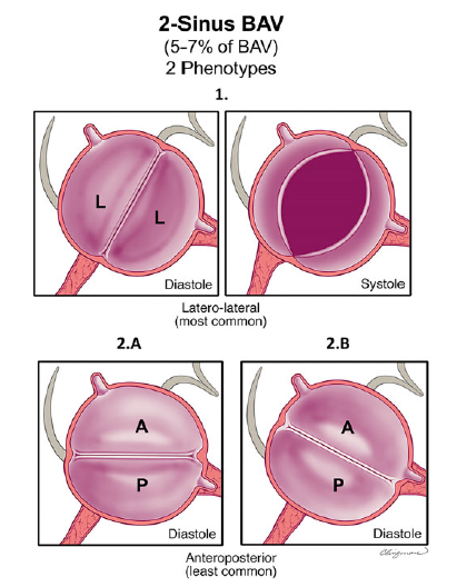
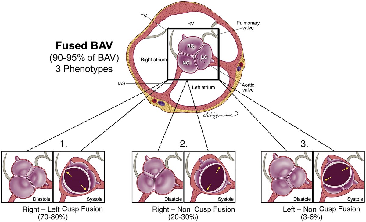
📚 Según el consenso internacional, existen tres tipos principales de Válvula aórtica bicúspide:
1️⃣ VAB con fusión completa de cúspides
2️⃣ VAB de dos senos
3️⃣ VAB con fusión parcial de cúspides, también llamada forma frustrada
📄 Can we finally measure RV volume accurately with simple echocardiography?
🔗 DOI: https://t.co/cHII0FgTLH
🫀 The right ventricle (RV) remains one of the most challenging chambers to assess—especially in congenital heart disease (CHD).
👉 Gold standard? Cardiac MRI
❗ But: expensive, time-consuming, and often requires sedation
✨ This study proposes a simple, fast, and accurate 2D echo-based method for RV volumetry—bringing us closer to true bedside quantification.
✨ The key idea:
👉 Model the RV using a cone-based geometric approach
➡️ Using only 2 standard echo views:
Apical 4-chamber (A4CH)
Parasternal short-axis (SAX)
📐 With just a few parameters:
✔ Cross-sectional areas (A4CH, ASAX)
✔ Tricuspid valve diameter
📊 Performance vs MRI:
🔥 Excellent agreement:
Systolic volume → ICC 0.98
Diastolic volume → ICC 0.96
📉 Minimal bias:
Δ systolic volume ≈ 0.1 mL
Δ diastolic volume ≈ 5.2 mL
➡️ Clearly outperforms traditional 2D models
💡 Why this matters clinically:
👉 Enables:
✔ Rapid bedside RV assessment
✔ Reduced need for repeated MRI
✔ Easier follow-up in paediatric & CHD patients
👉 Particularly valuable in:
Post-operative monitoring
Serial evaluations
Resource-limited settings
🚀 Key innovation:
👉 A mathematically robust yet practical model
➡️ Balancing accuracy + simplicity
➡️ Adaptable to different RV shapes
🚨 Bottom line:
2D echocardiography—when combined with smart modelling—can approach MRI-level accuracy for RV volumetry.
#Cardiology #Echocardiography #RightVentricle #CongenitalHeartDisease #CardiacImaging #CMR #Innovation #MedTech #PediatricCardiology 🫀📊
Cardiovascular considerations DURING cancer therapy🫀
The second of a 3 part #JACCCardioOnc Expert Panel series exploring cardiovascular care before, during, & after cancer therapy.
Recommendations for #CardioOncology care during cancer treatment:
Cardioprotection🛡️:
📍ACEI/ARB, beta-blockers, & statins may be considered to prevent ⬇️LVEF with anthracyclines 📍Consider dexrazoxane or liposomal doxorubicin for high cumulative anthracycline dose or LVEF <50% 📍Implement strategies to minimize cardiac radiation exposure
Surveillance🖥️:
📍Echo for anthracyclines & HER2-targeted therapies 📍Troponin & ECG for patients receiving ICIs
📍Prompt treatment & evaluation for high suspicion of ICI myocarditis 📍Blood pressure monitoring for VEGFi, BTKi, & ALKi therapies 📍Lipid monitoring for lorlatinib
Permissive cardiotoxicity⚖️:
📍Continue HER2-targeted therapy in selected patients with mild LV dysfunction, but need to evaluate risk & benefits 📍ACEi may be reasonable in patients with ⬆️ troponin during anthracycline therapy 📍Nitrates and/or calcium channel blockers can be considered to treat & rechallenge selected patients with 5-FU/capecitabine vasospasm, with close monitoring
Read more in @JACCJournals:
https://t.co/gxcpXpcDP2
🫀⚡ 𝗙𝗶𝗯𝗿𝗶𝗹𝗮𝗰𝗶𝗼́𝗻 𝗮𝘂𝗿𝗶𝗰𝘂𝗹𝗮𝗿: 𝘆𝗮 𝗻𝗼 𝘀𝗲 𝗺𝗮𝗻𝗲𝗷𝗮 𝘀𝗼𝗹𝗼 𝗰𝗼𝗻 “𝗰𝗼𝗻𝘁𝗿𝗼𝗹 𝗱𝗲 𝗳𝗿𝗲𝗰𝘂𝗲𝗻𝗰𝗶𝗮 𝘆 𝗮𝗻𝘁𝗶𝗰𝗼𝗮𝗴𝘂𝗹𝗮𝗰𝗶𝗼́𝗻” 🚨
@TheLancet
👇🏼👇🏼👇🏼👇🏼
📑🔗🔑🔓
https://t.co/89ht2mBRSA
⬇️⬇️⬇️⬇️
🧵👇
Este 𝘚𝘦𝘮𝘪𝘯𝘢𝘳 de 𝙇𝙖𝙣𝙘𝙚𝙩 𝟮𝟬𝟮𝟲 deja un mensaje central: la FA exige un enfoque 𝙞𝙣𝙩𝙚𝙜𝙧𝙖𝙙𝙤, 𝙙𝙞𝙣𝙖́𝙢𝙞𝙘𝙤 𝙮 𝙘𝙚𝙣𝙩𝙧𝙖𝙙𝙤 𝙚𝙣 𝙚𝙡 𝙥𝙖𝙘𝙞𝙚𝙣𝙩𝙚.
📌 Afecta a ~𝟯𝟳.𝟲 𝙢𝙞𝙡𝙡𝙤𝙣𝙚𝙨 de personas en el mundo
📌 y su prevalencia podría 𝙙𝙪𝙥𝙡𝙞𝙘𝙖𝙧𝙨𝙚 en los próximos 35 años.
🧠 𝙇𝙖 𝙁𝘼 𝙣𝙤 𝙚𝙨 𝙪𝙣𝙖 𝙖𝙧𝙧𝙞𝙩𝙢𝙞𝙖 “𝙗𝙚𝙣𝙞𝙜𝙣𝙖”
Se asocia con mayor riesgo de:
🧠 𝙞𝙘𝙩𝙪𝙨 / 𝙩𝙧𝙤𝙢𝙗𝙤𝙚𝙢𝙗𝙤𝙡𝙞𝙨𝙢𝙤
🫀 𝙞𝙣𝙨𝙪𝙛𝙞𝙘𝙞𝙚𝙣𝙘𝙞𝙖 𝙘𝙖𝙧𝙙𝙞𝙖𝙘𝙖
🧓 𝙙𝙚𝙩𝙚𝙧𝙞𝙤𝙧𝙤 𝙘𝙤𝙜𝙣𝙞𝙩𝙞𝙫𝙤 / 𝙙𝙚𝙢𝙚𝙣𝙘𝙞𝙖
☠️ 𝙢𝙪𝙚𝙧𝙩𝙚
Además, reduce calidad de vida y suele coexistir con multimorbilidad.
🔍 𝘿𝙞𝙖𝙜𝙣𝙤́𝙨𝙩𝙞𝙘𝙤: 𝙚𝙡 𝙀𝘾𝙂 𝙙𝙚 𝟭𝟮 𝙙𝙚𝙧𝙞𝙫𝙖𝙘𝙞𝙤𝙣𝙚𝙨 𝙨𝙞𝙜𝙪𝙚 𝙨𝙞𝙚𝙣𝙙𝙤 𝙚𝙡 𝙜𝙤𝙡𝙙 𝙨𝙩𝙖𝙣𝙙𝙖𝙧𝙙
La FA se confirma con:
📉 ausencia de ondas 𝙋
📉 intervalos 𝙍𝙍 𝙞𝙧𝙧𝙚𝙜𝙪𝙡𝙖𝙧𝙚𝙨
Pero en FA paroxística muchas veces hace falta monitorización adicional:
⌚ wearables
📱 ECG de una derivación
🎛️ Holter / patch / event recorder.
🛡️ 𝙇𝙖 𝙥𝙧𝙚𝙫𝙚𝙣𝙘𝙞𝙤́𝙣 𝙙𝙚𝙡 𝙞𝙘𝙩𝙪𝙨 𝙨𝙞𝙜𝙪𝙚 𝙨𝙞𝙚𝙣𝙙𝙤 𝙚𝙡 𝙥𝙞𝙡𝙖𝙧 𝙥𝙧𝙞𝙣𝙘𝙞𝙥𝙖𝙡
La revisión es clara:
✅ la 𝙖𝙣𝙩𝙞𝙘𝙤𝙖𝙜𝙪𝙡𝙖𝙘𝙞𝙤́𝙣 𝙤𝙧𝙖𝙡 sigue siendo el estándar para la mayoría
✅ los 𝘿𝙊𝘼𝘾𝙨 son preferidos sobre warfarina en FA no valvular
🚫 la 𝙖𝙨𝙥𝙞𝙧𝙞𝙣𝙖 𝙣𝙤 𝙨𝙚 𝙧𝙚𝙘𝙤𝙢𝙞𝙚𝙣𝙙𝙖 para prevención de ictus en FA.
📊 𝙀𝙡 𝙧𝙞𝙚𝙨𝙜𝙤 𝙚𝙨 𝙙𝙞𝙣𝙖́𝙢𝙞𝙘𝙤: 𝙣𝙤 𝙗𝙖𝙨𝙩𝙖 𝙘𝙤𝙣 𝙘𝙖𝙡𝙘𝙪𝙡𝙖𝙧𝙡𝙤 𝙪𝙣𝙖 𝙫𝙚𝙯
El artículo insiste en reevaluar periódicamente:
🧠 riesgo tromboembólico
🩸 riesgo de sangrado
📌 CHA₂DS₂-VASc sigue ampliamente aceptado
📌 HAS-BLED ayuda a identificar riesgo hemorrágico
⚠️ Un score alto de sangrado 𝙣𝙤 𝙙𝙚𝙗𝙚 𝙪𝙨𝙖𝙧𝙨𝙚 𝙘𝙤𝙢𝙤 𝙚𝙭𝙘𝙪𝙨𝙖 𝙥𝙖𝙧𝙖 𝙣𝙚𝙜𝙖𝙧 𝙖𝙣𝙩𝙞𝙘𝙤𝙖𝙜𝙪𝙡𝙖𝙘𝙞𝙤́𝙣.
🎯 𝘾𝙖𝙢𝙗𝙞𝙤 𝙞𝙢𝙥𝙤𝙧𝙩𝙖𝙣𝙩𝙚: 𝙚𝙡 𝙘𝙤𝙣𝙩𝙧𝙤𝙡 𝙩𝙚𝙢𝙥𝙧𝙖𝙣𝙤 𝙙𝙚𝙡 𝙧𝙞𝙩𝙢𝙤 𝙜𝙖𝙣𝙖 𝙥𝙧𝙤𝙩𝙖𝙜𝙤𝙣𝙞𝙨𝙢𝙤
Antes, el control de frecuencia era el enfoque por defecto.
Ahora, el paper resalta que el 𝙘𝙤𝙣𝙩𝙧𝙤𝙡 𝙩𝙚𝙢𝙥𝙧𝙖𝙣𝙤 𝙙𝙚𝙡 𝙧𝙞𝙩𝙢𝙤 se asocia con menos:
🫀 muerte cardiovascular
🧠 ictus
🏥 hospitalización por insuficiencia cardiaca
y mejora síntomas y calidad de vida.
🔥 𝘼𝙗𝙡𝙖𝙘𝙞𝙤́𝙣: 𝙘𝙖𝙙𝙖 𝙫𝙚𝙯 𝙢𝙖́𝙨 𝙛𝙪𝙚𝙧𝙩𝙚, 𝙞𝙣𝙘𝙡𝙪𝙨𝙤 𝙘𝙤𝙢𝙤 𝙚𝙨𝙩𝙧𝙖𝙩𝙚𝙜𝙞𝙖 𝙞𝙣𝙞𝙘𝙞𝙖𝙡 𝙚𝙣 𝙥𝙖𝙘𝙞𝙚𝙣𝙩𝙚𝙨 𝙨𝙚𝙡𝙚𝙘𝙘𝙞𝙤𝙣𝙖𝙙𝙤𝙨
La revisión destaca que la ablación con catéter:
✅ supera a los antiarrítmicos para mantener ritmo sinusal
✅ mejora síntomas y calidad de vida
✅ hoy puede considerarse incluso como 𝙩𝙧𝙖𝙩𝙖𝙢𝙞𝙚𝙣𝙩𝙤 𝙞𝙣𝙞𝙘𝙞𝙖𝙡 en algunos pacientes
📌 Las complicaciones graves contemporáneas son <𝟮%.
🫶 𝙀𝙡 𝙜𝙧𝙖𝙣 𝙢𝙚𝙣𝙨𝙖𝙟𝙚 𝙛𝙞𝙣𝙖𝙡: 𝙩𝙧𝙖𝙩𝙖𝙧 𝙁𝘼 𝙚𝙨 𝙢𝙪𝙘𝙝𝙤 𝙢𝙖́𝙨 𝙦𝙪𝙚 “𝙦𝙪𝙞𝙩𝙖𝙧 𝙡𝙖 𝙖𝙧𝙧𝙞𝙩𝙢𝙞𝙖”
El artículo cierra reforzando un modelo integral:
✅ prevenir ictus
✅ aliviar síntomas
✅ tratar comorbilidades
✅ corregir factores de riesgo:
⚖️ obesidad
😴 apnea del sueño
🚬 tabaquismo
🍺 alcohol
🏃 inactividad física
👉 En FA, el mejor tratamiento combina 𝙖𝙣𝙩𝙞𝙘𝙤𝙖𝙜𝙪𝙡𝙖𝙘𝙞𝙤́𝙣 + 𝙚𝙨𝙩𝙧𝙖𝙩𝙚𝙜𝙞𝙖 𝙙𝙚 𝙧𝙞𝙩𝙢𝙤/𝙛𝙧𝙚𝙘𝙪𝙚𝙣𝙘𝙞𝙖 + 𝙢𝙖𝙣𝙚𝙟𝙤 𝙞𝙣𝙩𝙚𝙣𝙨𝙞𝙫𝙤 𝙙𝙚 𝙘𝙤𝙢𝙤𝙧𝙗𝙞𝙡𝙞𝙙𝙖𝙙𝙚𝙨 𝙮 𝙚𝙨𝙩𝙞𝙡𝙤 𝙙𝙚 𝙫𝙞𝙙𝙖.
‼️Si te sirve: ❤️ Me gusta | 🔁 Repost | ➕ Follow para más👇🏼👇🏼👇🏼👇🏼
📚📖#ClubCrit👨🏻⚕️👨🏻🏫🧠🫶
📚📖 Más en el blog 👉 [https://t.co/HUCnQ3Pi7l]
#AFib #ClubCrit #FibrilaciónAuricular #CardioTwitter #Electrofisiología #PrevenciónCV #POCUS #FOAMed #FOAMcc #CriticalCare #CriticalCare #CuidadoCrítico #MedTwitter #CritCare #icu #intensivecare #diagnosis #management #UCI #MedicinaBasadaEnEvidencia #MedEd #MedX #IntensiveCare #MedIntensiva #MedXCommunity #MedED #ICUmanagement #MustRead #LecturaRecomendada
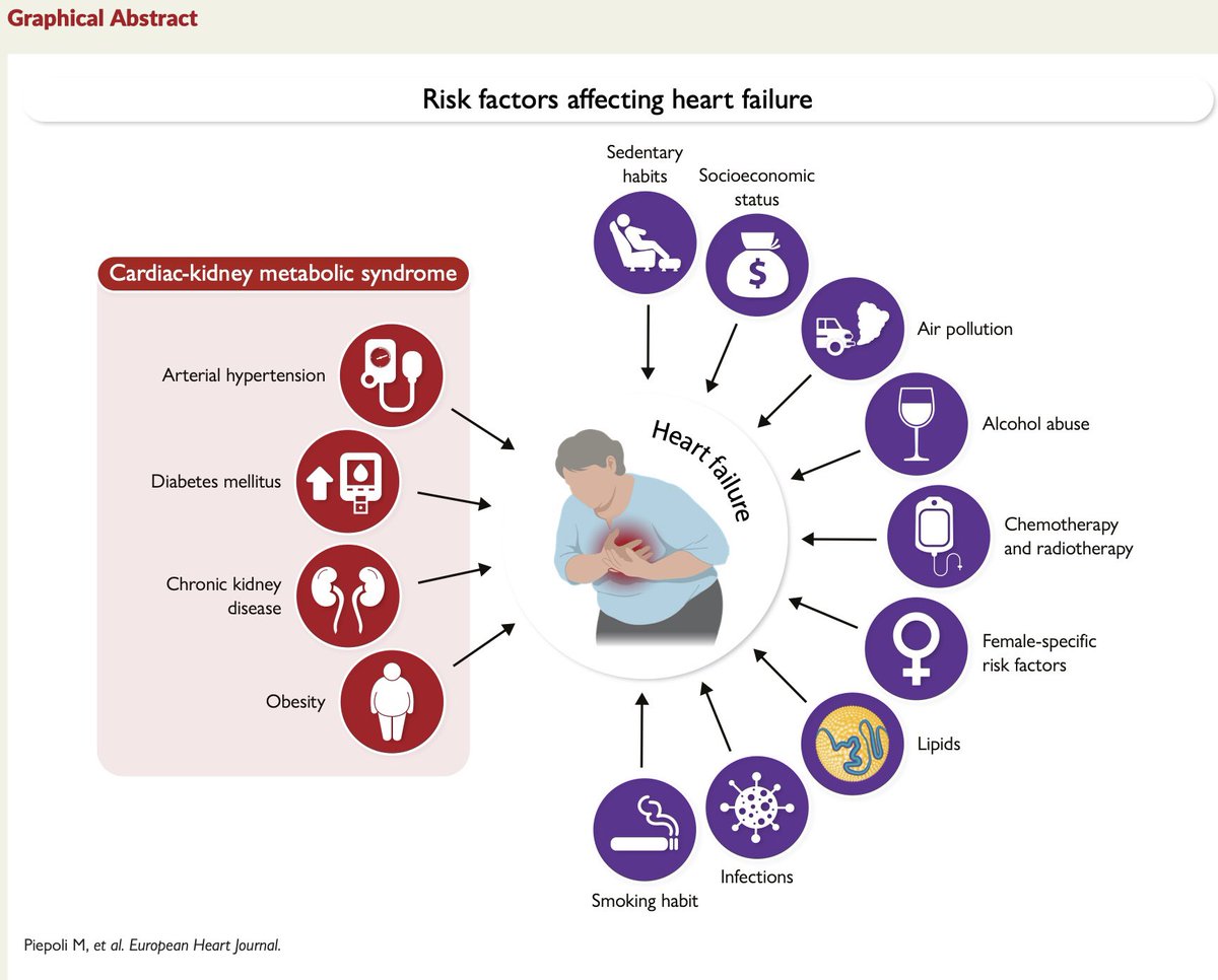
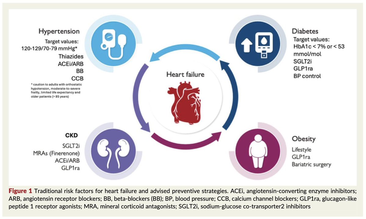
🫀What drives the development of heart failure?
The new #HFA_ESC scientific statement highlights the broad spectrum of risk factors contributing to #HeartFailure including:
📍Cardiac-kidney-metabolic syndrome
📍Lifestyle factors
📍Cancer therapies
📍Female-specific risk factors
📍Environmental & socioeconomic determinants
Preventing HF requires a holistic approach focused on early identification & management of these interconnected risks
Read more in #EJHF @EJHFEiC : https://t.co/EqOZ6zRLG7
@escardio@MarcoMetra@m4ggiesimpson






![MarlonVFZR's tweet photo. 🫀⚡ 𝗙𝗶𝗯𝗿𝗶𝗹𝗮𝗰𝗶𝗼́𝗻 𝗮𝘂𝗿𝗶𝗰𝘂𝗹𝗮𝗿: 𝘆𝗮 𝗻𝗼 𝘀𝗲 𝗺𝗮𝗻𝗲𝗷𝗮 𝘀𝗼𝗹𝗼 𝗰𝗼𝗻 “𝗰𝗼𝗻𝘁𝗿𝗼𝗹 𝗱𝗲 𝗳𝗿𝗲𝗰𝘂𝗲𝗻𝗰𝗶𝗮 𝘆 𝗮𝗻𝘁𝗶𝗰𝗼𝗮𝗴𝘂𝗹𝗮𝗰𝗶𝗼́𝗻” 🚨
@TheLancet
👇🏼👇🏼👇🏼👇🏼
📑🔗🔑🔓
https://t.co/89ht2mBRSA
⬇️⬇️⬇️⬇️
🧵👇
Este 𝘚𝘦𝘮𝘪𝘯𝘢𝘳 de 𝙇𝙖𝙣𝙘𝙚𝙩 𝟮𝟬𝟮𝟲 deja un mensaje central: la FA exige un enfoque 𝙞𝙣𝙩𝙚𝙜𝙧𝙖𝙙𝙤, 𝙙𝙞𝙣𝙖́𝙢𝙞𝙘𝙤 𝙮 𝙘𝙚𝙣𝙩𝙧𝙖𝙙𝙤 𝙚𝙣 𝙚𝙡 𝙥𝙖𝙘𝙞𝙚𝙣𝙩𝙚.
📌 Afecta a ~𝟯𝟳.𝟲 𝙢𝙞𝙡𝙡𝙤𝙣𝙚𝙨 de personas en el mundo
📌 y su prevalencia podría 𝙙𝙪𝙥𝙡𝙞𝙘𝙖𝙧𝙨𝙚 en los próximos 35 años.
🧠 𝙇𝙖 𝙁𝘼 𝙣𝙤 𝙚𝙨 𝙪𝙣𝙖 𝙖𝙧𝙧𝙞𝙩𝙢𝙞𝙖 “𝙗𝙚𝙣𝙞𝙜𝙣𝙖”
Se asocia con mayor riesgo de:
🧠 𝙞𝙘𝙩𝙪𝙨 / 𝙩𝙧𝙤𝙢𝙗𝙤𝙚𝙢𝙗𝙤𝙡𝙞𝙨𝙢𝙤
🫀 𝙞𝙣𝙨𝙪𝙛𝙞𝙘𝙞𝙚𝙣𝙘𝙞𝙖 𝙘𝙖𝙧𝙙𝙞𝙖𝙘𝙖
🧓 𝙙𝙚𝙩𝙚𝙧𝙞𝙤𝙧𝙤 𝙘𝙤𝙜𝙣𝙞𝙩𝙞𝙫𝙤 / 𝙙𝙚𝙢𝙚𝙣𝙘𝙞𝙖
☠️ 𝙢𝙪𝙚𝙧𝙩𝙚
Además, reduce calidad de vida y suele coexistir con multimorbilidad.
🔍 𝘿𝙞𝙖𝙜𝙣𝙤́𝙨𝙩𝙞𝙘𝙤: 𝙚𝙡 𝙀𝘾𝙂 𝙙𝙚 𝟭𝟮 𝙙𝙚𝙧𝙞𝙫𝙖𝙘𝙞𝙤𝙣𝙚𝙨 𝙨𝙞𝙜𝙪𝙚 𝙨𝙞𝙚𝙣𝙙𝙤 𝙚𝙡 𝙜𝙤𝙡𝙙 𝙨𝙩𝙖𝙣𝙙𝙖𝙧𝙙
La FA se confirma con:
📉 ausencia de ondas 𝙋
📉 intervalos 𝙍𝙍 𝙞𝙧𝙧𝙚𝙜𝙪𝙡𝙖𝙧𝙚𝙨
Pero en FA paroxística muchas veces hace falta monitorización adicional:
⌚ wearables
📱 ECG de una derivación
🎛️ Holter / patch / event recorder.
🛡️ 𝙇𝙖 𝙥𝙧𝙚𝙫𝙚𝙣𝙘𝙞𝙤́𝙣 𝙙𝙚𝙡 𝙞𝙘𝙩𝙪𝙨 𝙨𝙞𝙜𝙪𝙚 𝙨𝙞𝙚𝙣𝙙𝙤 𝙚𝙡 𝙥𝙞𝙡𝙖𝙧 𝙥𝙧𝙞𝙣𝙘𝙞𝙥𝙖𝙡
La revisión es clara:
✅ la 𝙖𝙣𝙩𝙞𝙘𝙤𝙖𝙜𝙪𝙡𝙖𝙘𝙞𝙤́𝙣 𝙤𝙧𝙖𝙡 sigue siendo el estándar para la mayoría
✅ los 𝘿𝙊𝘼𝘾𝙨 son preferidos sobre warfarina en FA no valvular
🚫 la 𝙖𝙨𝙥𝙞𝙧𝙞𝙣𝙖 𝙣𝙤 𝙨𝙚 𝙧𝙚𝙘𝙤𝙢𝙞𝙚𝙣𝙙𝙖 para prevención de ictus en FA.
📊 𝙀𝙡 𝙧𝙞𝙚𝙨𝙜𝙤 𝙚𝙨 𝙙𝙞𝙣𝙖́𝙢𝙞𝙘𝙤: 𝙣𝙤 𝙗𝙖𝙨𝙩𝙖 𝙘𝙤𝙣 𝙘𝙖𝙡𝙘𝙪𝙡𝙖𝙧𝙡𝙤 𝙪𝙣𝙖 𝙫𝙚𝙯
El artículo insiste en reevaluar periódicamente:
🧠 riesgo tromboembólico
🩸 riesgo de sangrado
📌 CHA₂DS₂-VASc sigue ampliamente aceptado
📌 HAS-BLED ayuda a identificar riesgo hemorrágico
⚠️ Un score alto de sangrado 𝙣𝙤 𝙙𝙚𝙗𝙚 𝙪𝙨𝙖𝙧𝙨𝙚 𝙘𝙤𝙢𝙤 𝙚𝙭𝙘𝙪𝙨𝙖 𝙥𝙖𝙧𝙖 𝙣𝙚𝙜𝙖𝙧 𝙖𝙣𝙩𝙞𝙘𝙤𝙖𝙜𝙪𝙡𝙖𝙘𝙞𝙤́𝙣.
🎯 𝘾𝙖𝙢𝙗𝙞𝙤 𝙞𝙢𝙥𝙤𝙧𝙩𝙖𝙣𝙩𝙚: 𝙚𝙡 𝙘𝙤𝙣𝙩𝙧𝙤𝙡 𝙩𝙚𝙢𝙥𝙧𝙖𝙣𝙤 𝙙𝙚𝙡 𝙧𝙞𝙩𝙢𝙤 𝙜𝙖𝙣𝙖 𝙥𝙧𝙤𝙩𝙖𝙜𝙤𝙣𝙞𝙨𝙢𝙤
Antes, el control de frecuencia era el enfoque por defecto.
Ahora, el paper resalta que el 𝙘𝙤𝙣𝙩𝙧𝙤𝙡 𝙩𝙚𝙢𝙥𝙧𝙖𝙣𝙤 𝙙𝙚𝙡 𝙧𝙞𝙩𝙢𝙤 se asocia con menos:
🫀 muerte cardiovascular
🧠 ictus
🏥 hospitalización por insuficiencia cardiaca
y mejora síntomas y calidad de vida.
🔥 𝘼𝙗𝙡𝙖𝙘𝙞𝙤́𝙣: 𝙘𝙖𝙙𝙖 𝙫𝙚𝙯 𝙢𝙖́𝙨 𝙛𝙪𝙚𝙧𝙩𝙚, 𝙞𝙣𝙘𝙡𝙪𝙨𝙤 𝙘𝙤𝙢𝙤 𝙚𝙨𝙩𝙧𝙖𝙩𝙚𝙜𝙞𝙖 𝙞𝙣𝙞𝙘𝙞𝙖𝙡 𝙚𝙣 𝙥𝙖𝙘𝙞𝙚𝙣𝙩𝙚𝙨 𝙨𝙚𝙡𝙚𝙘𝙘𝙞𝙤𝙣𝙖𝙙𝙤𝙨
La revisión destaca que la ablación con catéter:
✅ supera a los antiarrítmicos para mantener ritmo sinusal
✅ mejora síntomas y calidad de vida
✅ hoy puede considerarse incluso como 𝙩𝙧𝙖𝙩𝙖𝙢𝙞𝙚𝙣𝙩𝙤 𝙞𝙣𝙞𝙘𝙞𝙖𝙡 en algunos pacientes
📌 Las complicaciones graves contemporáneas son <𝟮%.
🫶 𝙀𝙡 𝙜𝙧𝙖𝙣 𝙢𝙚𝙣𝙨𝙖𝙟𝙚 𝙛𝙞𝙣𝙖𝙡: 𝙩𝙧𝙖𝙩𝙖𝙧 𝙁𝘼 𝙚𝙨 𝙢𝙪𝙘𝙝𝙤 𝙢𝙖́𝙨 𝙦𝙪𝙚 “𝙦𝙪𝙞𝙩𝙖𝙧 𝙡𝙖 𝙖𝙧𝙧𝙞𝙩𝙢𝙞𝙖”
El artículo cierra reforzando un modelo integral:
✅ prevenir ictus
✅ aliviar síntomas
✅ tratar comorbilidades
✅ corregir factores de riesgo:
⚖️ obesidad
😴 apnea del sueño
🚬 tabaquismo
🍺 alcohol
🏃 inactividad física
👉 En FA, el mejor tratamiento combina 𝙖𝙣𝙩𝙞𝙘𝙤𝙖𝙜𝙪𝙡𝙖𝙘𝙞𝙤́𝙣 + 𝙚𝙨𝙩𝙧𝙖𝙩𝙚𝙜𝙞𝙖 𝙙𝙚 𝙧𝙞𝙩𝙢𝙤/𝙛𝙧𝙚𝙘𝙪𝙚𝙣𝙘𝙞𝙖 + 𝙢𝙖𝙣𝙚𝙟𝙤 𝙞𝙣𝙩𝙚𝙣𝙨𝙞𝙫𝙤 𝙙𝙚 𝙘𝙤𝙢𝙤𝙧𝙗𝙞𝙡𝙞𝙙𝙖𝙙𝙚𝙨 𝙮 𝙚𝙨𝙩𝙞𝙡𝙤 𝙙𝙚 𝙫𝙞𝙙𝙖.
‼️Si te sirve: ❤️ Me gusta | 🔁 Repost | ➕ Follow para más👇🏼👇🏼👇🏼👇🏼
📚📖#ClubCrit👨🏻⚕️👨🏻🏫🧠🫶
📚📖 Más en el blog 👉 [https://t.co/HUCnQ3Pi7l]
#AFib #ClubCrit #FibrilaciónAuricular #CardioTwitter #Electrofisiología #PrevenciónCV #POCUS #FOAMed #FOAMcc #CriticalCare #CriticalCare #CuidadoCrítico #MedTwitter #CritCare #icu #intensivecare #diagnosis #management #UCI #MedicinaBasadaEnEvidencia #MedEd #MedX #IntensiveCare #MedIntensiva #MedXCommunity #MedED #ICUmanagement #MustRead #LecturaRecomendada](https://pbs.twimg.com/media/HJz3zyqXsAARTAW.jpg)
![MarlonVFZR's tweet photo. 🫀⚡ 𝗙𝗶𝗯𝗿𝗶𝗹𝗮𝗰𝗶𝗼́𝗻 𝗮𝘂𝗿𝗶𝗰𝘂𝗹𝗮𝗿: 𝘆𝗮 𝗻𝗼 𝘀𝗲 𝗺𝗮𝗻𝗲𝗷𝗮 𝘀𝗼𝗹𝗼 𝗰𝗼𝗻 “𝗰𝗼𝗻𝘁𝗿𝗼𝗹 𝗱𝗲 𝗳𝗿𝗲𝗰𝘂𝗲𝗻𝗰𝗶𝗮 𝘆 𝗮𝗻𝘁𝗶𝗰𝗼𝗮𝗴𝘂𝗹𝗮𝗰𝗶𝗼́𝗻” 🚨
@TheLancet
👇🏼👇🏼👇🏼👇🏼
📑🔗🔑🔓
https://t.co/89ht2mBRSA
⬇️⬇️⬇️⬇️
🧵👇
Este 𝘚𝘦𝘮𝘪𝘯𝘢𝘳 de 𝙇𝙖𝙣𝙘𝙚𝙩 𝟮𝟬𝟮𝟲 deja un mensaje central: la FA exige un enfoque 𝙞𝙣𝙩𝙚𝙜𝙧𝙖𝙙𝙤, 𝙙𝙞𝙣𝙖́𝙢𝙞𝙘𝙤 𝙮 𝙘𝙚𝙣𝙩𝙧𝙖𝙙𝙤 𝙚𝙣 𝙚𝙡 𝙥𝙖𝙘𝙞𝙚𝙣𝙩𝙚.
📌 Afecta a ~𝟯𝟳.𝟲 𝙢𝙞𝙡𝙡𝙤𝙣𝙚𝙨 de personas en el mundo
📌 y su prevalencia podría 𝙙𝙪𝙥𝙡𝙞𝙘𝙖𝙧𝙨𝙚 en los próximos 35 años.
🧠 𝙇𝙖 𝙁𝘼 𝙣𝙤 𝙚𝙨 𝙪𝙣𝙖 𝙖𝙧𝙧𝙞𝙩𝙢𝙞𝙖 “𝙗𝙚𝙣𝙞𝙜𝙣𝙖”
Se asocia con mayor riesgo de:
🧠 𝙞𝙘𝙩𝙪𝙨 / 𝙩𝙧𝙤𝙢𝙗𝙤𝙚𝙢𝙗𝙤𝙡𝙞𝙨𝙢𝙤
🫀 𝙞𝙣𝙨𝙪𝙛𝙞𝙘𝙞𝙚𝙣𝙘𝙞𝙖 𝙘𝙖𝙧𝙙𝙞𝙖𝙘𝙖
🧓 𝙙𝙚𝙩𝙚𝙧𝙞𝙤𝙧𝙤 𝙘𝙤𝙜𝙣𝙞𝙩𝙞𝙫𝙤 / 𝙙𝙚𝙢𝙚𝙣𝙘𝙞𝙖
☠️ 𝙢𝙪𝙚𝙧𝙩𝙚
Además, reduce calidad de vida y suele coexistir con multimorbilidad.
🔍 𝘿𝙞𝙖𝙜𝙣𝙤́𝙨𝙩𝙞𝙘𝙤: 𝙚𝙡 𝙀𝘾𝙂 𝙙𝙚 𝟭𝟮 𝙙𝙚𝙧𝙞𝙫𝙖𝙘𝙞𝙤𝙣𝙚𝙨 𝙨𝙞𝙜𝙪𝙚 𝙨𝙞𝙚𝙣𝙙𝙤 𝙚𝙡 𝙜𝙤𝙡𝙙 𝙨𝙩𝙖𝙣𝙙𝙖𝙧𝙙
La FA se confirma con:
📉 ausencia de ondas 𝙋
📉 intervalos 𝙍𝙍 𝙞𝙧𝙧𝙚𝙜𝙪𝙡𝙖𝙧𝙚𝙨
Pero en FA paroxística muchas veces hace falta monitorización adicional:
⌚ wearables
📱 ECG de una derivación
🎛️ Holter / patch / event recorder.
🛡️ 𝙇𝙖 𝙥𝙧𝙚𝙫𝙚𝙣𝙘𝙞𝙤́𝙣 𝙙𝙚𝙡 𝙞𝙘𝙩𝙪𝙨 𝙨𝙞𝙜𝙪𝙚 𝙨𝙞𝙚𝙣𝙙𝙤 𝙚𝙡 𝙥𝙞𝙡𝙖𝙧 𝙥𝙧𝙞𝙣𝙘𝙞𝙥𝙖𝙡
La revisión es clara:
✅ la 𝙖𝙣𝙩𝙞𝙘𝙤𝙖𝙜𝙪𝙡𝙖𝙘𝙞𝙤́𝙣 𝙤𝙧𝙖𝙡 sigue siendo el estándar para la mayoría
✅ los 𝘿𝙊𝘼𝘾𝙨 son preferidos sobre warfarina en FA no valvular
🚫 la 𝙖𝙨𝙥𝙞𝙧𝙞𝙣𝙖 𝙣𝙤 𝙨𝙚 𝙧𝙚𝙘𝙤𝙢𝙞𝙚𝙣𝙙𝙖 para prevención de ictus en FA.
📊 𝙀𝙡 𝙧𝙞𝙚𝙨𝙜𝙤 𝙚𝙨 𝙙𝙞𝙣𝙖́𝙢𝙞𝙘𝙤: 𝙣𝙤 𝙗𝙖𝙨𝙩𝙖 𝙘𝙤𝙣 𝙘𝙖𝙡𝙘𝙪𝙡𝙖𝙧𝙡𝙤 𝙪𝙣𝙖 𝙫𝙚𝙯
El artículo insiste en reevaluar periódicamente:
🧠 riesgo tromboembólico
🩸 riesgo de sangrado
📌 CHA₂DS₂-VASc sigue ampliamente aceptado
📌 HAS-BLED ayuda a identificar riesgo hemorrágico
⚠️ Un score alto de sangrado 𝙣𝙤 𝙙𝙚𝙗𝙚 𝙪𝙨𝙖𝙧𝙨𝙚 𝙘𝙤𝙢𝙤 𝙚𝙭𝙘𝙪𝙨𝙖 𝙥𝙖𝙧𝙖 𝙣𝙚𝙜𝙖𝙧 𝙖𝙣𝙩𝙞𝙘𝙤𝙖𝙜𝙪𝙡𝙖𝙘𝙞𝙤́𝙣.
🎯 𝘾𝙖𝙢𝙗𝙞𝙤 𝙞𝙢𝙥𝙤𝙧𝙩𝙖𝙣𝙩𝙚: 𝙚𝙡 𝙘𝙤𝙣𝙩𝙧𝙤𝙡 𝙩𝙚𝙢𝙥𝙧𝙖𝙣𝙤 𝙙𝙚𝙡 𝙧𝙞𝙩𝙢𝙤 𝙜𝙖𝙣𝙖 𝙥𝙧𝙤𝙩𝙖𝙜𝙤𝙣𝙞𝙨𝙢𝙤
Antes, el control de frecuencia era el enfoque por defecto.
Ahora, el paper resalta que el 𝙘𝙤𝙣𝙩𝙧𝙤𝙡 𝙩𝙚𝙢𝙥𝙧𝙖𝙣𝙤 𝙙𝙚𝙡 𝙧𝙞𝙩𝙢𝙤 se asocia con menos:
🫀 muerte cardiovascular
🧠 ictus
🏥 hospitalización por insuficiencia cardiaca
y mejora síntomas y calidad de vida.
🔥 𝘼𝙗𝙡𝙖𝙘𝙞𝙤́𝙣: 𝙘𝙖𝙙𝙖 𝙫𝙚𝙯 𝙢𝙖́𝙨 𝙛𝙪𝙚𝙧𝙩𝙚, 𝙞𝙣𝙘𝙡𝙪𝙨𝙤 𝙘𝙤𝙢𝙤 𝙚𝙨𝙩𝙧𝙖𝙩𝙚𝙜𝙞𝙖 𝙞𝙣𝙞𝙘𝙞𝙖𝙡 𝙚𝙣 𝙥𝙖𝙘𝙞𝙚𝙣𝙩𝙚𝙨 𝙨𝙚𝙡𝙚𝙘𝙘𝙞𝙤𝙣𝙖𝙙𝙤𝙨
La revisión destaca que la ablación con catéter:
✅ supera a los antiarrítmicos para mantener ritmo sinusal
✅ mejora síntomas y calidad de vida
✅ hoy puede considerarse incluso como 𝙩𝙧𝙖𝙩𝙖𝙢𝙞𝙚𝙣𝙩𝙤 𝙞𝙣𝙞𝙘𝙞𝙖𝙡 en algunos pacientes
📌 Las complicaciones graves contemporáneas son <𝟮%.
🫶 𝙀𝙡 𝙜𝙧𝙖𝙣 𝙢𝙚𝙣𝙨𝙖𝙟𝙚 𝙛𝙞𝙣𝙖𝙡: 𝙩𝙧𝙖𝙩𝙖𝙧 𝙁𝘼 𝙚𝙨 𝙢𝙪𝙘𝙝𝙤 𝙢𝙖́𝙨 𝙦𝙪𝙚 “𝙦𝙪𝙞𝙩𝙖𝙧 𝙡𝙖 𝙖𝙧𝙧𝙞𝙩𝙢𝙞𝙖”
El artículo cierra reforzando un modelo integral:
✅ prevenir ictus
✅ aliviar síntomas
✅ tratar comorbilidades
✅ corregir factores de riesgo:
⚖️ obesidad
😴 apnea del sueño
🚬 tabaquismo
🍺 alcohol
🏃 inactividad física
👉 En FA, el mejor tratamiento combina 𝙖𝙣𝙩𝙞𝙘𝙤𝙖𝙜𝙪𝙡𝙖𝙘𝙞𝙤́𝙣 + 𝙚𝙨𝙩𝙧𝙖𝙩𝙚𝙜𝙞𝙖 𝙙𝙚 𝙧𝙞𝙩𝙢𝙤/𝙛𝙧𝙚𝙘𝙪𝙚𝙣𝙘𝙞𝙖 + 𝙢𝙖𝙣𝙚𝙟𝙤 𝙞𝙣𝙩𝙚𝙣𝙨𝙞𝙫𝙤 𝙙𝙚 𝙘𝙤𝙢𝙤𝙧𝙗𝙞𝙡𝙞𝙙𝙖𝙙𝙚𝙨 𝙮 𝙚𝙨𝙩𝙞𝙡𝙤 𝙙𝙚 𝙫𝙞𝙙𝙖.
‼️Si te sirve: ❤️ Me gusta | 🔁 Repost | ➕ Follow para más👇🏼👇🏼👇🏼👇🏼
📚📖#ClubCrit👨🏻⚕️👨🏻🏫🧠🫶
📚📖 Más en el blog 👉 [https://t.co/HUCnQ3Pi7l]
#AFib #ClubCrit #FibrilaciónAuricular #CardioTwitter #Electrofisiología #PrevenciónCV #POCUS #FOAMed #FOAMcc #CriticalCare #CriticalCare #CuidadoCrítico #MedTwitter #CritCare #icu #intensivecare #diagnosis #management #UCI #MedicinaBasadaEnEvidencia #MedEd #MedX #IntensiveCare #MedIntensiva #MedXCommunity #MedED #ICUmanagement #MustRead #LecturaRecomendada](https://pbs.twimg.com/media/HJz3zu6WwAA5tX6.jpg)
![MarlonVFZR's tweet photo. 🫀⚡ 𝗙𝗶𝗯𝗿𝗶𝗹𝗮𝗰𝗶𝗼́𝗻 𝗮𝘂𝗿𝗶𝗰𝘂𝗹𝗮𝗿: 𝘆𝗮 𝗻𝗼 𝘀𝗲 𝗺𝗮𝗻𝗲𝗷𝗮 𝘀𝗼𝗹𝗼 𝗰𝗼𝗻 “𝗰𝗼𝗻𝘁𝗿𝗼𝗹 𝗱𝗲 𝗳𝗿𝗲𝗰𝘂𝗲𝗻𝗰𝗶𝗮 𝘆 𝗮𝗻𝘁𝗶𝗰𝗼𝗮𝗴𝘂𝗹𝗮𝗰𝗶𝗼́𝗻” 🚨
@TheLancet
👇🏼👇🏼👇🏼👇🏼
📑🔗🔑🔓
https://t.co/89ht2mBRSA
⬇️⬇️⬇️⬇️
🧵👇
Este 𝘚𝘦𝘮𝘪𝘯𝘢𝘳 de 𝙇𝙖𝙣𝙘𝙚𝙩 𝟮𝟬𝟮𝟲 deja un mensaje central: la FA exige un enfoque 𝙞𝙣𝙩𝙚𝙜𝙧𝙖𝙙𝙤, 𝙙𝙞𝙣𝙖́𝙢𝙞𝙘𝙤 𝙮 𝙘𝙚𝙣𝙩𝙧𝙖𝙙𝙤 𝙚𝙣 𝙚𝙡 𝙥𝙖𝙘𝙞𝙚𝙣𝙩𝙚.
📌 Afecta a ~𝟯𝟳.𝟲 𝙢𝙞𝙡𝙡𝙤𝙣𝙚𝙨 de personas en el mundo
📌 y su prevalencia podría 𝙙𝙪𝙥𝙡𝙞𝙘𝙖𝙧𝙨𝙚 en los próximos 35 años.
🧠 𝙇𝙖 𝙁𝘼 𝙣𝙤 𝙚𝙨 𝙪𝙣𝙖 𝙖𝙧𝙧𝙞𝙩𝙢𝙞𝙖 “𝙗𝙚𝙣𝙞𝙜𝙣𝙖”
Se asocia con mayor riesgo de:
🧠 𝙞𝙘𝙩𝙪𝙨 / 𝙩𝙧𝙤𝙢𝙗𝙤𝙚𝙢𝙗𝙤𝙡𝙞𝙨𝙢𝙤
🫀 𝙞𝙣𝙨𝙪𝙛𝙞𝙘𝙞𝙚𝙣𝙘𝙞𝙖 𝙘𝙖𝙧𝙙𝙞𝙖𝙘𝙖
🧓 𝙙𝙚𝙩𝙚𝙧𝙞𝙤𝙧𝙤 𝙘𝙤𝙜𝙣𝙞𝙩𝙞𝙫𝙤 / 𝙙𝙚𝙢𝙚𝙣𝙘𝙞𝙖
☠️ 𝙢𝙪𝙚𝙧𝙩𝙚
Además, reduce calidad de vida y suele coexistir con multimorbilidad.
🔍 𝘿𝙞𝙖𝙜𝙣𝙤́𝙨𝙩𝙞𝙘𝙤: 𝙚𝙡 𝙀𝘾𝙂 𝙙𝙚 𝟭𝟮 𝙙𝙚𝙧𝙞𝙫𝙖𝙘𝙞𝙤𝙣𝙚𝙨 𝙨𝙞𝙜𝙪𝙚 𝙨𝙞𝙚𝙣𝙙𝙤 𝙚𝙡 𝙜𝙤𝙡𝙙 𝙨𝙩𝙖𝙣𝙙𝙖𝙧𝙙
La FA se confirma con:
📉 ausencia de ondas 𝙋
📉 intervalos 𝙍𝙍 𝙞𝙧𝙧𝙚𝙜𝙪𝙡𝙖𝙧𝙚𝙨
Pero en FA paroxística muchas veces hace falta monitorización adicional:
⌚ wearables
📱 ECG de una derivación
🎛️ Holter / patch / event recorder.
🛡️ 𝙇𝙖 𝙥𝙧𝙚𝙫𝙚𝙣𝙘𝙞𝙤́𝙣 𝙙𝙚𝙡 𝙞𝙘𝙩𝙪𝙨 𝙨𝙞𝙜𝙪𝙚 𝙨𝙞𝙚𝙣𝙙𝙤 𝙚𝙡 𝙥𝙞𝙡𝙖𝙧 𝙥𝙧𝙞𝙣𝙘𝙞𝙥𝙖𝙡
La revisión es clara:
✅ la 𝙖𝙣𝙩𝙞𝙘𝙤𝙖𝙜𝙪𝙡𝙖𝙘𝙞𝙤́𝙣 𝙤𝙧𝙖𝙡 sigue siendo el estándar para la mayoría
✅ los 𝘿𝙊𝘼𝘾𝙨 son preferidos sobre warfarina en FA no valvular
🚫 la 𝙖𝙨𝙥𝙞𝙧𝙞𝙣𝙖 𝙣𝙤 𝙨𝙚 𝙧𝙚𝙘𝙤𝙢𝙞𝙚𝙣𝙙𝙖 para prevención de ictus en FA.
📊 𝙀𝙡 𝙧𝙞𝙚𝙨𝙜𝙤 𝙚𝙨 𝙙𝙞𝙣𝙖́𝙢𝙞𝙘𝙤: 𝙣𝙤 𝙗𝙖𝙨𝙩𝙖 𝙘𝙤𝙣 𝙘𝙖𝙡𝙘𝙪𝙡𝙖𝙧𝙡𝙤 𝙪𝙣𝙖 𝙫𝙚𝙯
El artículo insiste en reevaluar periódicamente:
🧠 riesgo tromboembólico
🩸 riesgo de sangrado
📌 CHA₂DS₂-VASc sigue ampliamente aceptado
📌 HAS-BLED ayuda a identificar riesgo hemorrágico
⚠️ Un score alto de sangrado 𝙣𝙤 𝙙𝙚𝙗𝙚 𝙪𝙨𝙖𝙧𝙨𝙚 𝙘𝙤𝙢𝙤 𝙚𝙭𝙘𝙪𝙨𝙖 𝙥𝙖𝙧𝙖 𝙣𝙚𝙜𝙖𝙧 𝙖𝙣𝙩𝙞𝙘𝙤𝙖𝙜𝙪𝙡𝙖𝙘𝙞𝙤́𝙣.
🎯 𝘾𝙖𝙢𝙗𝙞𝙤 𝙞𝙢𝙥𝙤𝙧𝙩𝙖𝙣𝙩𝙚: 𝙚𝙡 𝙘𝙤𝙣𝙩𝙧𝙤𝙡 𝙩𝙚𝙢𝙥𝙧𝙖𝙣𝙤 𝙙𝙚𝙡 𝙧𝙞𝙩𝙢𝙤 𝙜𝙖𝙣𝙖 𝙥𝙧𝙤𝙩𝙖𝙜𝙤𝙣𝙞𝙨𝙢𝙤
Antes, el control de frecuencia era el enfoque por defecto.
Ahora, el paper resalta que el 𝙘𝙤𝙣𝙩𝙧𝙤𝙡 𝙩𝙚𝙢𝙥𝙧𝙖𝙣𝙤 𝙙𝙚𝙡 𝙧𝙞𝙩𝙢𝙤 se asocia con menos:
🫀 muerte cardiovascular
🧠 ictus
🏥 hospitalización por insuficiencia cardiaca
y mejora síntomas y calidad de vida.
🔥 𝘼𝙗𝙡𝙖𝙘𝙞𝙤́𝙣: 𝙘𝙖𝙙𝙖 𝙫𝙚𝙯 𝙢𝙖́𝙨 𝙛𝙪𝙚𝙧𝙩𝙚, 𝙞𝙣𝙘𝙡𝙪𝙨𝙤 𝙘𝙤𝙢𝙤 𝙚𝙨𝙩𝙧𝙖𝙩𝙚𝙜𝙞𝙖 𝙞𝙣𝙞𝙘𝙞𝙖𝙡 𝙚𝙣 𝙥𝙖𝙘𝙞𝙚𝙣𝙩𝙚𝙨 𝙨𝙚𝙡𝙚𝙘𝙘𝙞𝙤𝙣𝙖𝙙𝙤𝙨
La revisión destaca que la ablación con catéter:
✅ supera a los antiarrítmicos para mantener ritmo sinusal
✅ mejora síntomas y calidad de vida
✅ hoy puede considerarse incluso como 𝙩𝙧𝙖𝙩𝙖𝙢𝙞𝙚𝙣𝙩𝙤 𝙞𝙣𝙞𝙘𝙞𝙖𝙡 en algunos pacientes
📌 Las complicaciones graves contemporáneas son <𝟮%.
🫶 𝙀𝙡 𝙜𝙧𝙖𝙣 𝙢𝙚𝙣𝙨𝙖𝙟𝙚 𝙛𝙞𝙣𝙖𝙡: 𝙩𝙧𝙖𝙩𝙖𝙧 𝙁𝘼 𝙚𝙨 𝙢𝙪𝙘𝙝𝙤 𝙢𝙖́𝙨 𝙦𝙪𝙚 “𝙦𝙪𝙞𝙩𝙖𝙧 𝙡𝙖 𝙖𝙧𝙧𝙞𝙩𝙢𝙞𝙖”
El artículo cierra reforzando un modelo integral:
✅ prevenir ictus
✅ aliviar síntomas
✅ tratar comorbilidades
✅ corregir factores de riesgo:
⚖️ obesidad
😴 apnea del sueño
🚬 tabaquismo
🍺 alcohol
🏃 inactividad física
👉 En FA, el mejor tratamiento combina 𝙖𝙣𝙩𝙞𝙘𝙤𝙖𝙜𝙪𝙡𝙖𝙘𝙞𝙤́𝙣 + 𝙚𝙨𝙩𝙧𝙖𝙩𝙚𝙜𝙞𝙖 𝙙𝙚 𝙧𝙞𝙩𝙢𝙤/𝙛𝙧𝙚𝙘𝙪𝙚𝙣𝙘𝙞𝙖 + 𝙢𝙖𝙣𝙚𝙟𝙤 𝙞𝙣𝙩𝙚𝙣𝙨𝙞𝙫𝙤 𝙙𝙚 𝙘𝙤𝙢𝙤𝙧𝙗𝙞𝙡𝙞𝙙𝙖𝙙𝙚𝙨 𝙮 𝙚𝙨𝙩𝙞𝙡𝙤 𝙙𝙚 𝙫𝙞𝙙𝙖.
‼️Si te sirve: ❤️ Me gusta | 🔁 Repost | ➕ Follow para más👇🏼👇🏼👇🏼👇🏼
📚📖#ClubCrit👨🏻⚕️👨🏻🏫🧠🫶
📚📖 Más en el blog 👉 [https://t.co/HUCnQ3Pi7l]
#AFib #ClubCrit #FibrilaciónAuricular #CardioTwitter #Electrofisiología #PrevenciónCV #POCUS #FOAMed #FOAMcc #CriticalCare #CriticalCare #CuidadoCrítico #MedTwitter #CritCare #icu #intensivecare #diagnosis #management #UCI #MedicinaBasadaEnEvidencia #MedEd #MedX #IntensiveCare #MedIntensiva #MedXCommunity #MedED #ICUmanagement #MustRead #LecturaRecomendada](https://pbs.twimg.com/media/HJz3zuXXEAASEM-.jpg)









![MarlonVFZR's tweet photo. 🫀⚡ 𝗙𝗶𝗯𝗿𝗶𝗹𝗮𝗰𝗶𝗼́𝗻 𝗮𝘂𝗿𝗶𝗰𝘂𝗹𝗮𝗿: 𝘆𝗮 𝗻𝗼 𝘀𝗲 𝗺𝗮𝗻𝗲𝗷𝗮 𝘀𝗼𝗹𝗼 𝗰𝗼𝗻 “𝗰𝗼𝗻𝘁𝗿𝗼𝗹 𝗱𝗲 𝗳𝗿𝗲𝗰𝘂𝗲𝗻𝗰𝗶𝗮 𝘆 𝗮𝗻𝘁𝗶𝗰𝗼𝗮𝗴𝘂𝗹𝗮𝗰𝗶𝗼́𝗻” 🚨
@TheLancet
👇🏼👇🏼👇🏼👇🏼
📑🔗🔑🔓
https://t.co/89ht2mBRSA
⬇️⬇️⬇️⬇️
🧵👇
Este 𝘚𝘦𝘮𝘪𝘯𝘢𝘳 de 𝙇𝙖𝙣𝙘𝙚𝙩 𝟮𝟬𝟮𝟲 deja un mensaje central: la FA exige un enfoque 𝙞𝙣𝙩𝙚𝙜𝙧𝙖𝙙𝙤, 𝙙𝙞𝙣𝙖́𝙢𝙞𝙘𝙤 𝙮 𝙘𝙚𝙣𝙩𝙧𝙖𝙙𝙤 𝙚𝙣 𝙚𝙡 𝙥𝙖𝙘𝙞𝙚𝙣𝙩𝙚.
📌 Afecta a ~𝟯𝟳.𝟲 𝙢𝙞𝙡𝙡𝙤𝙣𝙚𝙨 de personas en el mundo
📌 y su prevalencia podría 𝙙𝙪𝙥𝙡𝙞𝙘𝙖𝙧𝙨𝙚 en los próximos 35 años.
🧠 𝙇𝙖 𝙁𝘼 𝙣𝙤 𝙚𝙨 𝙪𝙣𝙖 𝙖𝙧𝙧𝙞𝙩𝙢𝙞𝙖 “𝙗𝙚𝙣𝙞𝙜𝙣𝙖”
Se asocia con mayor riesgo de:
🧠 𝙞𝙘𝙩𝙪𝙨 / 𝙩𝙧𝙤𝙢𝙗𝙤𝙚𝙢𝙗𝙤𝙡𝙞𝙨𝙢𝙤
🫀 𝙞𝙣𝙨𝙪𝙛𝙞𝙘𝙞𝙚𝙣𝙘𝙞𝙖 𝙘𝙖𝙧𝙙𝙞𝙖𝙘𝙖
🧓 𝙙𝙚𝙩𝙚𝙧𝙞𝙤𝙧𝙤 𝙘𝙤𝙜𝙣𝙞𝙩𝙞𝙫𝙤 / 𝙙𝙚𝙢𝙚𝙣𝙘𝙞𝙖
☠️ 𝙢𝙪𝙚𝙧𝙩𝙚
Además, reduce calidad de vida y suele coexistir con multimorbilidad.
🔍 𝘿𝙞𝙖𝙜𝙣𝙤́𝙨𝙩𝙞𝙘𝙤: 𝙚𝙡 𝙀𝘾𝙂 𝙙𝙚 𝟭𝟮 𝙙𝙚𝙧𝙞𝙫𝙖𝙘𝙞𝙤𝙣𝙚𝙨 𝙨𝙞𝙜𝙪𝙚 𝙨𝙞𝙚𝙣𝙙𝙤 𝙚𝙡 𝙜𝙤𝙡𝙙 𝙨𝙩𝙖𝙣𝙙𝙖𝙧𝙙
La FA se confirma con:
📉 ausencia de ondas 𝙋
📉 intervalos 𝙍𝙍 𝙞𝙧𝙧𝙚𝙜𝙪𝙡𝙖𝙧𝙚𝙨
Pero en FA paroxística muchas veces hace falta monitorización adicional:
⌚ wearables
📱 ECG de una derivación
🎛️ Holter / patch / event recorder.
🛡️ 𝙇𝙖 𝙥𝙧𝙚𝙫𝙚𝙣𝙘𝙞𝙤́𝙣 𝙙𝙚𝙡 𝙞𝙘𝙩𝙪𝙨 𝙨𝙞𝙜𝙪𝙚 𝙨𝙞𝙚𝙣𝙙𝙤 𝙚𝙡 𝙥𝙞𝙡𝙖𝙧 𝙥𝙧𝙞𝙣𝙘𝙞𝙥𝙖𝙡
La revisión es clara:
✅ la 𝙖𝙣𝙩𝙞𝙘𝙤𝙖𝙜𝙪𝙡𝙖𝙘𝙞𝙤́𝙣 𝙤𝙧𝙖𝙡 sigue siendo el estándar para la mayoría
✅ los 𝘿𝙊𝘼𝘾𝙨 son preferidos sobre warfarina en FA no valvular
🚫 la 𝙖𝙨𝙥𝙞𝙧𝙞𝙣𝙖 𝙣𝙤 𝙨𝙚 𝙧𝙚𝙘𝙤𝙢𝙞𝙚𝙣𝙙𝙖 para prevención de ictus en FA.
📊 𝙀𝙡 𝙧𝙞𝙚𝙨𝙜𝙤 𝙚𝙨 𝙙𝙞𝙣𝙖́𝙢𝙞𝙘𝙤: 𝙣𝙤 𝙗𝙖𝙨𝙩𝙖 𝙘𝙤𝙣 𝙘𝙖𝙡𝙘𝙪𝙡𝙖𝙧𝙡𝙤 𝙪𝙣𝙖 𝙫𝙚𝙯
El artículo insiste en reevaluar periódicamente:
🧠 riesgo tromboembólico
🩸 riesgo de sangrado
📌 CHA₂DS₂-VASc sigue ampliamente aceptado
📌 HAS-BLED ayuda a identificar riesgo hemorrágico
⚠️ Un score alto de sangrado 𝙣𝙤 𝙙𝙚𝙗𝙚 𝙪𝙨𝙖𝙧𝙨𝙚 𝙘𝙤𝙢𝙤 𝙚𝙭𝙘𝙪𝙨𝙖 𝙥𝙖𝙧𝙖 𝙣𝙚𝙜𝙖𝙧 𝙖𝙣𝙩𝙞𝙘𝙤𝙖𝙜𝙪𝙡𝙖𝙘𝙞𝙤́𝙣.
🎯 𝘾𝙖𝙢𝙗𝙞𝙤 𝙞𝙢𝙥𝙤𝙧𝙩𝙖𝙣𝙩𝙚: 𝙚𝙡 𝙘𝙤𝙣𝙩𝙧𝙤𝙡 𝙩𝙚𝙢𝙥𝙧𝙖𝙣𝙤 𝙙𝙚𝙡 𝙧𝙞𝙩𝙢𝙤 𝙜𝙖𝙣𝙖 𝙥𝙧𝙤𝙩𝙖𝙜𝙤𝙣𝙞𝙨𝙢𝙤
Antes, el control de frecuencia era el enfoque por defecto.
Ahora, el paper resalta que el 𝙘𝙤𝙣𝙩𝙧𝙤𝙡 𝙩𝙚𝙢𝙥𝙧𝙖𝙣𝙤 𝙙𝙚𝙡 𝙧𝙞𝙩𝙢𝙤 se asocia con menos:
🫀 muerte cardiovascular
🧠 ictus
🏥 hospitalización por insuficiencia cardiaca
y mejora síntomas y calidad de vida.
🔥 𝘼𝙗𝙡𝙖𝙘𝙞𝙤́𝙣: 𝙘𝙖𝙙𝙖 𝙫𝙚𝙯 𝙢𝙖́𝙨 𝙛𝙪𝙚𝙧𝙩𝙚, 𝙞𝙣𝙘𝙡𝙪𝙨𝙤 𝙘𝙤𝙢𝙤 𝙚𝙨𝙩𝙧𝙖𝙩𝙚𝙜𝙞𝙖 𝙞𝙣𝙞𝙘𝙞𝙖𝙡 𝙚𝙣 𝙥𝙖𝙘𝙞𝙚𝙣𝙩𝙚𝙨 𝙨𝙚𝙡𝙚𝙘𝙘𝙞𝙤𝙣𝙖𝙙𝙤𝙨
La revisión destaca que la ablación con catéter:
✅ supera a los antiarrítmicos para mantener ritmo sinusal
✅ mejora síntomas y calidad de vida
✅ hoy puede considerarse incluso como 𝙩𝙧𝙖𝙩𝙖𝙢𝙞𝙚𝙣𝙩𝙤 𝙞𝙣𝙞𝙘𝙞𝙖𝙡 en algunos pacientes
📌 Las complicaciones graves contemporáneas son <𝟮%.
🫶 𝙀𝙡 𝙜𝙧𝙖𝙣 𝙢𝙚𝙣𝙨𝙖𝙟𝙚 𝙛𝙞𝙣𝙖𝙡: 𝙩𝙧𝙖𝙩𝙖𝙧 𝙁𝘼 𝙚𝙨 𝙢𝙪𝙘𝙝𝙤 𝙢𝙖́𝙨 𝙦𝙪𝙚 “𝙦𝙪𝙞𝙩𝙖𝙧 𝙡𝙖 𝙖𝙧𝙧𝙞𝙩𝙢𝙞𝙖”
El artículo cierra reforzando un modelo integral:
✅ prevenir ictus
✅ aliviar síntomas
✅ tratar comorbilidades
✅ corregir factores de riesgo:
⚖️ obesidad
😴 apnea del sueño
🚬 tabaquismo
🍺 alcohol
🏃 inactividad física
👉 En FA, el mejor tratamiento combina 𝙖𝙣𝙩𝙞𝙘𝙤𝙖𝙜𝙪𝙡𝙖𝙘𝙞𝙤́𝙣 + 𝙚𝙨𝙩𝙧𝙖𝙩𝙚𝙜𝙞𝙖 𝙙𝙚 𝙧𝙞𝙩𝙢𝙤/𝙛𝙧𝙚𝙘𝙪𝙚𝙣𝙘𝙞𝙖 + 𝙢𝙖𝙣𝙚𝙟𝙤 𝙞𝙣𝙩𝙚𝙣𝙨𝙞𝙫𝙤 𝙙𝙚 𝙘𝙤𝙢𝙤𝙧𝙗𝙞𝙡𝙞𝙙𝙖𝙙𝙚𝙨 𝙮 𝙚𝙨𝙩𝙞𝙡𝙤 𝙙𝙚 𝙫𝙞𝙙𝙖.
‼️Si te sirve: ❤️ Me gusta | 🔁 Repost | ➕ Follow para más👇🏼👇🏼👇🏼👇🏼
📚📖#ClubCrit👨🏻⚕️👨🏻🏫🧠🫶
📚📖 Más en el blog 👉 [https://t.co/HUCnQ3Pi7l]
#AFib #ClubCrit #FibrilaciónAuricular #CardioTwitter #Electrofisiología #PrevenciónCV #POCUS #FOAMed #FOAMcc #CriticalCare #CriticalCare #CuidadoCrítico #MedTwitter #CritCare #icu #intensivecare #diagnosis #management #UCI #MedicinaBasadaEnEvidencia #MedEd #MedX #IntensiveCare #MedIntensiva #MedXCommunity #MedED #ICUmanagement #MustRead #LecturaRecomendada](https://pbs.twimg.com/media/HJz30H6XEAE16RO.jpg)