Assistant Professor of Medical Physics, Interested in Medical Image Processing and CAD System Studies
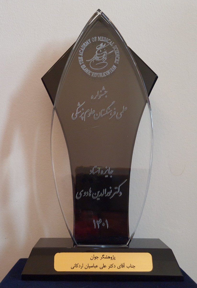
Top Scholar of The Iranian Academy of Medical Sciences
Our #AI model incorporating peritumoral regions in #ultrasound imaging outperformed radiologists in diagnosing thyroid nodule malignancy. A step towards more accurate, accessible, and efficient diagnostics. #Radiomics#ThyroidResearch#MedicalImaging
🔗 https://t.co/1sudsrM1Jk
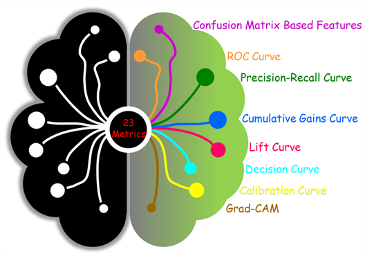
Attention physicians without #AI expertise! Our review article in #JUM provides a comprehensive guide for you to interpret the outputs of diagnostic #AI_models in healthcare! Shed light on real-world AI models with our guide! https://t.co/bJuV4HuJyT
@AIUMultrasound@nj_bureau
I'm delighted to share that our work, published in Journal of Ultrasound in Medicine on behalf of American Institute of Ultrasound in Medicine, is one of the top most-cited papers published 2022-2023. A special thanks to @nj_bureau and @MoMirzaAA for their help.
@AIUMultrasound
Our proposed #denoising model filtered out noises on #ultrasound images effectively. It can also preserve the #texture of tissues and can be used to enhance the diagnostic profile of #CAD systems and radiologists in malignancy diagnosis. See more on JCU: https://t.co/UIjst4Wl6W
#ChatGPT passed 2023's Iranian Medical Residency Entrance Exam in Persian, English, French, and Spanish! The type of input language did not significantly affect both perception and performance of ChatGPT. See our article on Informatics in Medicine Unlocked:https://t.co/j3enaIcR9v
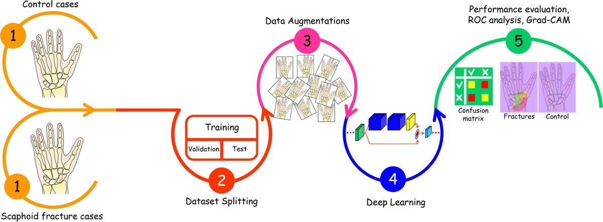
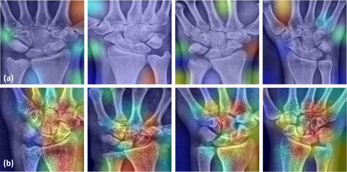
Our proposed #AI model detected #ScaphoidFractures with 90% accuracy! It can be a beneficial alternative to MRI for early fracture detection (especially occult) on radiographs. See more on Engineering Applications of Artificial Intelligence: https://t.co/H2ItIwElDb.
@comp_science
Motivated!
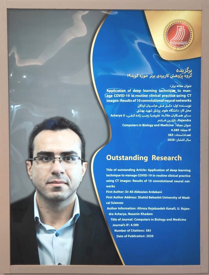
I am deeply honored and humbled to have been recognized as the "Top Young Researcher" by The Iranian Academy of Medical Sciences in 2022! Excited!
Please do not listen to the "rejection" and just go on! 😊
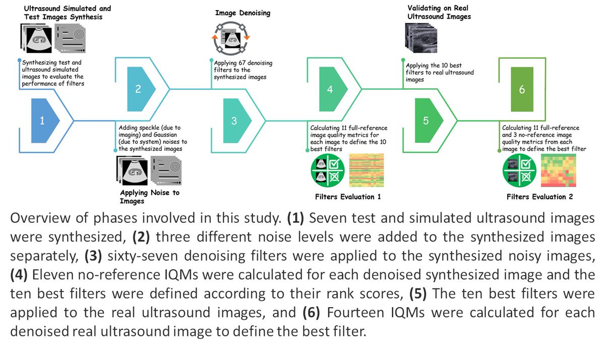
What is the best denoising filter in ultrasound images? Our new article sought to find the best one among 67 filters. See more on International Journal of Imaging Systems and Technology (IMA): https://t.co/RhdR84tnxG
@WileyEngineer
@WileyHealth
Looking for a #BreastUltrasound image #Database for your #AI studies? Find one in our new publication in Computers in Biology and Medicine: https://t.co/4u2GFKN2af
@comp_science
@ELS_Radiology
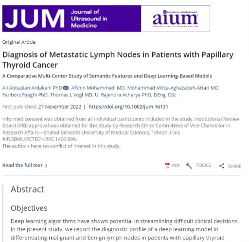
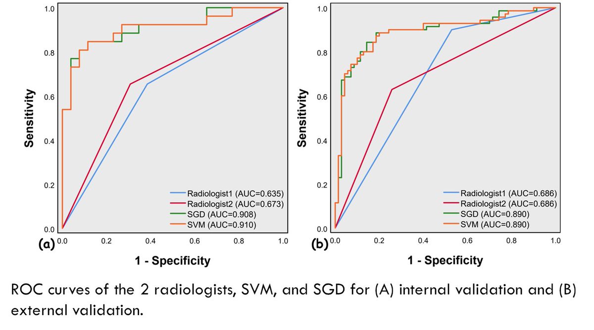
Our #DeepLearning based model was able to diagnose metastatic #LymphNodes with an AUC of 0.948 using all cohorts, which was superior to three #MachineLearning models with common imaging features used by radiologists.
See more on JUM: https://t.co/LMW3KRKMPT
@AIUMultrasound
Our new multi-center international study indicated that the proposed reproducible #AI model could significantly help radiologists diagnose #breastcancer using #ultrasonography based on ACR #BIRADS interpretations and morphometry. See our paper in #EJR via https://t.co/IudA7PhGz9
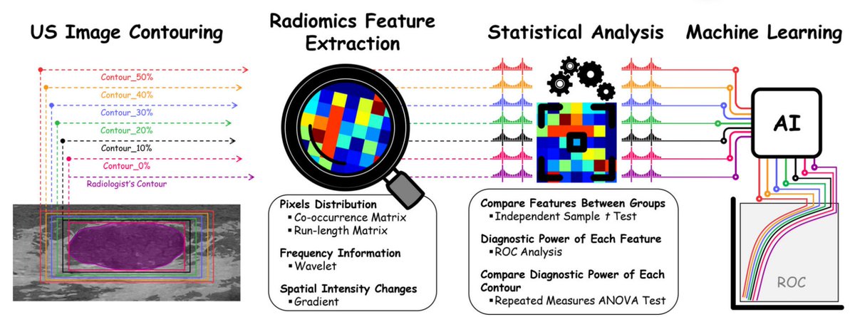
Excited to share our article that indicates tumor periphery should be considered for differentiating benign and malignant lesions in images quantification studies! 😊@AIUMultrasound
How vital are Tumor Periphery and TME in medical imaging diagnosis? Check out this article from the AIUM's Journal of Ultrasound in Medicine.
Click here to read more: https://t.co/0hJSNVCLDZ.
#JUM#Ultrasound#FOAMed
The question is "Should we consider tumor periphery in diagnostic procedures?"
The answer is YES. See our article for more details on Journal of Ultrasound in Medicine:
https://t.co/Vlyc2MRyTW
Thanks @nj_bureau for collaboration on this project!
@AIUMultrasound@WileyHealth
Excited to share our multi-center study on #BreastCancer diagnosis using #AI.
Analysis of 1259 lesions indicated that #Radiomics features diagnosed breast cancer with an AUC of 0.890 for all cohorts. See more on Biocybernetics and Biomedical Engineering:
https://t.co/jEq4iq0tOa
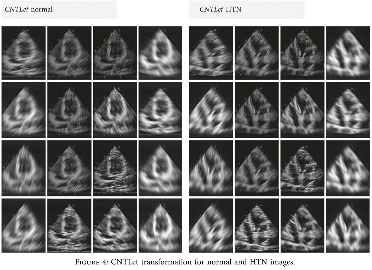
The proposed #multi_resolution#CAD system can be utilized for the detection of early #hypertension-induced left ventricular heart muscle changes using #ultrasound imaging. See more on Contrast Media & Molecular Imaging: https://t.co/rXt9nVqmDr
@HindawiMedicine
@HindawiMECS
The new ability of #Radiomics! It could diagnose cardiac abnormality in patients with #pacemaker using #Ultrasonography
See more on Computational and Mathematical Methods in Medicine:
https://t.co/lWTzWbVg9m
@HindawiMedicine
@HindawiMECS
#Radiomics is not an incomprehensive and vague topic! Radiomics features can be interpreted easily! See our pictorial review in Computer Methods and Programs in Biomedicine via https://t.co/3MIidF35Hy
Thanks @EdwardCiaccio
and @nj_bureau for collaboration.
@comp_science